Review
. 2020 Nov;1865(11):158617.
doi: 10.1016/j.bbalip.2020.158617. Epub 2020 Jan 10.

The macular carotenoids: A biochemical overview

Affiliations
Review

The macular carotenoids: A biochemical overview

Ranganathan Arunkumar et al. Biochim Biophys Acta Mol Cell Biol Lipids. 2020 Nov.

Abstract

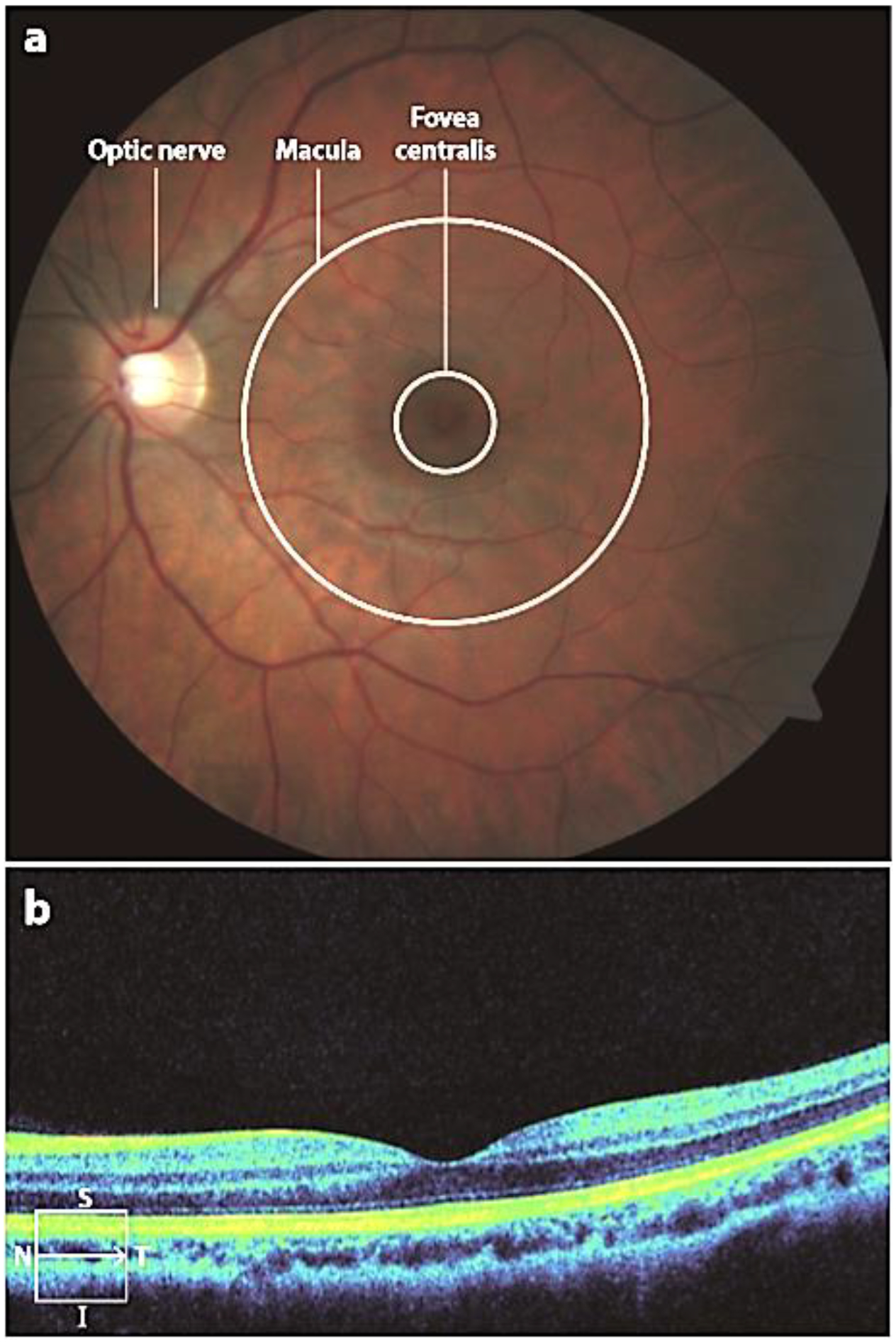
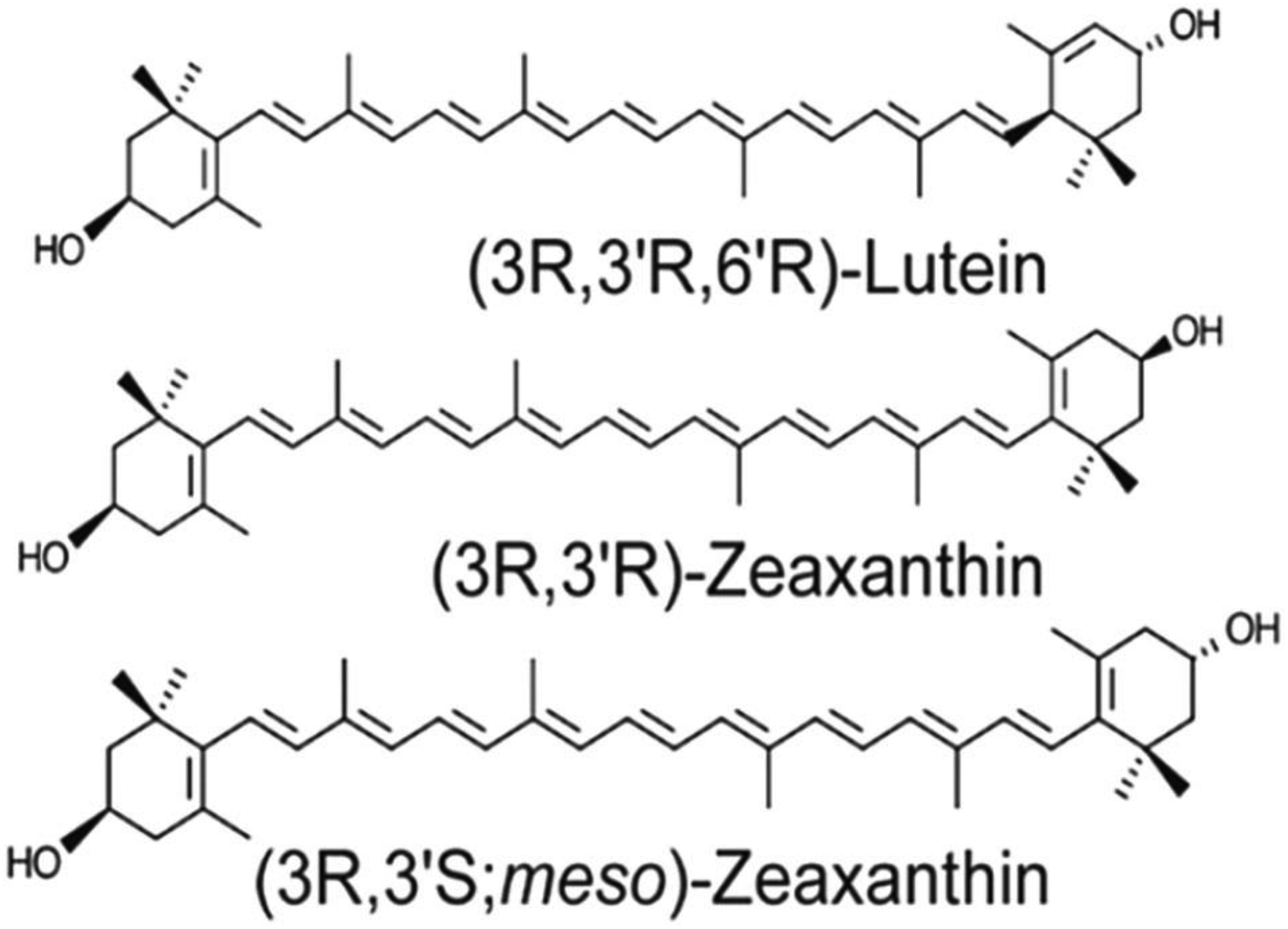
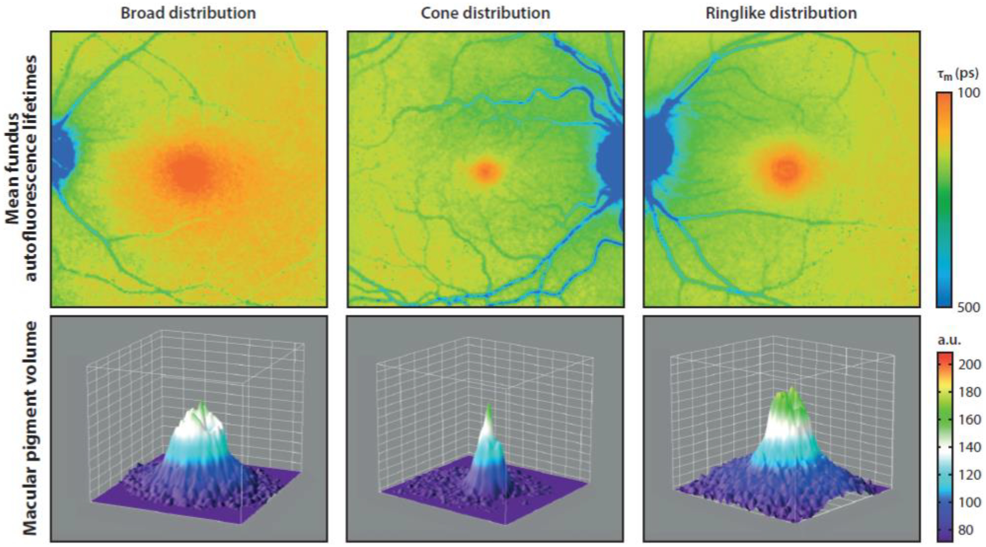
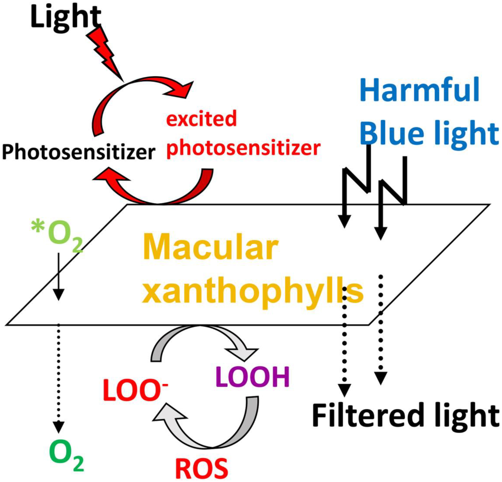
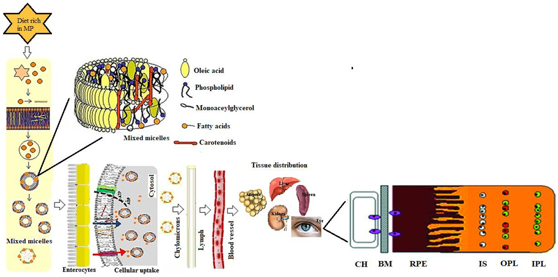
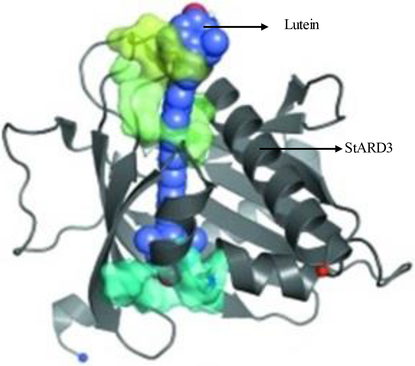
Among the more than 750 carotenoids identified in nature, only lutein, zeaxanthin, meso-zeaxanthin, and their oxidative metabolites are selectively accumulated in the macula lutea region of the human retina. These retinal carotenoids are collectively referred to as the macular pigment (MP) and are obtained only through dietary sources such as green leafy vegetables and yellow and orange fruits and vegetables. Lutein- and zeaxanthin-specific binding proteins (StARD3 and GSTP1, respectively) mediate the highly selective uptake of MP into the retina. Meso-zeaxanthin is rarely present in the diet, and its unique presence in the human eye results from metabolic conversion from dietary lutein by the RPE65 enzyme. The MP carotenoids filter high-intensity, short-wavelength visible light and are powerful antioxidants in a region vulnerable to light-induced oxidative stress. This review focuses on MP chemistry, absorption, metabolism, transport, and distribution with special emphasis on animal models used for MP study. This article is part of a Special Issue entitled Carotenoids recent advances in cell and molecular biology edited by Johannes von Lintig and Loredana Quadro.

Keywords: Age-related macular degeneration; Carotenoid; Lutein; Macular pigment; Nutrition; Zeaxanthin.


Conflict of interest statement

Declaration of competing interest The authors declare that they have no known competing financial interests or personal relationships that could have appeared to influence the work reported in this paper.

Figures

Figure 1.
Figure 1.
Anatomy of a healthy eye. Images from (a) fundus photography and (b) optical coherence tomography. The line in the lower left corner of panel b indicates the position of the scan within the macula. Abbreviations: I, inferior; N, nasal; S, superior; T, temporal.
Figure 2.
Figure 2.
Structures of the macular carotenoids: lutein, zeaxanthin, and meso-zeaxanthin.
Figure 3.
Figure 3.
Absorption spectra of lutein (red) and zeaxanthin (blue) in olive oil. A mixture of lutein plus zeaxanthin (dashed black line) closely approximates the absorption spectrum of the macular pigment in the living human eye.
Figure 4.
Figure 4.
Distribution of macular pigment in healthy individuals. Abbreviations: a.u., arbitrary units; τm, amplitude-weighted mean fluorescence lifetime. Fluorescent life time imaging (FLIO) was used to obtain pigment distribution data and the figure was published in Lydia et al. Investigative Ophthalmology & Visual Science (2018); 59: 3094–3103
Figure 5.
Figure 5.
Protective roles of lutein and zeaxanthin as absorbers of harmful blue light and as antioxidants reacting with reactive oxygen species (ROS). *O2, singlet oxygen; LOO, lipid peroxyl radicals; LOOH, lipid peroxides.
Figure 6.
Figure 6.
Possible pathways for MP carotenoid uptake, transport, and accumulation in the human retina. Choriocapillaris (CH); Bruch’s membrane (BM); Retinal pigment epithelium (RPE); Inner segments (IS); Outer plexiform layer (OPL); Inner plexiform layer (IPL).
Figure 7.
Figure 7.
The retinal distribution of macular pigment binding proteins. (a) GSTP1 labeling of foveal cones in the macula of a 3-year-old monkey. This montage shows strongest labeling by antibody against GSTP1 (red) over the myoid and ellipsoid regions of cones identified by monoclonal antibody (7G6, green). (b) A low-magnification view of a near-foveal retina section in which N-62 StAR (red) identifies StARD3, an anti-cone arrestin monoclonal antibody (7G6, green) identifies monkey cones. Images courtesy of Dr. Jeanne M. Frederick and the figure was published in Bernstein et al. 2016 Prog. Retin. Eye Res.; 50:34–36. The layers are labelled as Inner segments of photoreceptors (IS), Outer plexiform layer (OPL), Ganglion cell layer (GCL), Retinal pigment epithelium (RPE), Photoreceptor layer (PL), Outer nuclear layer (ONL), Outer plexiform layer (OPL), Inner nuclear layer (INL), Inner plexiform layer (IPL).
Figure 8.
Figure 8.
Overview of lutein molecule docked with StARD3 structure shows the best-scoring lutein candidate from the one portal set and the figure was published in Horvath et al. Acta Cryst. (2016); 72:609–618.

References

    1. Landrum JT, Bone RA, Lutein, zeaxanthin, and the macular pigment, Arch. Biochem. Biophys 385 (2001) 28–40. 10.1006/abbi.2000.2171. - DOI - PubMed
    1. Bone RA, Landrum JT, Hime GW, Cains A, Zamor J, Stereochemistry of the human macular carotenoids, Invest. Ophthalmol. Vis. Sci 34 (1993) 2033–2040. - PubMed
    1. Krinsky NI, Landrum JT, Bone RA, Biologic mechanisms of the protective role of lutein and zeaxanthin in the eye, Annu. Rev. Nutr 23 (2003) 171–201. 10.1146/annurev.nutr.23.011702.073307. - DOI - PubMed
    1. Sauer L, Li B, Bernstein PS, Ocular Carotenoid Status in Health and Disease, Annual Review of Nutrition. 39 (2019) 95–120. 10.1146/annurev-nutr-082018-124555. - DOI - PubMed
    1. Woodall AA, Britton G, Jackson MJ, Carotenoids and protection of phospholipids in solution or in liposomes against oxidation by peroxyl radicals: Relationship between carotenoid structure and protective ability, Biochimica et Biophysica Acta (BBA) - General Subjects. 1336 (1997) 575–586. 10.1016/S0304-4165(97)00007-X. - DOI - PubMed

Publication types

MeSH terms