. 2020 Jan 13;15(1):e0227642.
doi: 10.1371/journal.pone.0227642. eCollection 2020.

Post-translational modifications of Drosophila melanogaster HOX protein, Sex combs reduced

Affiliations

Post-translational modifications of Drosophila melanogaster HOX protein, Sex combs reduced

Anirban Banerjee et al. PLoS One. .

Abstract

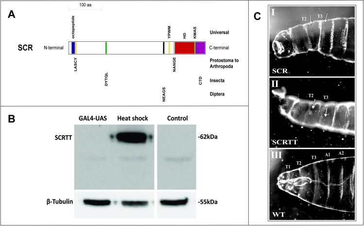
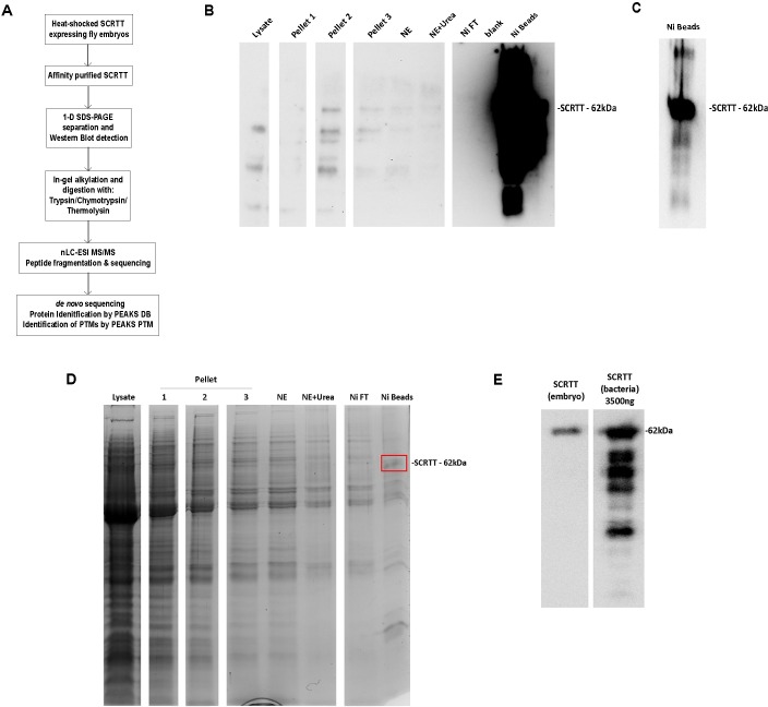
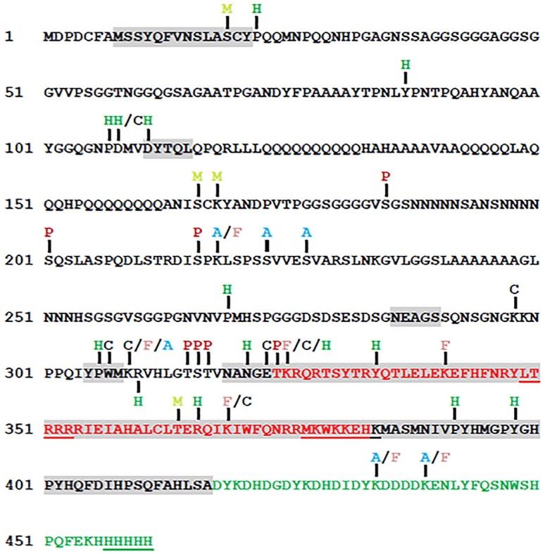
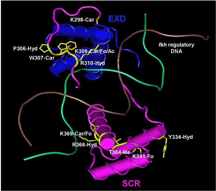
Homeotic selector (HOX) transcription factors (TFs) regulate gene expression that determines the identity of Drosophila segments along the anterior-posterior (A-P) axis. The current challenge with HOX proteins is understanding how they achieve their functional specificity while sharing a highly conserved homeodomain (HD) that recognize the same DNA binding sites. One mechanism proposed to regulate HOX activity is differential post-translational modification (PTM). As a first step in investigating this hypothesis, the sites of PTM on a Sex combs reduced protein fused to a triple tag (SCRTT) extracted from developing embryos were identified by Tandem Mass Spectrometry (MS/MS). The PTMs identified include phosphorylation at S185, S201, T315, S316, T317 and T324, acetylation at K218, S223, S227, K309, K434 and K439, formylation at K218, K309, K325, K341, K369, K434 and K439, methylation at S19, S166, K168 and T364, carboxylation at D108, K298, W307, K309, E323, K325 and K369, and hydroxylation at P22, Y87, P107, D108, D111, P269, P306, R310, N321, K325, Y334, R366, P392 and Y398. Of the 44 modifications, 18 map to functionally important regions of SCR. Besides a highly conserved DNA-binding HD, HOX proteins also have functionally important, evolutionarily conserved small motifs, which may be Short Linear Motifs (SLiMs). SLiMs are proposed to be preferential sites of phosphorylation. Although 6 of 7 phosphosites map to regions of predicted SLiMs, we find no support for the hypothesis that the individual S, T and Y residues of predicted SLiMs are phosphorylated more frequently than S, T and Y residues outside of predicted SLiMs.


Conflict of interest statement

The authors have declared that no competing interests exist.

Figures

Fig 1
Fig 1. Sex combs reduced functional domains/motifs, expression and phenotype.
(A) Schematic of SCR protein showing highly conserved, functional domains/motifs. The block diagram is drawn to scale and the domains/motifs are color-coded. The octapeptide motif is labeled in blue, LASCY motif in orange, DYTQL motif in dark green, NEAGS motif in black, YPWM motif in yellow, NANGE motif in grey, HD in dark red, KMAS motif in light green and CTD in purple. The taxonomic level of conservation of the domains/motifs is indicated on the right. (B) Comparison of the expression of SCRTT protein from heat-shock and UAS promoters. The method of expression is indicated above the lanes of the Western Blot of protein extracted from embryos. The position of SCRTT is indicated on the left, and the positions of molecular weight markers are indicated on the right. The level of β-tubulin expression is shown at the bottom. y w is the untransformed control. (C) First instar larval cuticular phenotypes of embryonic ectopic expression of SCR and SCRTT proteins. The anterior half of the larva is shown. The untagged SCR protein expressed with the GAL4-UAS system using a ubiquitous armadillo-GAL4 is shown in panel (I); whereas, the SCRTT protein expressed from a heat-shock promoter is shown in panel (II). T1, T2 and T3 refer to first, second and third thoracic segments. A1 and A2 refer to first and second abdominal segments. (I) and (II) Ectopic expression of SCR and SCRTT, respectively (T2 and T3 beards marked with arrows). (III) Control wild-type (WT) first instar larval cuticle.
Fig 2
Fig 2. Schematic of SCR showing total and conserved SLiMs.
The schematic is drawn to scale. The ordered region of the homeodomain is labeled by dark red and SLiMs are labeled blue. SLiMs that are conserved are red. The numbers beside SLiMs correspond to the SLiM data tables in the supplement (S4 and S5 Tables). The PTMs which map to SCR SLiMs are above the modified residue and color-coded. Phosphorylation sites are indicated by P in dark red, acetylation by A in blue, formylation by F in orange, methylation by M in light green, carboxylation by C in grey and hydroxylation by H in green. SLiMs underlined are candidate phosphorylation sites.
Fig 3
Fig 3. Overall approach for PTM mapping of SCRTT.
(A) Analytical workflow for affinity purification, digestion and sequence mapping of PTMs in SCRTT expressed from developing embryos. (B) Overexposed Western Blot showing SCRTT at 62 kDa in the Ni beads fraction. Purification fraction is indicated at top of the lane. (C) Autoexposed Western Blot showing SCRTT at 62 kDa in the Ni beads fraction. (D) Coomasie-stained 1-D SDS-polyacrylamide gel of affinity purified SCRTT. Purification fraction is indicated at top of the lane. A band of SCRTT at 62 kDa (marked with a red box) was observed in the Ni beads fraction. (E) Comparison of Drosophila SCRTT vs. bacterial SCRTT to estimate the amount of protein to be analyzed by MS/MS. The Western Blot shows signals for SCRTT at 62 kDa indicated on the right. The source of SCRTT is at the top of the lane. NE Nuclear Extract, Ni Nickel and Ni FT Nickel Flow-Through. For each of the panels (B), (D) and (E), all lanes shown are from the same gel and the white space between lanes indicate the splicing out of irrelevant lanes.
Fig 4
Fig 4. A summary map of post-translational modifications in SCRTT identified by MS/MS.
Phosphorylation sites are indicated by P in dark red, acetylation by A in light blue, formylation by F in pink, methylation by M in yellowish green, carboxylation by C in black and hydroxylation by H in green. The homeodomain is highlighted in red and the triple tag is highlighted in green. The functional regions of SCR are shaded in grey (Fig 1A). The amino acids underlined were not detected by MS/MS. Phosphorylation at S216 was identified by a bulk proteomic analysis [80].
Fig 5
Fig 5. The structure of SCR-EXD-DNA complex determined by crystallography.
SCR is shown in pink and EXD in blue. The two strands of fkh regulatory DNA is shown in brown and green. The modified amino acids of SCR along with their side chains are shown in yellow. Ac–acetylation, Car–carboxylation, Fo–formylation, Hyd–hydroxylation and Me–methylation. The structure coordinates with accession code 2R5Z (fkh250) were retrieved from RCSB Protein Data Bank [16]. Cn3D 4.3.1 (NCBI) was used to annotate the 3-D structure.

Similar articles

Cited by

References

    1. Akam M. Hox genes, homeosis and the evolution of segmental identity: no need for hopeless monsters. Int J Dev Biol. 1998;42: 445–451. 10.1387/ijdb.9654030 - DOI - PubMed
    1. Lewis EB. A gene complex controlling segmentation in Drosophila. Nature. 1978;276: 565–570. 10.1038/276565a0 - DOI - PubMed
    1. Gehring WJ, Qian YQ, Billeter M, Furukubo-Tokunaga K, Schier AF, Resendez-Perez D, et al. Homeodomain-DNA recognition. Cell. 1994;78: 211–223. 10.1016/0092-8674(94)90292-5 - DOI - PubMed
    1. Primon M, Hunter KD, Pandha HS, Morgan R. Kinase Regulation of HOX Transcription Factors. Cancers. 2019;11: E508 10.3390/cancers11040508 - DOI - PMC - PubMed
    1. Draime A, Bridoux L, Graba Y, Rezsohazy R. Post-translational modifications of HOX proteins, an underestimated issue. Int J Dev Biol. 2018;62: 733–744. 10.1387/ijdb.180178rr - DOI - PubMed

Publication types

MeSH terms

LinkOut - more resources