. 2020 Feb 25;141(8):667-677.
doi: 10.1161/CIRCULATIONAHA.119.044582. Epub 2020 Jan 14.

Cytokine mRNA Degradation in Cardiomyocytes Restrains Sterile Inflammation in Pressure-Overloaded Hearts

Affiliations

Cytokine mRNA Degradation in Cardiomyocytes Restrains Sterile Inflammation in Pressure-Overloaded Hearts

Shigemiki Omiya et al. Circulation. .

Erratum in

Abstract

Background: Proinflammatory cytokines play an important role in the pathogenesis of heart failure. The mechanisms responsible for maintaining sterile inflammation within failing hearts remain poorly defined. Although transcriptional control is important for proinflammatory cytokine gene expression, the stability of mRNA also contributes to the kinetics of immune responses. Regnase-1 is an RNase involved in the degradation of a set of proinflammatory cytokine mRNAs in immune cells. The role of Regnase-1 in nonimmune cells such as cardiomyocytes remains to be elucidated.

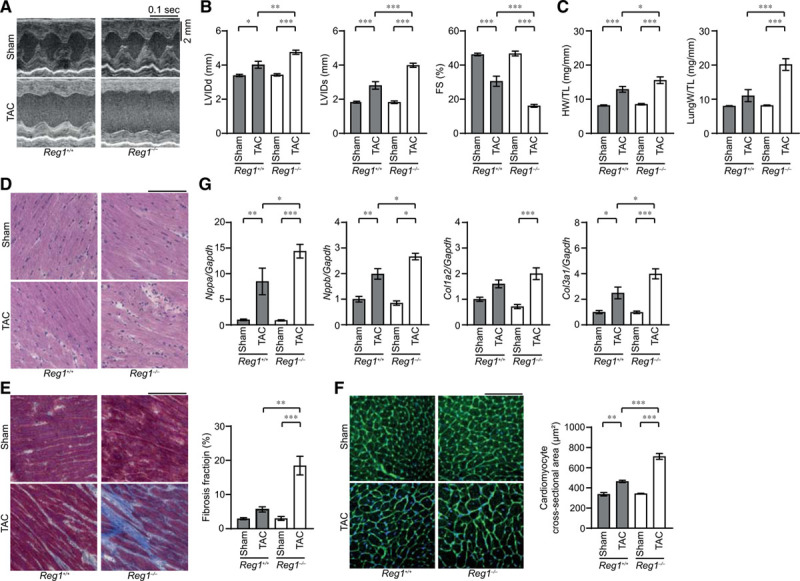
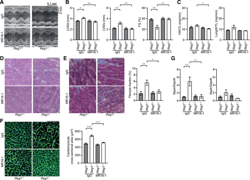
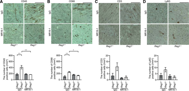
Methods: To examine the role of proinflammatory cytokine degradation by Regnase-1 in cardiomyocytes, cardiomyocyte-specific Regnase-1-deficient mice were generated. The mice were subjected to pressure overload by means of transverse aortic constriction to induce heart failure. Cardiac remodeling was assessed by echocardiography as well as histological and molecular analyses 4 weeks after operation. Inflammatory cell infiltration was examined by immunostaining. Interleukin-6 signaling was inhibited by administration with its receptor antibody. Overexpression of Regnase-1 in the heart was performed by adeno-associated viral vector-mediated gene transfer.

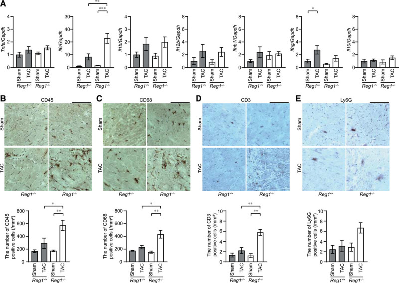
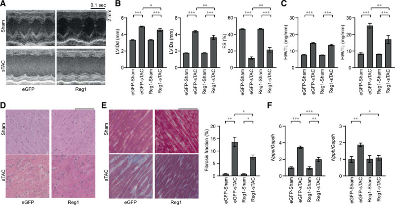
Results: Cardiomyocyte-specific Regnase-1-deficient mice showed no cardiac phenotypes under baseline conditions, but exhibited severe inflammation and dilated cardiomyopathy after 4 weeks of pressure overload compared with control littermates. Four weeks after transverse aortic constriction, the Il6 mRNA level was upregulated, but not other cytokine mRNAs, including tumor necrosis factor-α, in Regnase-1-deficient hearts. Although the Il6 mRNA level increased 1 week after operation in both Regnase-1-deficient and control hearts, it showed no increase in control hearts 4 weeks after operation. Administration of anti-interleukin-6 receptor antibody attenuated the development of inflammation and cardiomyopathy in cardiomyocyte-specific Regnase-1-deficient mice. In severe pressure overloaded wild-type mouse hearts, sustained induction of Il6 mRNA was observed, even though the protein level of Regnase-1 increased. Adeno-associated virus 9-mediated cardiomyocyte-targeted gene delivery of Regnase-1 or administration of anti-interleukin-6 receptor antibody attenuated the development of cardiomyopathy induced by severe pressure overload in wild-type mice.

Conclusions: The degradation of cytokine mRNA by Regnase-1 in cardiomyocytes plays an important role in restraining sterile inflammation in failing hearts and the Regnase-1-mediated pathway might be a therapeutic target to treat patients with heart failure.

Keywords: RNA stability; heart failure; inflammation; interleukin-6.


Figures

Figure 1.
Figure 1.
Pressure overload–induced cardiomyopathy in Reg1-/- mice. The Reg1+/+ and Reg1−/− mice were subjected to pressure overload by means of transverse aortic constriction (TAC). The mice were analyzed 4 weeks after TAC. Data were evaluated by 1-way analysis of variance with the Bonferroni post hoc test. Data are mean ± SEM. *P<0.05, **P<0.01, ***P<0.001. A, M-mode echocardiographic tracings from sham- or TAC-operated Reg1+/+ or Reg1−/− mice. B and C, Echocardiographic (B) and physiologic (C) parameters. Total n=7 (sham–Reg1+/+), 7 (TAC–Reg1+/+), 7 (sham–Reg1−/−), or 6 (TAC–Reg1−/−) per group. D through F, Hematoxylin-eosin–stained (D), Masson trichrome–stained (E), and wheat germ agglutinin–stained (F) heart sections. Scale bar, 100 µm. Fibrosis fraction was measured (n=3). Cardiomyocyte cross-sectional area was measured by tracing the outline of 70 myocytes in the nonfibrotic area in each section (n=3). G, mRNA expression of Nppa, Nppb, Col1a2, and Col3a1. Total n=5 (sham–Reg1+/+), 4 (TAC–Reg1+/+), 5 (sham–Reg1−/−), or 5 (TAC–Reg1−/−) per group. Gapdh mRNA was used as the loading control. The averaged value in sham-operated Reg1+/+ hearts was set equal to 1. FS indicates fractional shortening; HW/TL, heart weight/tibia length; LungW/TL, lung weight/TL; LVIDd, end-diastolic left ventricular internal dimension; and LVIDs, end-systolic left ventricular internal dimension.
Figure 2.
Figure 2.
Inflammatory responses in pressure-overloaded Reg1-/- hearts. The Reg1+/+ and Reg1−/− mice subjected to transverse aortic constriction (TAC) were analyzed 4 weeks after TAC. Data were evaluated by 1-way analysis of variance with the Bonferroni post hoc test. Data are mean ± SEM. *P<0.05, **P<0.01, ***P<0.001. A, Inflammatory cytokine mRNAs including Tnfa, Il6, Il1b, Il12b, Ifnb1, Ifng, and Il10. Total n=5 (sham–Reg1+/+), 4 (TAC–Reg1+/+), 5 (sham–Reg1−/−), or 5 (TAC–Reg1−/−) per group. Gapdh mRNA was used as the loading control. The averaged value in sham-operated Reg1+/+ mice was set equal to 1. B through E, Immunohistochemical analysis for CD45 (B), CD68 (C), CD3 (D), and Ly6G (E). Scale bar, 100 µm. Bottom graphs show quantitative analysis of each infiltrating inflammatory cell type (n=3).
Figure 3.
Figure 3.
Production of I16 mRNA in Reg1-/- hearts under pressure overload. Reg1+/+ and Reg1−/− hearts 4 weeks (A through D) or 1 week (E) after transverse aortic constriction (TAC) were analyzed. Data were evaluated by the Student t test (C and D) or 1-way analysis of variance with the Bonferroni post hoc test (E). Data are mean ± SEM. *P<0.05, ***P<0.001. A, Double staining of TAC-operated Reg1−/− heart sections with anti-CD68 (red) and anti-CD11c (green) antibodies. B, Double staining of TAC-operated Reg1−/− heart sections with anti-CD68 (red) and anti-CD206 (green) antibodies. Scale bar, 100 µm. C, The ratio of CD11c-positive or CD206-positive to CD68-postive cell numbers (n=3). D, In situ hybridization for Il6 mRNA (red) in Reg1+/+ or Reg1−/− hearts, followed by immunostaining with α-sarcomeric action antibody (green). Scale bar, 100 µm. Right graph shows the number of red dots in cardiomyocytes. E, Tnfa and Il6 mRNA levels 1 week after TAC. Total n=5 (sham–Reg1+/+), 5 (TAC–Reg1+/+), 5 (sham–Reg1−/−), or 6 (TAC–Reg1−/−) per group. Gapdh mRNA was used as the loading control. The averaged value in sham-operated Reg1+/+ mice was set equal to 1.
Figure 4.
Figure 4.
Interleukin-6 blockade ameliorated transverse aortic constriction–induced cardiomyopathy in Reg1-/- mice. After transverse aortic constriction (TAC) operation, Reg1+/+ and Reg1−/− mice received an intraperitoneal injection of anti-mouse interleukin-6 receptor antibody MR16-1 or control immunoglobulin G (IgG). Afterwards, they were injected intraperitoneally once a week with a total of 3 injections with either MR16-1 or IgG. The mice were analyzed 4 weeks after TAC. Data were evaluated by 1-way analysis of variance with the Bonferroni post hoc test. Data are mean ± SEM. *P<0.05, **P<0.01, ***P<0.001. A, M-mode echocardiographic tracings from IgG-treated or MR16-1-treated Reg1+/+ or Reg1−/− mice. B and C, Echocardiographic (B) and physiologic (C) parameters. Total n=7 (IgG–Reg1+/+), 8 (IgG–Reg1−/−), 6 (MR16-1–Reg1+/+), or 8 (MR16-1–Reg1−/−) per group. D through F, Hematoxylin-eosin–stained (D), Masson trichrome–stained (E), and wheat germ agglutinin–stained (F) heart sections. Scale bar, 100 µm. Fibrosis fraction (n=5) and cross-sectional area of cardiomyocytes (n=3) were measured. G, mRNA expression of Nppa and Nppb. Total n=6 (IgG–Reg1+/+), 5 (IgG–Reg1−/−), 5 (MR16-1–Reg1+/+), or 5 (MR16-1–Reg1−/−) per group. Gapdh mRNA was used as the loading control. The averaged value in TAC-operated Reg1+/+ hearts treated with IgG was set equal to 1. FS indicates fractional shortening; HW/TL, heart weight/tibia length; LungW/TL, lung weight/TL; LVIDd, end-diastolic left ventricular internal dimension; and LVIDs, end-systolic left ventricular internal dimension.
Figure 5.
Figure 5.
Interleukin-6 blockade inhibited infiltration of inflammatory cells. Transverse aortic constriction (TAC)–operated Reg1+/+ and Reg1−/− hearts treated with anti-mouse interleukin-6 receptor antibody MR16-1 or control immunoglobulin G (IgG) were analyzed. Data were evaluated by 1-way analysis of variance with the Bonferroni post hoc test. Data are mean ± SEM. *P<0.05, **P<0.01. A through D, Immunohistochemical analysis for CD45 (A), CD68 (B), CD3 (C), and Ly6G (D). Scale bar, 100 µm. Bottom graphs show quantitative analysis of each infiltrating inflammatory cell type (n=3).
Figure 6.
Figure 6.
Overexpression of Regnase-1 protein attenuated severe transverse aortic constriction–induced heart failure. Wild-type C57BL/6 mice were intraperitoneally injected with adeno-associated virus expressing enhanced green fluorescent protein (eGFP-AAV9) or Regnase-1 (Reg1-AAV9) and were subjected to severe transverse aortic constriction (sTAC) 1 week after infection. Sham- or sTAC-operated wild-type mice infected with eGFP-AAV9 (eGFP–sham or eGFP–sTAC) or Reg1-AAV9 (Reg1–sham or Reg1–sTAC) were analyzed 4 weeks after operation. Data were evaluated by 1-way analysis of variance with the Bonferroni post hoc test. Data are mean ± SEM. *P<0.05, **P<0.01, ***P<0.001. A, M-mode echocardiographic tracings from eGFP–sham, eGFP–sTAC, Reg1–sham, or Reg1–sTAC wild-type mice. B and C, Echocardiographic (B) and physiologic (C) parameters. Total n=16 (eGFP–sham), 11 (eGFP–sTAC), 16 (Reg1–sham), or 15 (Reg1–sTAC). D and E, Hematoxylin-eosin–stained (D) and Masson trichrome–stained (E) heart sections. Scale bar, 100 µm. Fibrosis fraction was evaluated. Total n=3 (eGFP–sham or Reg1–sham) or 4 (eGFP–sTAC or Reg1–sTAC). F, mRNA expressions of Nppa and Nppb. Total n=7 (eGFP–sham), 6 (eGFP–sTAC), 7 (Reg1–sham), or 7 (Reg1–sTAC). Gapdh mRNA was used as the loading control. The averaged value in the eGFP–sham group was set equal to 1. FS indicates fractional shortening; HW/TL, heart weight/tibia length; LungW/TL, lung weight/TL; LVIDd, end-diastolic left ventricular internal dimension; and LVIDs, end-systolic left ventricular internal dimension.
Figure 7.
Figure 7.
Induction of Regnase-1 protein suppressed the extent of inflammatory responses in severe transverse aortic constriction–induced heart failure. Severe transverse aortic constriction (sTAC)–operated wild-type C57BL/6 mice infected with adeno-associated virus expressing enhanced green fluorescent protein(eGFP–sTAC) or Regnase-1 (Reg1–sTAC) were analyzed. Data were evaluated by 1-way analysis of variance with the Bonferroni post hoc test. Data are mean ± SEM. *P<0.05, ***P<0.001. A and B, Immunohistochemical analysis for CD45 (A) and CD68 (B). Scale bar, 100 µm. Bottom graphs show quantitative analysis of each infiltrating inflammatory cell type in eGFP–sham, eGFP–sTAC, Reg1–sham, or Reg1–sTAC hearts (n=3). C, Il6 mRNA expressions. Total n=7 (eGFP–sham), 6 (eGFP–sTAC), 7 (Reg1–sham), or 7 (Reg1–sTAC). Gapdh mRNA was used as the loading control. The averaged value in the eGFP–sham group was set equal to 1.

References

    1. Mann DL. Innate immunity and the failing heart: the cytokine hypothesis revisited. Circ Res. 2015;116:1254–1268. doi: 10.1161/CIRCRESAHA.116.302317. - PMC - PubMed
    1. Mann DL, McMurray JJ, Packer M, Swedberg K, Borer JS, Colucci WS, Djian J, Drexler H, Feldman A, Kober L, et al. Targeted anticytokine therapy in patients with chronic heart failure: results of the Randomized Etanercept Worldwide Evaluation (RENEWAL). Circulation. 2004;109:1594–1602. doi: 10.1161/01.CIR.0000124490.27666.B2. - PubMed
    1. Oka T, Hikoso S, Yamaguchi O, Taneike M, Takeda T, Tamai T, Oyabu J, Murakawa T, Nakayama H, Nishida K, et al. Mitochondrial DNA that escapes from autophagy causes inflammation and heart failure. Nature. 2012;485:251–255. doi: 10.1038/nature10992. - PMC - PubMed
    1. Hao S, Baltimore D. The stability of mRNA influences the temporal order of the induction of genes encoding inflammatory molecules. Nat Immunol. 2009;10:281–288. doi: 10.1038/ni.1699. - PMC - PubMed
    1. Matsushita K, Takeuchi O, Standley DM, Kumagai Y, Kawagoe T, Miyake T, Satoh T, Kato H, Tsujimura T, Nakamura H, et al. Zc3h12a is an RNase essential for controlling immune responses by regulating mRNA decay. Nature. 2009;458:1185–1190. doi: 10.1038/nature07924. - PubMed

Publication types

MeSH terms