. 2020 Jan 13;18(1):22.
doi: 10.1186/s12967-019-02202-0.

Translational research of temporomandibular joint pathology: a preliminary biomarker and fMRI study

Affiliations

Translational research of temporomandibular joint pathology: a preliminary biomarker and fMRI study

Andre Barkhordarian et al. J Transl Med. .

Abstract

Background: The temporomandibular joint (TMJ) is well innervated by braches of the trigeminal nerve. The temporomandibular joint disorders (TMD) can cause neural-inflammation in the peripheral nervous system (PNS) at the site of injury, or compression, and may have systemic effects on the central nervous system (CNS). Neural-inflammation causes elevations in cytokine expression and microglia activation. When the site of injury, or compression is treated, or relieved, neural inflammation is reduced. These changes can be seen and measured with fMRI brain activities.

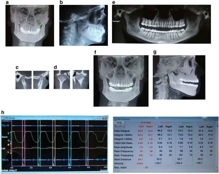
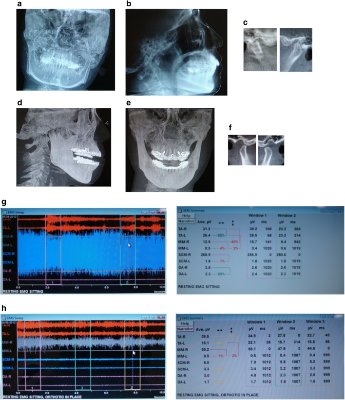
Methods: For this study, patients with comorbid TMD and systemic/neurologic conditions were compared using clinical diagnostic markers, inflammatory, pain, tissue destruction enzymatic biomarkers, and functional magnetic resonance imaging (fMRI) activity of the brain, with and without a custom-made dental orthotic.

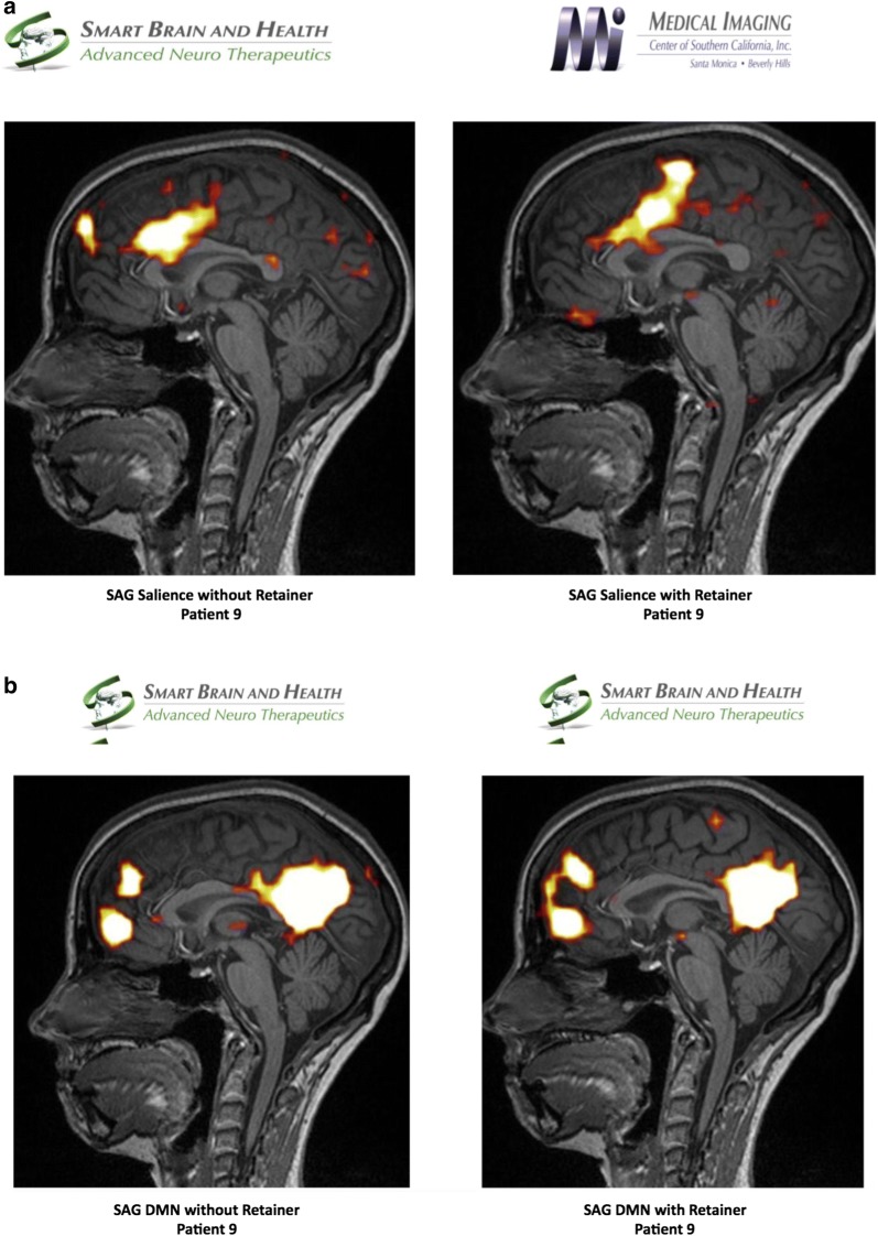
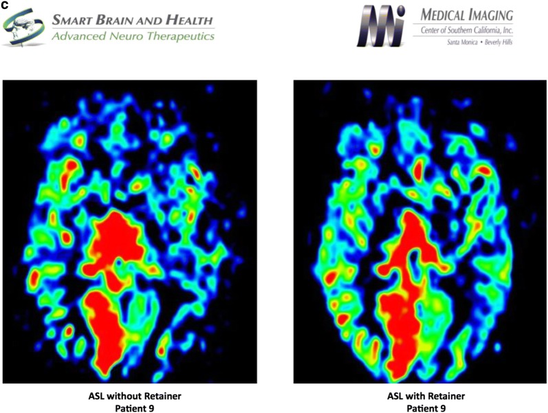
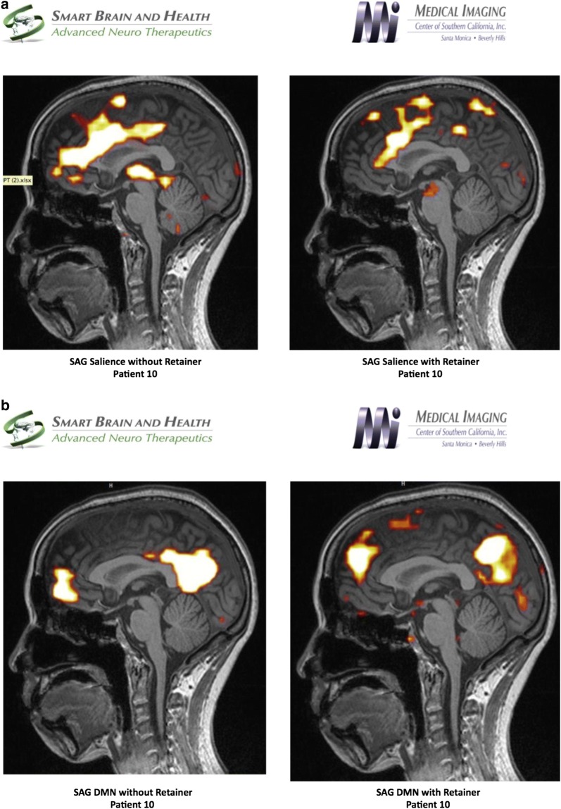
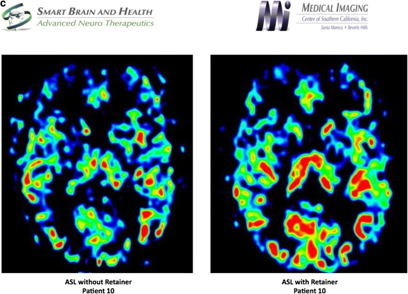
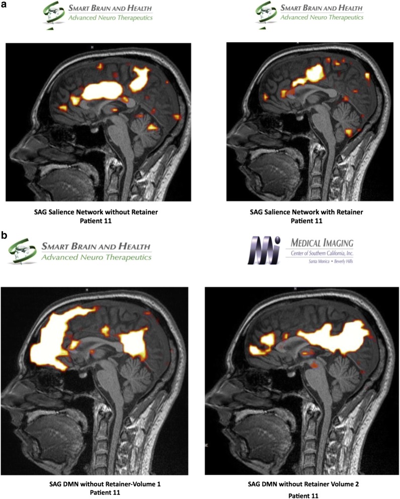
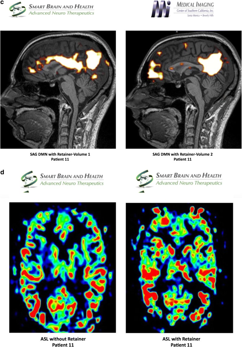
Results: Our results showed a correlation between the clinical diagnosis of the pathological TMJ, biomarkers and the fMRI study. There was a marked elevation of biomarkers in samples taken from TMJ of patients who were clinically diagnosed with TMD. The fMRI study of TMD patients showed an abnormal hyper-connected salience network and a diminished blood flow to the anterior frontal lobes when they did not wear their customized dental orthotics.

Conclusions: Our findings highlight the importance of TMJ-CNS connections and use of fMRI as an investigative tool for understanding TMD and its related neurological pathologies.

Keywords: Arterial spin labeling (ASL); Blood oxygenation level dependent (BOLD); Default mode network (DMN); Dental orthotic; Fractional anisotropy (FA); Functional magnetic resonance imaging (fMRI); Microglia cells; Temporomandibular joint (TMJ); Temporomandibular joint disorders (TMD); Trigeminal ganglion (TG).


Conflict of interest statement

The authors declare that they have no competing interests.

Figures

Fig. 1
Fig. 1
a Pre-treatment anterior/posterior cephalometric, b pre-treatment lateral cephalometric, c sagittal view right and left TMJ, d anteroposterior view of right and left TMJ, notice the Bifid shape of the right TMJ, e panographic film shows the joint position with the splint in the mouth, f frontal view with the splint in the mouth, g lateral view with the dental orthotic in the mouth, h joint vibration analysis indicating left TMJ disc displacement with reduction
Fig. 2
Fig. 2
a Salient network analysis of patient 9 without and with dental orthotic, b default Mode Network analysis of patient 9 without and with dental orthotic, c arterial spin labeling analysis of patient 9 without and with dental orthotic
Fig. 2
Fig. 2
a Salient network analysis of patient 9 without and with dental orthotic, b default Mode Network analysis of patient 9 without and with dental orthotic, c arterial spin labeling analysis of patient 9 without and with dental orthotic
Fig. 3
Fig. 3
a Frontal cephalometric before treatment, demonstrating the head position. b lateral cephalometric before treatment, c right and Left tomography before treatment, d lateral cephalometric demonstrating dental orthotic in the mouth, e frontal View, notice the head is more leveled with the dental orthotic, f Tomography of the Right and Left TMJ showing condylar position with the dental orthotic in the mouth. g EMG pre-treatment, notice the high activity of the right SCM at 286 micro-volts. h EMG with splint in, notice the decrease in activity of the SCM muscles. The temporalis and right masseter are slightly elevated and the patient is gently clenching on the dental orthotic
Fig. 4
Fig. 4
a Salient network analysis of patient 10 without and with dental orthotic, b default mode network analysis of patient 10 without and with dental orthotic, c arterial Spin Labeling analysis of patient 10 without and with dental orthotic
Fig. 4
Fig. 4
a Salient network analysis of patient 10 without and with dental orthotic, b default mode network analysis of patient 10 without and with dental orthotic, c arterial Spin Labeling analysis of patient 10 without and with dental orthotic
Fig. 5
Fig. 5
a Panoramic, b AP cephalometric, c lateral cephalometric, d right and left TMJ tomography
Fig. 6
Fig. 6
a Salient network analysis of patient 11 without and with dental orthotic, b default mode network analysis of patient 11 without dental orthotic volume 1 and 2, c default mode network analysis of patient 11 with dental orthotic volume 1 and 2, d arterial spin labeling analysis of patient 11 without and with dental orthotic
Fig. 6
Fig. 6
a Salient network analysis of patient 11 without and with dental orthotic, b default mode network analysis of patient 11 without dental orthotic volume 1 and 2, c default mode network analysis of patient 11 with dental orthotic volume 1 and 2, d arterial spin labeling analysis of patient 11 without and with dental orthotic

References

    1. Alomar X, Medrano J, Cabratosa J, Clavero JA, Lorente M, Serra I, Monill JM, Salvador A. Anatomy of the temporomandibular joint. Semin Ultrasound CT MR. 2007;28:170–183. doi: 10.1053/j.sult.2007.02.002. - DOI - PubMed
    1. Gülekon N, Anil A, Poyraz A, Peker T, Basri Turgut H, Karakose M. Variation in the anatomy of the auriculotemporal nerve. Clin Anatom. 2005;18:15–22. doi: 10.1002/ca.20068. - DOI - PubMed
    1. De Kanter RJ, Truin GJ, Burgersdijk RC, van’t Hof MA, Battis-tuzzi PG, Kalsbeek H, et al. Prevalence in the Dutch adult population and a meta-analysis of signs and symptoms of temporomandibular disorder. J Dent Res. 1993;72:1509–1518. doi: 10.1177/00220345930720110901. - DOI - PubMed
    1. De Leeuw R, Klasser GD, Albuquerque RJ. Are female patients with orofacial pain medically compromised? JADA. 2005;136(4):459–468. - PubMed
    1. Korszun A, Papadopoulos E, Demitrack M, Engleberg C, Crofford L. The relationship between temporomandibular disorders and stress-associated syndromes. Oral Surg Oral Med Oral Pathol Oral Radiol Endod. 1998;86(4):416–420. doi: 10.1016/S1079-2104(98)90366-3. - DOI - PubMed

Publication types