. 2020 Jan 13;20(1):6.
doi: 10.1186/s40644-020-0286-5.

Quantification of tumor burden in multiple myeloma by atlas-based semi-automatic segmentation of WB-DWI

Affiliations

Quantification of tumor burden in multiple myeloma by atlas-based semi-automatic segmentation of WB-DWI

Sílvia D Almeida et al. Cancer Imaging. .

Abstract

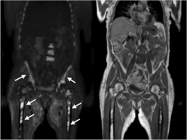
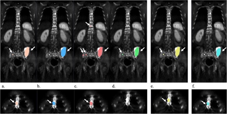
Background: Whole-body diffusion weighted imaging (WB-DWI) has proven value to detect multiple myeloma (MM) lesions. However, the large volume of imaging data and the presence of numerous lesions makes the reading process challenging. The aim of the current study was to develop a semi-automatic lesion segmentation algorithm for WB-DWI images in MM patients and to evaluate this smart-algorithm (SA) performance by comparing it to the manual segmentations performed by radiologists.

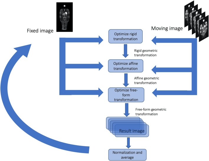
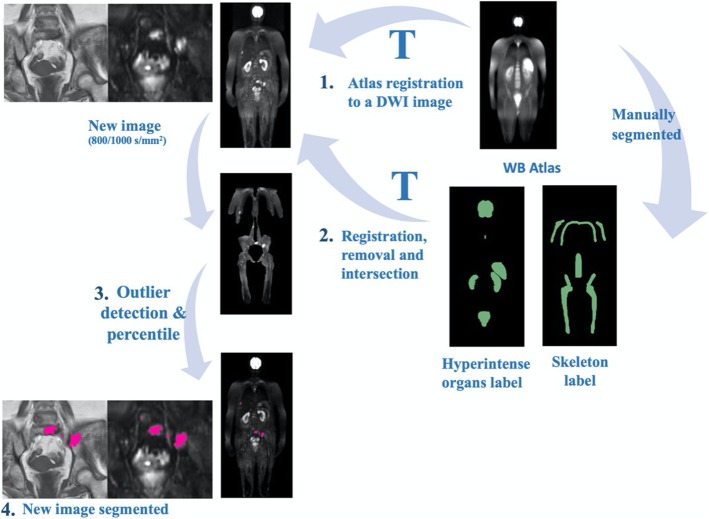
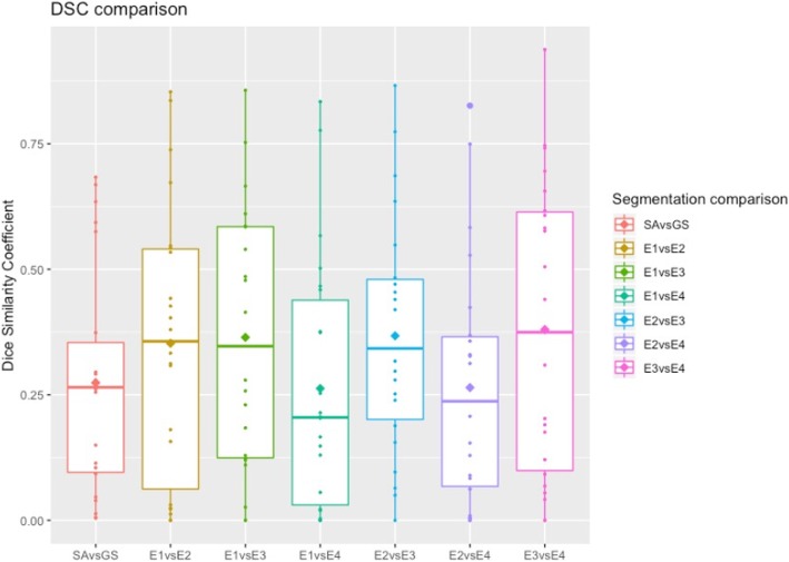
Methods: An atlas-based segmentation was developed to remove the high-signal intensity normal tissues on WB-DWI and to restrict the lesion area to the skeleton. Then, an outlier threshold-based segmentation was applied to WB-DWI images, and the segmented area's signal intensity was compared to the average signal intensity of a low-fat muscle on T1-weighted images. This method was validated in 22 whole-body DWI images of patients diagnosed with MM. Dice similarity coefficient (DSC), sensitivity and positive predictive value (PPV) were computed to evaluate the SA performance against the gold standard (GS) and to compare with the radiologists. A non-parametric Wilcoxon test was also performed. Apparent diffusion coefficient (ADC) histogram metrics and lesion volume were extracted for the GS segmentation and for the correctly identified lesions by SA and their correlation was assessed.

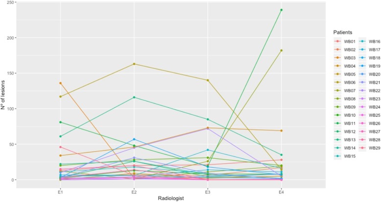
Results: The mean inter-radiologists DSC was 0.323 ± 0.268. The SA vs GS achieved a DSC of 0.274 ± 0.227, sensitivity of 0.764 ± 0.276 and PPV 0.217 ± 0.207. Its distribution was not significantly different from the mean DSC of inter-radiologist segmentation (p = 0.108, Wilcoxon test). ADC and lesion volume intraclass correlation coefficient (ICC) of the GS and of the correctly identified lesions by the SA was 0.996 for the median and 0.894 for the lesion volume (p < 0.001). The duration of the lesion volume segmentation by the SA was, on average, 10.22 ± 0.86 min, per patient.

Conclusions: The SA provides equally reproducible segmentation results when compared to the manual segmentation of radiologists. Thus, the proposed method offers robust and efficient segmentation of MM lesions on WB-DWI. This method may aid accurate assessment of tumor burden and therefore provide insights to treatment response assessment.

Keywords: Atlas-based segmentation; Diffusion weighted imaging; Multiple myeloma; Semi-automatic segmentation; Total lesion burden.


Conflict of interest statement

The authors declare that they have no competing interests.

Figures

Fig. 1
Fig. 1
Representative coronal slice of a WB-T1w (left) and WB-DWI (right) of the same MM patient. MM focal lesions visible on left femoral head, right iliac wing and lumbar vertebral bodies, show hyperintense signal on b800/1000 image (right) and hypointense signal on T1w image (left)
Fig. 2
Fig. 2
Atlas building scheme. Initially, an image is selected as the reference (fixed) and the others are moving images. Each moving image is registered to the fixed image, by applying an optimal rigid transformation, followed by an optimal affine and free-form transformations (B-spline), which maximizes the similarity between the fixed and moving images. Then, the registered moving images are averaged, resulting in a mean image. Then, this mean image is selected as the new fixed image and all the original moving images are again registered to it, following the same process. This was repeated until no meaningful changes were found between successive mean images
Fig. 3
Fig. 3
Semi-automatic lesion detection in DWI scheme. (1) The atlas is registered to a DWI image; (2) The transformation found is used to register the organs and skeleton atlas to the DWI image. Then the lesion search area is obtained by removing the organs and selecting the skeleton and nearby areas; (3) Afterwards, an automatic threshold is applied to the image, enhancing possible lesions. The T1w intensities of these possible lesions are then compared to the average of the psoas muscle to remove possible false detections; (4) The final result is shown in pink
Fig. 4
Fig. 4
Parallel coordinate plot for the number of manually segmented lesions by each radiologist (E1, E2, E3, E4), per DWI (WB 01–22)
Fig. 5
Fig. 5
Representative coronal slices of the segmentation of the spine, pelvis and sternum. Segmentation from E1 (a), E2 (b), E3 (c), E4 (d), GS (e) and SA (f) on one image example. Segmentations are color-coded for easy reference. Identified lesions are highlighted by a white arrow. Radiologists segmented 7, 6, 42, 16 different lesions, of which 6 were identified by at least three (GS). SA identified 19 different lesions, from which 4 were correctly identified as such, which results in a sensitivity of 0.666 and PPV of 0.211. Overall, the segmentation of the lesions is consistent between the manually, GS and SA segmented images, with a greater focus on the iliac lesion
Fig. 6
Fig. 6
Boxplots depicting the distribution of DSC of SA vs GS and inter-radiologist. The horizontal line represents the median of the distribution while the diamond symbolizes the average

References

    1. Lacognata C, Crimì F, Guolo A, Varin C, De March E, Vio S, et al. Diffusion-weighted whole-body MRI for evaluation of early response in multiple myeloma. Clin Radiol. 2017;72:850–857. doi: 10.1016/j.crad.2017.05.004. - DOI - PubMed
    1. Rajkumar SV, Dimopoulos MA, Palumbo A, Blade J, Merlini G, Mateos M-V, et al. International myeloma working group updated criteria for the diagnosis of multiple myeloma. Lancet Oncol. 2014;15:e538–e548. doi: 10.1016/S1470-2045(14)70442-5. - DOI - PubMed
    1. Messiou C, Kaiser M. Whole body diffusion weighted MRI - a new view of myeloma. Br J Haematol. 2015;171:29–37. doi: 10.1111/bjh.13509. - DOI - PMC - PubMed
    1. Dimopoulos MA, Hillengass J, Usmani S, Zamagni E, Lentzsch S, Davies FE, et al. Role of magnetic resonance imaging in the management of patients with multiple myeloma: a consensus statement. J Clin Oncol. 2015;33:657–664. doi: 10.1200/JCO.2014.57.9961. - DOI - PubMed
    1. Messiou C, Hillengass J, Delorme S, Lecouvet FE, Moulopoulos LA, Collins DJ, et al. Guidelines for acquisition, interpretation, and reporting of whole-body MRI in myeloma: myeloma response assessment and diagnosis system (MY-RADS) Radiology. 2019;291:5–13. doi: 10.1148/radiol.2019181949. - DOI - PubMed