. 2020 Feb 20;77(4):709-722.e7.
doi: 10.1016/j.molcel.2019.12.009. Epub 2020 Jan 10.

HORMA Domain Proteins and a Trip13-like ATPase Regulate Bacterial cGAS-like Enzymes to Mediate Bacteriophage Immunity

Affiliations

HORMA Domain Proteins and a Trip13-like ATPase Regulate Bacterial cGAS-like Enzymes to Mediate Bacteriophage Immunity

Qiaozhen Ye et al. Mol Cell. .

Abstract

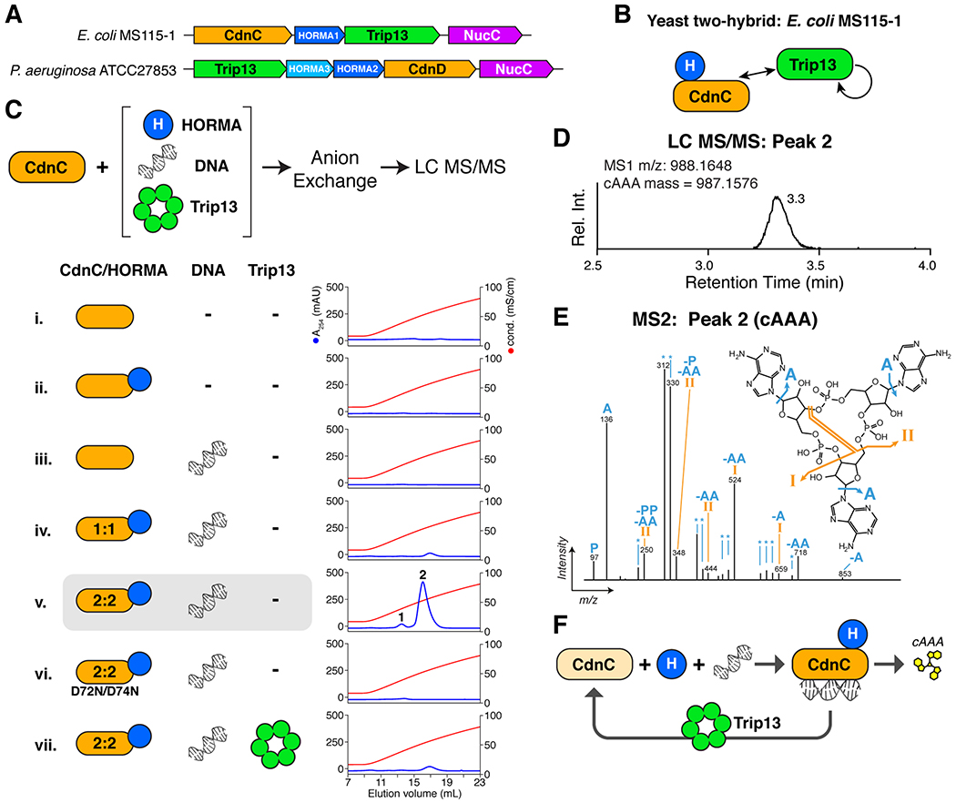
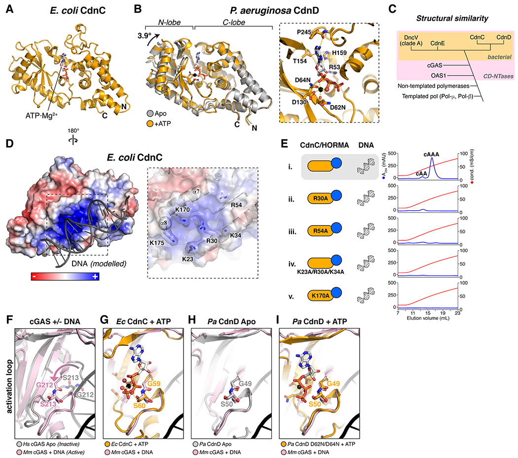
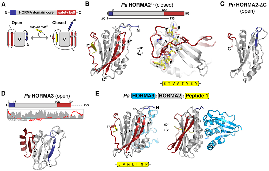
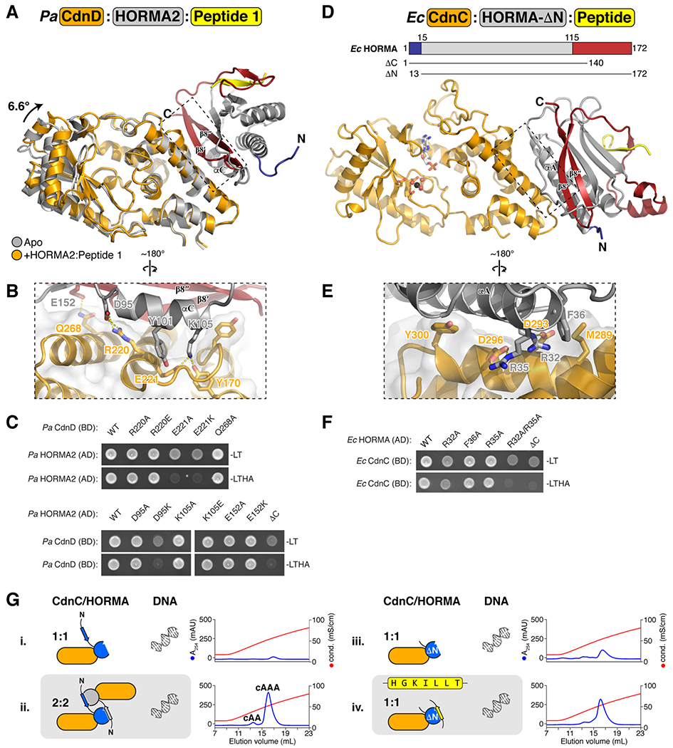
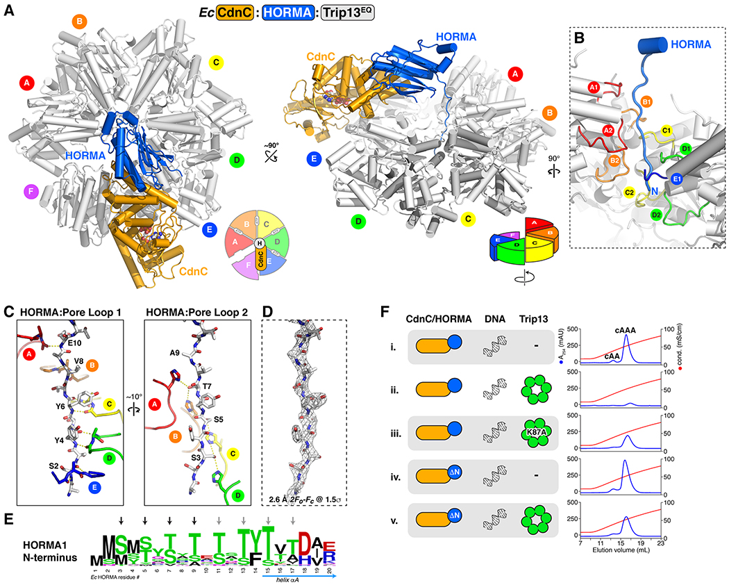
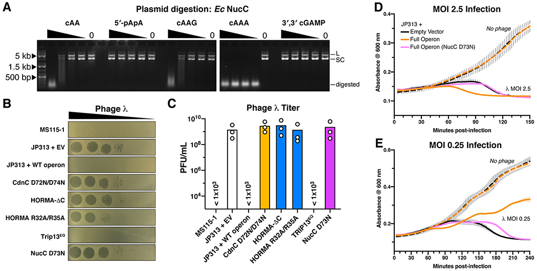
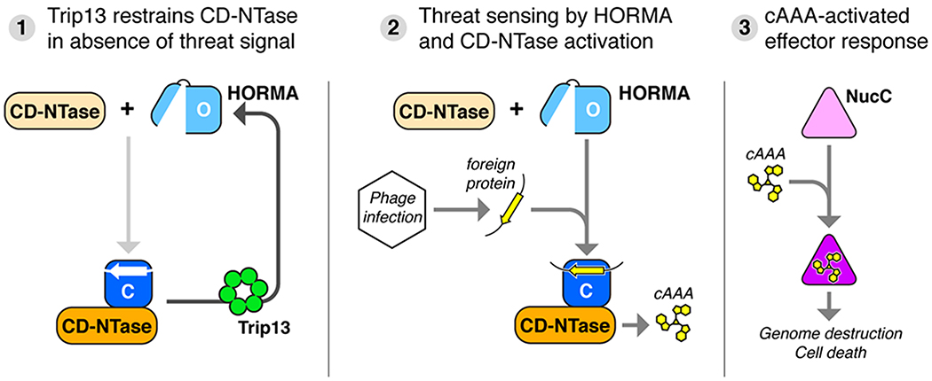
Bacteria are continually challenged by foreign invaders, including bacteriophages, and have evolved a variety of defenses against these invaders. Here, we describe the structural and biochemical mechanisms of a bacteriophage immunity pathway found in a broad array of bacteria, including E. coli and Pseudomonas aeruginosa. This pathway uses eukaryotic-like HORMA domain proteins that recognize specific peptides, then bind and activate a cGAS/DncV-like nucleotidyltransferase (CD-NTase) to generate a cyclic triadenylate (cAAA) second messenger; cAAA in turn activates an endonuclease effector, NucC. Signaling is attenuated by a homolog of the AAA+ ATPase Pch2/TRIP13, which binds and disassembles the active HORMA-CD-NTase complex. When expressed in non-pathogenic E. coli, this pathway confers immunity against bacteriophage λ through an abortive infection mechanism. Our findings reveal the molecular mechanisms of a bacterial defense pathway integrating a cGAS-like nucleotidyltransferase with HORMA domain proteins for threat sensing through protein detection and negative regulation by a Trip13 ATPase.

Keywords: AAA+ ATPase remodeler; CD-NTase; HORMA domain; abortive infection; bacteriophage immunity; second messenger signaling.


Conflict of interest statement

Declaration of Interests The authors declare no competing interests.

Figures

Figure 1.
Figure 1.. HORMA+Trip13-associated CD-NTases synthesize cAAA
(A) Schematic of CD-NTase+HORMA+Trip13 operons from E. coli MS115-1 (top) and P. aeruginosa ATCC27853 (bottom). See Figure S1 for phylogenetic analysis of Trip13 and HORMA proteins. See Table S4 for protein sequences. (B) Summary of yeast two-hybrid and three-hybrid assays with the E. coli MS115-1 CdnC, HORMA, and Trip13. See Figure S2A for yeast two-hybrid and three-hybrid results, Figure S2B for purification and stoichiometry of a CdnC:HORMA complex, and Figure S2C for purification of a CdnC:HORMA:Trip13EQ complex. See Figure S2D–G for equivalent assays with the P. aeruginosa ATCC27853 operon. (C) Top: Schematic of second messenger synthesis assays. Ec CdnC was incubated with potential regulators (HORMA, plasmid DNA, and Trip13) plus ATP, then the products were separated by anion-exchange chromatography and analyzed by liquid chromatography with tandem mass spectrometry (LC-MS/MS). Bottom: Anion exchange elution profiles from second messenger synthesis assays using Ec CdnC, HORMA, DNA, and Trip13. Blue lines show absorbance at 254 nm, and red lines show conductivity for the 0.2-2.0 M ammonium acetate gradient elution. CdnC D72N/D74N contains aspartate-to-asparagine mutations in the putative active-site residues 72 and 74. See Figure S3A for assays with Ec CdnC and different DNAs, and Figure S3B for equivalent assays with Pa CdnD. (D) Liquid chromatography elution profile of the major product of Ec CdnC (peak 2 from panel C, sample v), with measured MS1 m/z and theoretical mass of cAAA. (E) MS2 fragmentation spectrum of the major product of Ec CdnC, annotated according to expected fragments of cAAA. The m/z of the extracted ion (corresponding to the [M+H] adduct of cAAA) was 988.1648, consistent with cAAA (monoisotopic mass = 987.1576 amu; molecular weight = 987.6263 amu). The minor product of Ec CdnC (peak 1 from panel C, sample v) was also analyzed by LC MS/MS and confirmed to be cAA (not shown). (F) Model for CdnC activation by HORMA and DNA binding, with negative regulation by Trip13.
Figure 2.
Figure 2.. Structures of HORMA+Trip13-associated CD-NTases
(A) Structure of Ec CdnC, with bound ATP·Mg2+ shown as sticks. (B) Overlay of Pa CdnD in the Apo state (gray) and bound to ATP (orange), with bound ATP·Mg2+ shown as sticks. Right: Closeup view of ATP·Mg2+ binding to Pa CdnD. See Figure S4A for ITC assays measuring nucleotide binding to Pa CdnD. (C) Schematic of structural similarity in Pol-β type nucleotidyltransferases. CD-NTases are shaded in pink, and bacterial CD-NTases are shaded in orange. (D) Reverse view of Ec CdnC, showing surface charge and DNA (gray) modelled from a superposition with M. musculus cGAS bound to DNA (PDB 4K9B) (Gao et al., 2013). For surface conservation of Ec CdnC, see Figure S4B. For surface conservation and charge of Pa CdnD, see Figure S4C–D. Closeup view (right) shows positively-charged residues, with helices α7 and α8 (equivalent to mammalian cGAS DNA-binding surface) labeled. (E) Anion exchange elution profiles from second messenger synthesis assays with wild-type Ec CdnC (sample i) and mutants to the putative DNA-binding surface (samples ii-v). (F) Overlay of inactive H. sapiens cGAS (gray; PDB ID 4O69 (Zhang et al., 2014)) with active, DNA-bound M. musculus cGAS (pink; PDB ID 4O6A (Zhang et al., 2014)) showing motion of the cGAS activation loop upon DNA binding. (G) Overlay of ATP-bound Ec CdnC (orange) with active cGAS (pink). (H) Overlay of Apo Pa CdnD (gray) with active cGAS (pink). (I) Overlay of ATP-bound Pa CdnD (orange) with active cGAS (pink).
Figure 3.
Figure 3.. Structure and closure motif binding of bacterial HORMA proteins
(A) Top: Schematic of HORMA domain primary structure, with the HORMA domain core gray, N-terminus blue, and C-terminal safety belt red. Bottom: Schematic of the open and closed states of the canonical HORMA domain protein MAD2. The transition from open to closed involves movement of both the N-terminus (blue) and C-terminal safety belt (red), and binding of a closure motif peptide (yellow) by the safety belt. (B) Left: Structure of Pa HORMA2 in the closed state, colored as in panel (A). Secondary-structure elements are labeled as in MAD2, except for strand β5′, which is not observed in other HORMA domain proteins. Right: Closeup view of Pa HORMA2 binding the extended N-terminus of a symmetry-related Pa HORMA2 molecule, which mimics a closure motif (yellow). (C) Structure of Pa HORMA2-ΔC (lacking residues 134–166) in the open state. (D) Top: Schematic of Pa HORMA3, with plot showing the Jalview alignment conservation score (3-point smoothed; gray) (Livingstone and Barton, 1993) and DISOPRED3 disorder propensity (red) for aligned bacterial HORMA3 proteins (Jones and Cozzetto, 2015). Bottom: Structure of Pa HORMA3 in the open state. (E) Structure of the Pa HORMA3:HORMA2:Peptide 1 complex. See Figure S5 for identification of closure motif sequences for Pa HORMA2 and Ec HORMA by phage display.
Figure 4.
Figure 4.. Structures of CD-NTase:HORMA complexes
(A) Overall structure of the Pa CdnD:HORMA2:Peptide 1 complex, with CdnD orange, HORMA2 gray with safety-belt red and N-terminus blue, and Peptide 1 yellow. CdnD is overlaid with Apo CdnD (gray), showing the 6.6° N-lobe rotation in the HORMA2-bound state. (B) Closeup view of the Pa CdnD:HORMA2 interface. (C) Yeast two-hybrid analysis of the Pa CdnD-HORMA2 interaction. BD: fusion to the Gal4 DNA-binding domain; AD: fusion to the Gal4 activation domain. -LT: media lacking leucine and tryptophan (non-selective); -LTHA: media lacking leucine, tryptophan, histidine, and adenine (stringent selection). (D) Overall structure of the Ec CdnC:HORMAΔN complex, with CdnC orange, HORMA gray with safety-belt red and N-terminus blue, and bound peptide yellow (see Figure S6B). (E) Closeup view of the Ec CdnC:HORMA interface. (F) Yeast two-hybrid analysis of the Ec CdnC:HORMA interaction. (G) Anion exchange elution profiles from second messenger synthesis assays with separately-purified 1:1 (sample i) and 2:2 (sample ii) samples of the full-length Ec CdnC:HORMA complex (with intact His6-tag on HORMA, same as Figure 1C, samples iv and v.) and Ec CdnC:HORMA-ΔN with His6-tag removed, without addition of peptide (sample iii) or with addition of consensus HORMA-binding closure motif peptide HGKILLT (sample iv). See Figure S6D for size exclusion elution profiles of Ec CdnC:HORMA-ΔN. Residual activity in sample iii is likely due to incomplete dissociation of the cleaved His6-tag from HORMA-ΔN.
Figure 5.
Figure 5.. Structure of the Ec CdnC:HORMA:Trip13EQ complex
(A) Top and side views of the Ec CdnC:HORMA:Trip13EQ complex, with CdnC orange, HORMA blue, and Trip13EQ white. See Figure S8 for structural analysis of Trip13EQ. Trip13 forms a spiral conformation, with its A subunit at top and F subunit at bottom (see schematics). CdnC binds the top surface of Trip13 subunits E and F (see Figure S8B for buried surface area). See Figure S6E–G for yeast two-hybrid analysis of the CdnC-Trip13 and HORMA-TRIP13 interfaces. (B) Closeup view of the Ec HORMA N-terminus (blue) engaged by Trip13 pore loops 1 (upper) and 2 (lower) from chains A-E. (C) Detail views of interactions between the Ec HORMA N-terminus (white sticks) with Trip13 pore loop 1 (left) and pore loop 2 (right). HORMA residues Ser4, Tyr4, Tyr6, Val8, and Glu10 form an extended backbone hydrogen-bonding network with Trip13 pore loop 1 residues Gly119 (main-chain carbonyl) and Val121 (main-chain amine) from monomers A-E (Trip13 Arg120 side-chains are not shown for clarity). HORMA residues Ser3, Ser5, Thr7, and Ala9 form an extended hydrogen-bond network with Trip13 pore loop 2 residue His173 from monomers A-D. (D) View equivalent to panel (C) showing 2Fo-Fc electron density for the HORMA N-terminus at 1.5 σ (2.6 Å resolution). (E) Sequence logo showing conservation of the N-termini of HORMA1 proteins. See Figure S7E–F for analysis of Trip13 pore loop 1 and 2 conservation, and equivalent logos of Trip13 and HORMA from two-HORMA operons. (F) Anion exchange elution profiles from second messenger synthesis assays with wild-type proteins (samples i-ii), Trip13 Walker A mutant K87A (sample iii), and HORMA-ΔN (missing N-terminal residues 1–12; samples iv-v). See Figure S3B for equivalent assays with Pa CdnD and Trip13 mutants.
Figure 6.
Figure 6.. The E. coli MS115-1 CBASS system confers bacteriophage immunity through activation of a DNA endonuclease.
(A) Plasmid digestion assay with Ec NucC (10 nM) and the indicated second messenger molecules at 400/100/25/6.25/0 nM. L: linear plasmid, SC: supercoiled plasmid. (B) Dilution of phage λ on E. coli MS115-1 (top) and JP313 (wild-type laboratory strain) with the plasmid-encoded E. coli MS115-1 CBASS system with wild-type proteins (WT) or the indicated mutations. EV: empty vector. Six 10-fold phage dilutions are shown. (C) Quantitation of phage λ infectivity. Shown is the average +/− standard deviation of three trials at a single bacteriophage dilution. The three strains marked “< 1x10³“ showed no plaques with the highest-tested bacteriophage concentration. Bars represent mean of three individual measurements (white circles). (D) Growth curves for E. coli JP313 transformed with the indicated plasmids and infected at 0 minutes with bacteriophage λ at an MOI of 2.45 (solid lines) or without phage addition (dashed lines). Lines represent the mean of four replicates, and error bars indicate standard deviation. (E) Growth curves of cell cultures infected at an MOI of 0.245.
Figure 7.
Figure 7.. Model for bacteriophage sensing and immunity by CD-NTase+HORMA+Trip13 operons
(1) In the absence of a bacteriophage threat, Trip13 maintains HORMA proteins in the open state (light blue) by disassembling complexes of closed-HORMA (dark blue) + CdnC, thereby restraining CD-NTase activation. (2) Upon infection, open HORMA proteins recognize closure motif sequences in foreign proteins (yellow), convert to the closed state, then bind their cognate CD-NTase (orange). The CD-NTase:HORMA complex (which further requires bound DNA in some cases; not shown) synthesizes a nucleotide-based second messenger (cAAA). (3) Second messengers bind and activate effector proteins including NucC (Lau et al., 2019), causing cell death and abortive infection.

References

    1. Afonine PV, Grosse-Kunstleve RW, Echols N, Headd JJ, Moriarty NW, Mustyakimov M, Terwilliger TC, Urzhumtsev A, Zwart PH, Adams PD, et al. (2012). Towards automated crystallographic structure refinement with phenix.refine. Acta Crystallogr. Sect. D, Biol. Crystallogr 68, 352–367. - PMC - PubMed
    1. Alfieri C, Chang L, and Barford D (2018). Mechanism for remodelling of the cell cycle checkpoint protein MAD2 by the ATPase TRIP13. Nature 559, 274–278. - PMC - PubMed
    1. Aravind L, and Koonin EV (1998). The HORMA domain: a common structural denominator in mitotic checkpoints, chromosome synapsis and DNA repair. Trends Biochem. Sci 23, 284–286. - PubMed
    1. Burdette DL, Monroe KM, Sotelo-Troha K, Iwig JS, Eckert B, Hyodo M, Hayakawa Y, and Vance RE (2011). STING is a direct innate immune sensor of cyclic di-GMP. Nature 478, 515–518. - PMC - PubMed
    1. Burroughs AM, Zhang D, Schäffer DE, Iyer LM, and Aravind L (2015). Comparative genomic analyses reveal a vast, novel network of nucleotide-centric systems in biological conflicts, immunity and signaling. Nucleic Acids Res. 43, gkv1267–10654. - PMC - PubMed

Publication types

MeSH terms