. 2020 Jan 28;117(4):2122-2132.
doi: 10.1073/pnas.1915152117. Epub 2020 Jan 13.

A molecularly engineered antiviral banana lectin inhibits fusion and is efficacious against influenza virus infection in vivo

Affiliations

A molecularly engineered antiviral banana lectin inhibits fusion and is efficacious against influenza virus infection in vivo

Evelyn M Covés-Datson et al. Proc Natl Acad Sci U S A. .

Abstract

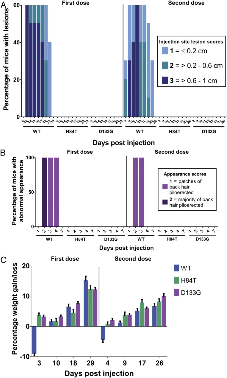
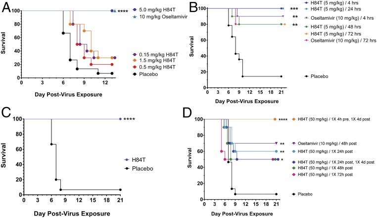
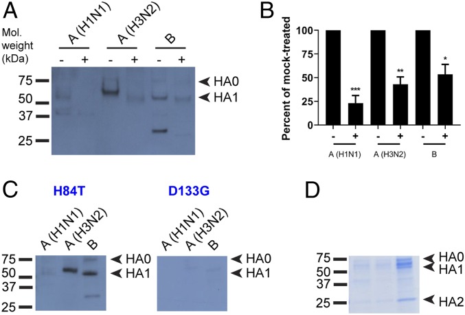
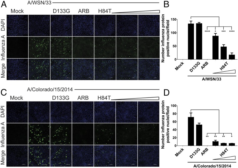
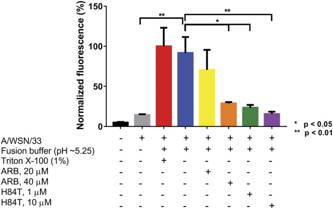
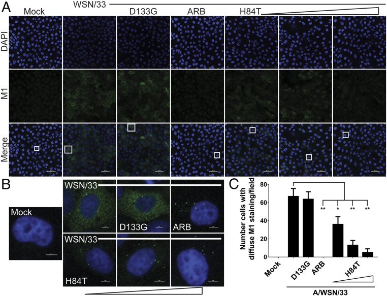
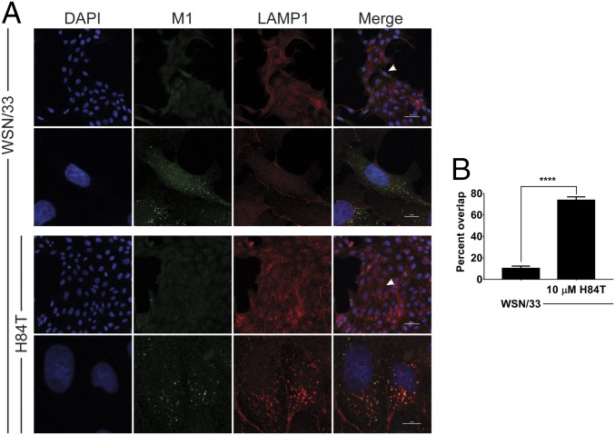
There is a strong need for a new broad-spectrum antiinfluenza therapeutic, as vaccination and existing treatments are only moderately effective. We previously engineered a lectin, H84T banana lectin (H84T), to retain broad-spectrum activity against multiple influenza strains, including pandemic and avian, while largely eliminating the potentially harmful mitogenicity of the parent compound. The amino acid mutation at position 84 from histidine to threonine minimizes the mitogenicity of the wild-type lectin while maintaining antiinfluenza activity in vitro. We now report that in a lethal mouse model H84T is indeed nonmitogenic, and both early and delayed therapeutic administration of H84T intraperitoneally are highly protective, as is H84T administered subcutaneously. Mechanistically, attachment, which we anticipated to be inhibited by H84T, was only somewhat decreased by the lectin. Instead, H84T is internalized into the late endosomal/lysosomal compartment and inhibits virus-endosome fusion. These studies reveal that H84T is efficacious against influenza virus in vivo, and that the loss of mitogenicity seen previously in tissue culture is also seen in vivo, underscoring the potential utility of H84T as a broad-spectrum antiinfluenza agent.

Keywords: antiviral; hemagglutinin; influenza virus; lectin; membrane fusion.


Conflict of interest statement

Competing interest statement: D.M.M. is an inventor on a patent for H84T BanLec and the founder of the company Virule, which aims to commercialize H84T BanLec.

Figures

Fig. 1.
Fig. 1.
Systemic H84T is very well tolerated in mice. BALB/c mice were injected intraperitoneally with two 50-mg/kg doses of WT, H84T, or D133G BanLec administered 1 mo apart (n = 10 per group). Injection site lesions (A) and appearances (B) were scored, and weight measured (C) for all mice for 59 d postinjection. In C, the data represent the mean for all mice in each group and error bars the SEM.
Fig. 2.
Fig. 2.
Systemic H84T treatment is highly efficacious against lethal influenza virus infection in mice. (AC) Therapeutic efficacy of H84T administered intraperitoneally in BALB/c mice challenged intranasally with two times the 50% mouse lethal dose of A/WSN/HA(NC/2099-N225G)/1933 (H1N1). (A) Survival of mice following intraperitoneal treatment with H84T (0.15–5 mg/kg) once daily for 5 d beginning 4 h postchallenge. (B) Survival of mice following intraperitoneal treatment with H84T (5 mg/kg) once daily for 5 d beginning 4, 24, 48, or 72 h postchallenge, or treatment with oseltamivir (10 mg/kg) beginning 4 or 72 h postchallenge. (C) Survival of mice following intraperitoneal treatment with H84T (50 mg/kg) on a single occasion 21 d before challenge, then once daily for 5 d (5 mg/kg/d) beginning 4 h postchallenge. Placebo mice received PBS 21 d prechallenge and once daily for 5 d beginning 4 h postchallenge. (D) Therapeutic efficacy of H84T administered subcutaneously in BALB/c mice challenged intranasally with a lethal dose of A/WSN/HA(NC/2099-N225G)/1933 (H1N1). Survival of mice following subcutaneous treatment with a regimen of either one or two 50-mg/kg doses of H84T within 4 d postinfection. *P < 0.05, **P < 0.01, ***P < 0.001, and ****P < 0.0001. Kaplan–Meier survival curves for A to D were compared by the log-rank (Mantel–Cox) test followed by pairwise comparison using the Gehan–Breslow–Wilcoxon test.
Fig. 3.
Fig. 3.
H84T binds to virus-derived HA. (A) Lectin blot. Virus particles from A/Florida/3/2006 (H1N1), A/Perth/16/2009 (H3N2), and B/Brisbane/60/2008 were lysed for protein extraction. Proteins were incubated with PNGaseF to remove N-glycans or with PBS (mock) overnight, then resolved by reducing SDS/PAGE and blotted. Blots were incubated with 100 nM H84T, followed by primary and secondary antibody incubations to detect BanLec bound to viral proteins. Note that the lower band seen in the influenza B lanes does not appear to be HA2 as it does not run at the correct size. Mol. weight, molecular weight. (B) Percent binding of H84T to PNGase- versus mock-treated HA1 in A, relative to mock-treated set at 100%. Data in A and B are representative of three independent experiments. *P < 0.05, **P < 0.01, ***P < 0.001, as compared to the mock-treated group. Error bars denote the SEM. +, PNGase F-treated; −, mock-treated. (C) Lectin blots were performed as in A, without PNGase F digestion. Blots were incubated with 100 nM H84T or D133G BanLec. Data are representative of three independent experiments. (D) Proteins were resolved as in C but not transferred and gels stained with a Coomassie-based reagent.
Fig. 4.
Fig. 4.
H84T prevents influenza virus protein expression in MDCK cells. MDCK cells were pretreated for 1 h with H84T (0.1, 1, or 10 μM in A; 0.05, 0.1, or 1 μM in C), 10 μM D133G, or 40 μM of the fusion inhibitor ARB and infected with A/WSN/1933 (H1N1) (A) or A/Colorado/15/2014 (H3N2) (C) (MOI = 0.5) for 5 h at 37 °C. (A and C) Representative immunofluorescent micrographs of influenza A antigen staining (green) and nuclei (blue) 5 h postinfection. Data for A and C are representative of three and four independent experiments, respectively. Scale bars indicate 100 μm. (B and D) Quantitation of the number of cells with influenza protein-positive nuclei per field, as assessed by a trained observer in a blinded fashion. Statistical analyses were performed by t test. *P < 0.05, **P < 0.01, ***P < 0.001, and ****P < 0.0001, as compared to the infected, untreated group. Error bars denote the SEM.
Fig. 5.
Fig. 5.
H84T inhibits influenza virus fusion. MDCK cells were pretreated for 1 h at 37 °C with 1 or 10 μM H84T or 20 or 40 μM of the fusion inhibitor ARB and incubated with octadecyl R18-labeled A/WSN/1933 for 1 h at 4 °C to allow attachment only. Excess virus was removed and virus fusion with the cell membrane was triggered at 37 °C using pH ∼5.25 fusion buffer. Fluorescence dequenching of R18 was monitored on a microplate fluorometer with excitation and emission wavelengths of 560 and 590 nm, respectively. Maximal dequenching was achieved with 1% Triton X-100 set at 100%. Bars represent the mean values from duplicate wells from two independent experiments and error bars represent the SEM. Statistical analyses were performed by t test. *P < 0.05, **P < 0.01, comparing groups indicated by brackets.
Fig. 6.
Fig. 6.
H84T reduces influenza virus uncoating. MDCK cells were pretreated for 1 h with 0.1, 1, or 10 μM H84T, 10 μM D133G, or 40 μM of the fusion inhibitor ARB and infected with A/WSN/1933 (MOI 0.5) for 2.5 h at 37 °C. (A) Representative immunofluorescent micrographs of M1 antigen staining (green) and nuclei (blue) 2.5 h postinfection. Scale bars indicate 50 μm. (B) Micrographs of single cells from A, as denoted by the boxes in A. Scale bars indicate 5 μm. Data for A and B are representative of 15 independent experiments. (C) Quantitation of the number of cells with diffuse cytoplasmic M1 staining per field, as assessed by a trained observer in a blinded fashion. Statistical analyses were performed by t test. *P < 0.05 and **P < 0.01, as compared to the infected, untreated group. Error bars represent the SEM.
Fig. 7.
Fig. 7.
H84T restricts influenza virus to the late endosomal/lysosomal compartment. (A) Representative immunofluorescent micrographs of M1 (green) and LAMP1 antigen staining (red) and nuclei (blue). Following 1 h pretreatment with H84T (10 μM), MDCK cells were incubated with A/WSN/1933 (H1N1) at 4 °C for 1 h to allow only for attachment, excess virus was removed, treatment-containing medium was replaced, and the cells were incubated at 37 °C for an additional 3 h. The second and fourth rows are micrographs of one or two cells from the first and third rows, respectively, indicated by the arrows. Data are representative of five independent experiments. Scale bars indicate 50 μm in the first and third rows and 10 μm in the second and fourth rows. (B) Quantitation of the percent of M1+ punctae that are also LAMP1+ (percent overlap). Statistical analyses were performed by t test. ****P < 0.0001, as compared to the infected, untreated group. Error bars represent the SEM.

References

    1. Osterholm M. T., Kelley N. S., Sommer A., Belongia E. A., Efficacy and effectiveness of influenza vaccines: A systematic review and meta-analysis. Lancet Infect. Dis. 12, 36–44 (2012). - PubMed
    1. Flannery B., et al. , Interim estimates of 2017-18 seasonal influenza vaccine effectiveness—United States, February 2018. MMWR Morb. Mortal. Wkly. Rep. 67, 180–185 (2018). - PMC - PubMed
    1. Koszalka P., Tilmanis D., Hurt A. C., Influenza antivirals currently in late-phase clinical trial. Influenza Other Respir. Viruses 11, 240–246 (2017). - PMC - PubMed
    1. Hayden F. G., et al. ; Baloxavir Marboxil Investigators Group , Baloxavir marboxil for uncomplicated influenza in adults and adolescents. N. Engl. J. Med. 379, 913–923 (2018). - PubMed
    1. Jefferson T., Jones M., Doshi P., Del Mar C., Neuraminidase inhibitors for preventing and treating influenza in healthy adults: Systematic review and meta-analysis. BMJ 339, b5106 (2009). - PMC - PubMed

Publication types

MeSH terms