. 2020 Mar 1;80(5):1171-1182.
doi: 10.1158/0008-5472.CAN-19-2348. Epub 2020 Jan 13.

Therapy-Induced Senescence Drives Bone Loss

Affiliations

Therapy-Induced Senescence Drives Bone Loss

Zhangting Yao et al. Cancer Res. .

Abstract

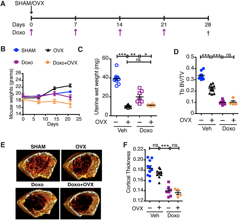
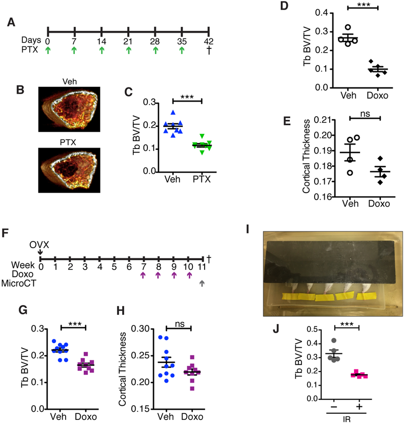
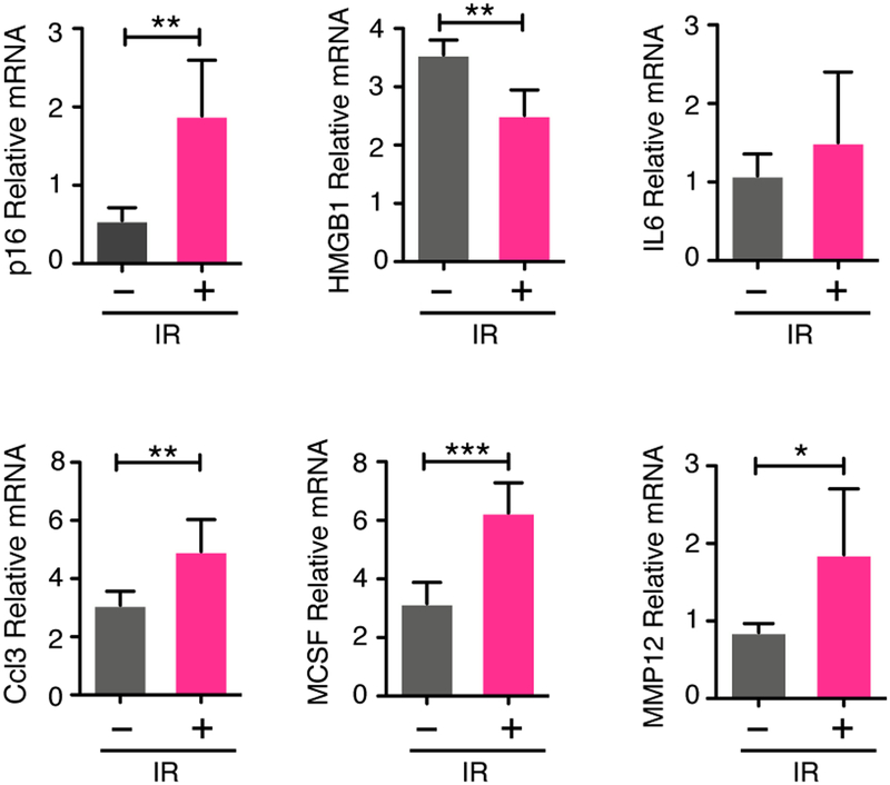
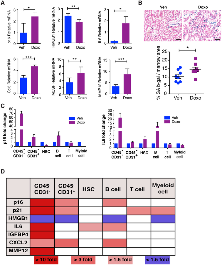
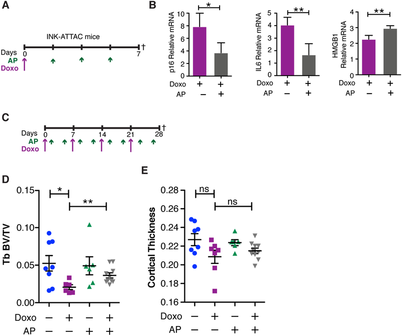
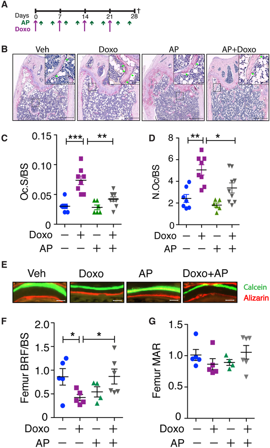
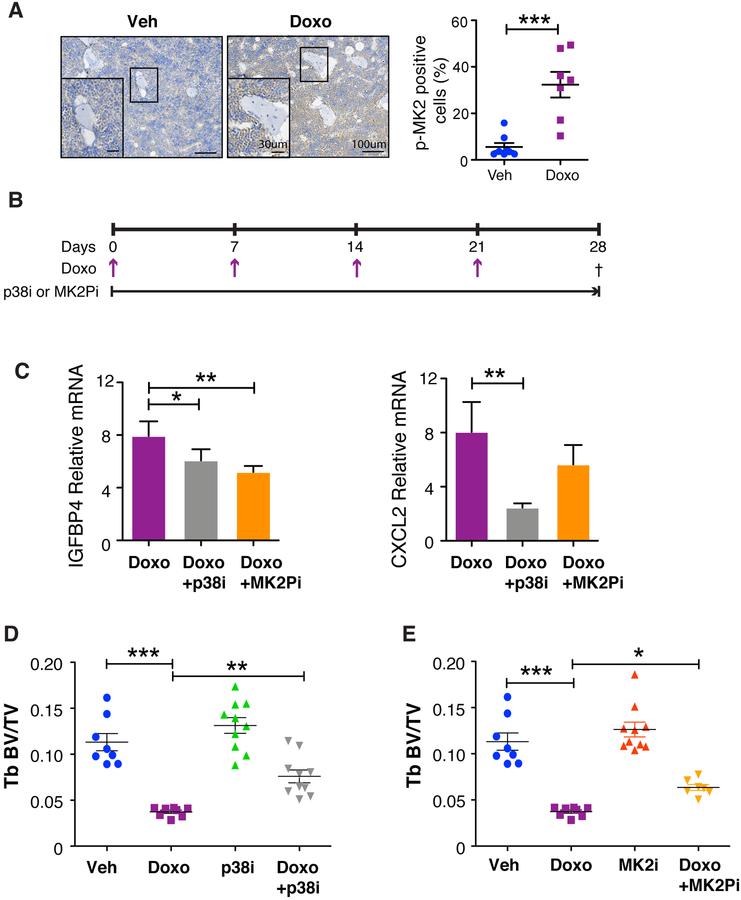
Chemotherapy is important for cancer treatment, however, toxicities limit its use. While great strides have been made to ameliorate the acute toxicities induced by chemotherapy, long-term comorbidities including bone loss remain a significant problem. Chemotherapy-driven estrogen loss is postulated to drive bone loss, but significant data suggests the existence of an estrogen-independent mechanism of bone loss. Using clinically relevant mouse models, we showed that senescence and its senescence-associated secretory phenotype (SASP) contribute to chemotherapy-induced bone loss that can be rescued by depleting senescent cells. Chemotherapy-induced SASP could be limited by targeting the p38MAPK-MK2 pathway, which resulted in preservation of bone integrity in chemotherapy-treated mice. These results transform our understanding of chemotherapy-induced bone loss by identifying senescent cells as major drivers of bone loss and the p38MAPK-MK2 axis as a putative therapeutic target that can preserve bone and improve a cancer survivor's quality of life. SIGNIFICANCE: Senescence drives chemotherapy-induced bone loss that is rescued by p38MAPK or MK2 inhibitors. These findings may lead to treatments for therapy-induced bone loss, significantly increasing quality of life for cancer survivors.


Conflict of interest statement

Conflict of Interest

JBM is the Executive Vice President of R&D of Aclaris Therapeutics, Inc. JvD and DJB are co-inventors on patent applications licensed to or filed by Unity Biotechnology, a company developing senolytic medicines, including small molecules that selectively eliminate senescent cells. JvD is a co-founder of Unity Biotechnology. The other authors have no competing interests to declare.

Figures

Figure 1:
Figure 1:. Chemotherapy-induced bone loss is more severe than estrogen-dependent bone loss.
(A) Schematic of the experimental timeline for ovariectomy (OVX) surgery and dosing regimen for doxorubicin (Doxo; 5 mg/kg) in 6-week old FVB/NJ mice. Dagger indicates time of sacrifice. (B) Mouse weights for the duration of the experiment on indicated days. (C) Uterine wet weights, measured as a surrogate for estrogen levels in SHAM or OVX mice treated with vehicle (Veh) or Doxo. (D) Trabecular (Tb) volume (TV) compared to bone volume (BV) of femurs, presented as Tb BV/TV. (E) Representative 3D reconstructions for each of the treatment groups. (F) Cortical thickness of treated bones. N≥4 per group. All data are presented as mean±SEM. *p≤0.05, **p≤0.01, ***p≤0.001, ns=not significant. Comparisons are indicated by capped lines.
Figure 2:
Figure 2:. Chemotherapy can induce bone loss independent of estrogen loss.
(A) Schematic of experimental setup and dosing regimen for Paclitaxel (PTX; 10 mg/kg) in 6-week old C57BL/6 female mice. Dagger indicates time of sacrifice. (B) Representative 3D reconstructions, generated using OsiriX, of 0.9 mm thick sections of femur right below the growth plate for each of the treatment groups. (C) Trabecular bone parameter of femurs after μCT presented as trabecular (Tb) bone volume (BV) to trabecular volume (Tb BV/TV). N=8 per group. (D) Tb BV/TV in 6-week old C57BL/6 male mice treated with 4 once weekly doses of Doxo (5 mg/mL) or vehicle (Veh) and (E) cortical thickness in the same male mice. N≥4 per group. (F) Schematic of experimental timeline showing dosing regimen for doxorubicin (Doxo; 5 mg/kg) after stabilization of ovariectomy (OVX)-induced bone loss. Dagger indicates time of sacrifice. (G) Trabecular (Tb) volume (TV) compared to bone volume (BV) of femurs, presented as Tb BV/TV, and (H) cortical thickness of femurs at 11 weeks post-OVX in mice treated with vehicle (Veh) or Doxo. N≥9 per group. (I) Picture of single-limb irradiation setup with only the right leg exposed and the rest of the body covered by a lead shield. The right hind legs of 6-week old FVB/NJ mice received 20 Gy radiation, and 4 weeks later the irradiated (IR) and contralateral femurs were isolated and scanned using μCT. (J) Trabecular (Tb) volume (TV) compared to bone volume (BV) of femurs, presented as Tb BV/TV for the irradiated leg (+) and contralateral control (–). N=5 per group. All data are presented as mean±SEM. ***p≤0.001, ns=not significant.
Figure 3:
Figure 3:. Radiation induces senescence in the bone.
6-week old FVB/NJ mice received 20 Gy radiation to the right hind limb and 48 h later the tibias were isolated, bone marrow was removed, and tibias were crushed and used to prepare RNA from bone-residing cells. (A) p16, HGMB1, IL6, IGFBP4, CXCL2, CCL3, MCSF, and MMP12 mRNA levels in crushed tibias (irradiated versus contralateral) devoid of marrow, as a measure of senescence induction. N=5 per group. All qRT-PCR data are presented as mean±SD. *p≤0.05, **p≤0.01, ***p≤0.001.
Figure 4:
Figure 4:. Chemotherapy induces senescence in the bone.
(A) Mice were treated with vehicle (Veh) or doxorubicin (Doxo) and 48 h later, legs were isolated, marrow was removed, and bones were crushed and used to make mRNA from bone-resident cells. Expression of p16, IL6 (6-week FVB, 25 mg/kg) and expression of HGMB1, CCL3, MCSF, and MMP12 (16-week C57Bl/6, 10 mg/kg) mRNA levels in crushed tibias was measured by qRT-PCR. N≥5 per group. Data are presented as mean±SD. (B) Upper: SA-β-Gal staining in femurs isolated 7 days after a single Doxo dose in 6-week old FVB/NJ mice. Bars = 100 μm. Lower: Quantification of SA-β-Gal. N=8 per group. Data are presented as mean±SEM. *p≤0.05, **p≤0.01, ***p≤0.001. (C) 6-week old C57BL/6 mice were treated with Veh of 25 mg/kg Doxo and 48 h later, legs were dissociated and bone resident cells were isolated and stained with antibodies against CD45, CD31, cKit, ScaI and various hematopeoitic markers followed by sorting for HSC, T cells, B cells, myeloid cells and CD45CD31+ and CD45CD31 populations. Expression of p16, IL6 mRNA were quantified in the indicated populations. (D) mRNA expression of other SASP factors were detected in the populations as indicated (graphs of expression can be found in Supplementary Figure S7). N=2. All qRT-PCR data are presented as mean±SD.
Figure 5:
Figure 5:. Clearance of senescent cells rescues chemotherapy-induced bone loss.
(A) Experimental schematic for 1-week time point showing a single dose of Doxorubicin (Doxo; 10 mg/kg) and 3 doses of AP20187 (AP; 2 mg/kg) in 16-week old C57BL/6 INK-ATTAC mice. Dagger indicates time of sacrifice. (B) qRT-PCR analysis of p16, IL6, and HGMB1 from mRNA obtained from bone-resident cells present in tibias devoid of marrow from INK-ATTAC mice 7 days after dosing with Doxo and AP. N≥3 per group. qRT-PCR data are presented as mean±SD. (C) Experimental schematic for 4-week time point with dosing frequency of Doxo (5 mg/kg) and AP20187 (2mg/kg) and symbols as in A. (D) Trabecular (Tb) BV/TV of femurs and (E) cortical thickness of femurs. N≥6 per group. Data are presented as mean±SEM. *p<0.05, **p≤0.01, and ns=not significant.
Figure 6:
Figure 6:. Clearance of senescent cells rescues osteoblast function and reduces osteoclasts.
(A) Experimental schematic as laid out in Figure 5. (B) Representative images of TRAP staining of bones from mice treated as indicated. Scale bars are 500 μm in main images and 50 μm in insets. (C-D) Histomorphometric analysis of TRAP-stained sections to quantify the ratio of osteoclast surface to bone surface (Oc.S/BS (/mm)) (C) and the number of osteoclasts per bone surface (N.Oc/BS (/mm)) (D). (E-G) Histomorphometric analysis of double bone labeling of femurs from mice injected with calcein (green) and alizarin (red) 7 days apart. (E) Representative images. Bottom: Quantification of bone formation rate (BFR/BS (mcm3/mcm2/d)) (F) and mineral apposition rate (MAR (mcm/d)) (G). Scale bar is 10 μm. N≥6 per group. All data are presented as mean±SEM. *p≤0.05, **p≤0.01, ***p≤0.001.
Figure 7:
Figure 7:. Inhibition of the p38MAPK-MK2 pathway limits SASP production and rescues chemotherapy-induced bone loss.
(A) Mice were sacrificed on day 28 and bone sections from vehicle and chemotherapy treated mice were stained for phosphor-MK2 and shows activation of the p38MAPK pathway following chemotherapy treatment. N≥7 per group. Data are presented as mean±SEM. (B) 16-week old C57BL/6 mice were treated with 4 once-weekly doses of vehicle (Veh) or doxorubicin (Doxo; 5 mg/kg) and provided ad libitum chow compounded with CDD-111 (p38i) or CDD-450 (MK2Pi). (C) Mice were sacrificed on day 28 and bone resident cells were isolated from tibias and the SASP factors IGFBB4 and CXCL12 were measured by qRT-PCR. qRT-PCR data are presented as mean±SD. (D-E) Femurs were isolated from animals treated with vehicle (Veh) or doxorubicin (Doxo) +/− p38i or +/− MK2Pi and measured by μCT to ascertain bone volume (BV) to trabecular (Tb) volume (BV/TV) of the p38i treatment cohort (D). Tb BV/TV of the MK2Pi treatment cohort (E). N≥8 per group. All data are presented as mean±SEM. *p≤0.05, **p≤0.01, ***p≤0.001.

References

    1. Chao C, Studts JL, Abell T, Hadley T, Roetzer L, Dineen S, et al. Adjuvant chemotherapy for breast cancer: how presentation of recurrence risk influences decision-making. J Clin Oncol 2003;21:4299–305 - PubMed
    1. Ishitobi M, Komoike Y, Motomura K, Koyama H, Inaji H. Early response to neo-adjuvant chemotherapy in carcinoma of the breast predicts both successful breast-conserving surgery and decreased risk of ipsilateral breast tumor recurrence. Breast J 2010;16:9–13 - PubMed
    1. Jones SE, Erban J, Overmoyer B, Budd GT, Hutchins L, Lower E, et al. Randomized phase III study of docetaxel compared with paclitaxel in metastatic breast cancer. J Clin Oncol 2005;23:5542–51 - PubMed
    1. Earl H, Provenzano E, Abraham J, Dunn J, Vallier AL, Gounaris I, et al. Neoadjuvant trials in early breast cancer: pathological response at surgery and correlation to longer term outcomes - what does it all mean? BMC Med 2015;13:234. - PMC - PubMed
    1. Bruning PF, Pit MJ, de Jong-Bakker M, van den Ende A, Hart A, van Enk A. Bone mineral density after adjuvant chemotherapy for premenopausal breast cancer. British journal of cancer 1990;61:308–10 - PMC - PubMed

Publication types

MeSH terms

Substances