. 2020 Mar 16;40(7):e00346-19.
doi: 10.1128/MCB.00346-19. Print 2020 Mar 16.

A Redox-Sensitive Thiol in Wis1 Modulates the Fission Yeast Mitogen-Activated Protein Kinase Response to H2O2 and Is the Target of a Small Molecule

Affiliations

A Redox-Sensitive Thiol in Wis1 Modulates the Fission Yeast Mitogen-Activated Protein Kinase Response to H2O2 and Is the Target of a Small Molecule

Johanna J Sjölander et al. Mol Cell Biol. .

Abstract

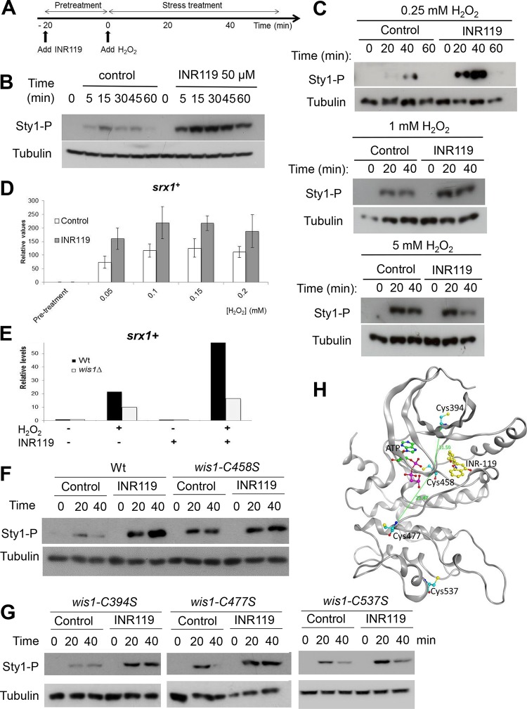
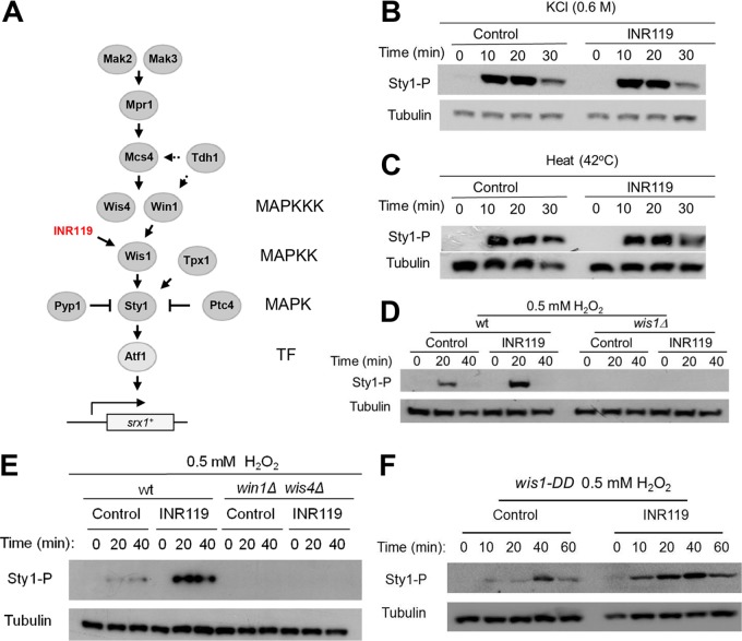
Oxidation of a highly conserved cysteine (Cys) residue located in the kinase activation loop of mitogen-activated protein kinase kinases (MAPKK) inactivates mammalian MKK6. This residue is conserved in the fission yeast Schizosaccharomyces pombe MAPKK Wis1, which belongs to the H2O2-responsive MAPK Sty1 pathway. Here, we show that H2O2 reversibly inactivates Wis1 through this residue (C458) in vitro We found that C458 is oxidized in vivo and that serine replacement of this residue significantly enhances Wis1 activation upon addition of H2O2 The allosteric MAPKK inhibitor INR119, which binds in a pocket next to the activation loop and C458, prevented the inhibition of Wis1 by H2O2in vitro and significantly increased Wis1 activation by low levels of H2O2in vivo We propose that oxidation of C458 inhibits Wis1 and that INR119 cancels out this inhibitory effect by binding close to this residue. Kinase inhibition through the oxidation of a conserved Cys residue in MKK6 (C196) is thus conserved in the S. pombe MAPKK Wis1.

Keywords: Schizosaccharomyces pombe; Sty1; cysteine oxidation; stress signaling.


Figures

FIG 1
FIG 1
Wis1 contains a conserved cysteine next to the DFG motif but lacks a cysteine homologous to MKK6C109. (A) Multiple alignment of the human, fission yeast, and budding yeast MAPKKs showing conservation of the cysteines involved in inhibition through disulfide formation in human MKK6. Cysteines are highlighted in red, the Wis1 sequence is marked with an asterisk, and the DFG motif is indicated by a green box. MKK6 Cys196/Wis1 Cys458 directly precedes the highly conserved DFG motif and is conserved in all MAPKKs, whereas the position of MKK6 Cys106 is less conserved. Wis1 has no cysteine corresponding to MKK6 Cys106. (B) Multiple alignment showing the degree of conservation of all cysteines in Wis1. Human, fission yeast, and budding yeast MAPKKs, MAPKs, and MAPKKKs are shown aligned with the homologous sequences flanking the positions of the six Wis1 cysteines. Conservation is restricted to MAPKKs, except for C458 which is also present in some MAPKs. aa, amino acid. (C) Schematic overview of the location of cysteines in Wis1 in relation to functional information within the Wis1 amino acid sequence. Five of the total six cysteines are found within the kinase domain, and one is found within the nuclear export signal (NES). The locations of functional domains in this figure are based on the work of Nguyen et al. (44). Hs, Homo sapiens; Sc, Saccharomyces cerevisiae; Sp, Schizosaccharomyces pombe.
FIG 2
FIG 2
Wis1 is inhibited by oxidation of Cys458 in vitro. (A) In vitro Wis1 kinase activity is constitutive upon coincubation of Wis1 and Sty1 purified in the absence of EDTA, suggesting that ATP copurifies with Wis1. Sty1 is not phosphorylated before (lane 1) or after (lane 2) assay incubation without Wis1 added. However, even without addition of ATP to the reaction buffer (lane 3), Wis1 phosphorylates Sty1 to the same degree as when ATP is included (lane 4). (B) Wis1 purified in the presence of EDTA phosphorylates Sty1 in an Mg2+- and ATP-dependent manner in vitro. Precipitates of Ni2+ beads from strain JJS7 expressing HA-Wis1-His6 or wis1Δ (JJS1) were tested for activity against the substrate Sty1. No phosphorylation was seen when everything except HA-Wis1-His6 from strain JJS7 (lane 2) was added or when the precipitates from strain JJS7 were exchanged with a precipitate from strain JJS1 (wis1Δ, lane 5). Note that no H2O2 was included in this assay. (C) Inactivation of Wis1 by low levels of H2O2 in vitro is caused by reversible thiol oxidation. Semipurified Wis1 (strain JJS7) was first incubated for 5 min in sample pairs with H2O2, giving the indicated final concentration, and subsequently for one tube in each pair for 5 min with 1 mM TCEP. Thereafter the Wis1 substrate Sty1 (expressed from pREP41MHNSTY1 in strain KS1598) was added, and the kinase reaction was started by addition of MgCl2-ATP. The kinase reaction was stopped after 20 min. (D) Wis1 is inactivated by low levels of H2O2 in vitro in a manner reversible by the reductant TCEP. In contrast, a Wis1C458S mutant enzyme is less inactivated, and activity lost could not be rescued by reductant. Wis1 was treated for 5 min with H2O2 before the kinase reaction was starting by the addition of MgCl2-ATP. This inactivation was in turn reversed upon a 5-min incubation with the reductant TCEP at 1 mM before the kinase assay. Wild-type Wis1 was expressed from JJS7, and Wis1C458 was expressed from JJS9. The bottom panel shows quantification of the relative levels of Sty1 phosphorylation in the different lanes.
FIG 3
FIG 3
Wis1 cysteines including C458 are oxidized in vivo, and Wis1 is rapidly sulfenylated upon H2O2 addition. (A) Schematic overview of the mPEG assay. NEM alkylates all presently reduced thiols. DTT reduces the remaining (reversibly oxidized [ox]) thiols, making them accessible for alkylation by mPEG. This adds approximately 5 kDa per oxidized thiol. Note that irreversibly oxidized thiols not reduced by DTT will not be mPEG labeled. MW, molecular weight. (B to G) The mPEG assay shows that Wis1 C458 as well as other cysteines in Wis1 are oxidized in vivo even without exogenous H2O2. Cells analyzed were from strain JJS15 expressing HA-tagged wt Wis1 from the endogenous promoter (B to D, F, and G), from strain JJS16 expressing cysteine-less Wis16CS from the endogenous promoter (C and D), or from strain JJS7 and JJS9 expression wt Wis1 and wis1C458S, respectively, from the nmt1 promoter (E). (B) Slower-migrating mPEG-modified Wis1 bands (marked with orange lines) appear in extracts of cells even without H2O2 treatment. Formation of these slower-migrating forms is blocked by an extra round of NEM before mPEG treatment. RED, reduced. (C) mPEG assay of wt and wis1C458S cell extracts without H2O2 added. The upper of the two bands representing oxidized Wis1 (orange markers) is absent in the strain expressing Wis1C458S mutant protein. (D and E) The constitutively mPEG-dependent altered migration of Wis1 is abolished in a cysteine-less mutant Wis1 protein (Wis16CS). A strain expressing cysteine-less Wis16CS was run on the blot together with wt Wis1 (B) or after the mPEG assay (C). Samples were divided in two equal aliquots and subjected to either NEM-DTT-NEM treatment or NEM-DTT-mPEG. (F) Time course of Wis1 cysteine oxidation in wt cells as assayed by the mPEG assay following the addition of 0.5 mM H2O2 for the indicated duration. (G) Wis1 cysteine oxidation as assayed by the mPEG assay at 20 min after the addition of H2O2 to different concentrations. (H and I) The use of a sulfenylation-specific probe shows rapid cysteine-dependent Wis1 sulfenylation following H2O2 addition. Cells expressing HA-tagged wt Wis1 (JJS15) (H and I) or Wis16CS (JJS16) (I) were labeled with the sulfenyl-binding reagent DYn-2 prior to exposure to 0.5 mM H2O2, and sample withdrawal was performed at 30 s and 1 and 2 min thereafter. Sulfenylated protein was immunoprecipitated as described in Materials and Methods. Following separation by SDS-PAGE, sulfenylated Wis1 was detected with anti-HA antibodies.
FIG 4
FIG 4
Cys458 inhibits Sty1 phosphorylation (Sty1-P) upon the addition of low levels of H2O2 and is essential for cellular resistance to H2O2 but not to hyperosmosis. (A) Lysates from exponentially growing wt (972 h), wis1C458S (JJS18), or wis1C537S (JJS20) strains, taken before and 20 min after exposure to H2O2 at the indicated concentrations, were subjected to Western blotting. (B) Exponentially growing cells of wt (972 h), wis1C11S (JJS20), wis1C394S (JJS17), wis1C458S (JJS18), wis1C477S (JJS19), wis1C537S (JJS20), wis1C559S (JJS21), wis16CS (JJS22), and wis1Δ (JJS1) strains were serially diluted and spotted on YES plates with and without 0.5 mM H2O2 or 1 M KCl.
FIG 5
FIG 5
The noncompetitive MAPKK inhibitor INR119 binds Wis1 near C458 and protects Wis1 from inactivation by low levels of H2O2 in vitro. (A) Homology model of Wis1 showing the predicted allosteric pocket with INR119 docked inside and its close proximity to the location of C458 and bound ATP. (B) Structures of INR119 and PD98059. (C) INR119 inhibits the MAPKK MEK1 in human MCF-7 breast cancer cells. Western blots of protein extract from MCF-7 human breast cancer cells exposed for 90 min to different concentrations of INR119 or PD98059 are shown. MEK1 activity was measured as the amount of phosphorylated Erk1/2 (Erk1/2-P). (D) Predictions of interactions between INR119 and the amino acids forming the allosteric pocket. (E) Western blotting of a thermal shift assay performed in vivo shows that INR119 stabilizes soluble Wis1 at elevated temperatures, indicating a direct in vivo interaction; however, the heat itself also strongly stabilizes soluble Wis1. Live S. pombe cells (JJS6) expressing tagged Wis1 were preincubated with 25 μM INR119 for 15 min and thereafter subjected to the indicated temperature for 3 min. The soluble fraction in lysates was thereafter obtained by centrifugation (see Materials and Methods). (F) Thermal shift assay of the INR119/Wis1 interaction. Cells (JJS6) expressing HA-tagged Wis1 from the nmt1 promoter were lysed. Cellular extracts were pretreated with 3 μM INR119 for 15 min, and the extracts were heated to the indicated temperatures for 3 min. Soluble protein was separated by centrifugation and analyzed by Western blotting. Quantification of soluble Wis1 is shown at temperatures from 30°C to 50°C. Values have been normalized relative to those of the untreated extract at 30°C and are the averages from four to seven independent experiments. (G) INR119 protects Wis1 from inactivation by low levels of H2O2 in vitro. Semipurified Wis1 (strain JJS7) was pretreated for 15 min with the indicated concentration of INR119 and subsequently for an additional 5 min with 0.05 mM H2O2. Thereafter the substrate Sty1 was added, as well as MgCl2-ATP. Kinase reactions were stopped after 20 min, and results were analyzed by Western blotting.
FIG 6
FIG 6
INR119 pretreatment strongly potentiates the Wis1 response to low H2O2 in vivo. (A) Schematic overview of the setup of experiments involving pretreatment with INR119 and H2O2 addition. (B) Western blot of Sty1 phosphorylation in cells pretreated with 50 μM INR119 and at the indicated time points following the addition 500 μM H2O2. The time line shows the design of experiment, where INR119 is added 20 min before the onset of peroxide stress. (C) The ability of INR119 to potentiate Wis1 signaling is more pronounced at low levels of H2O2. Western blotting was performed of cells (972 h) pretreated with 50 μM INR119 and following after the addition of different amounts of H2O2 (0.25 to 5 mM) at the indicated time points as in described for panel A. (D) The Atf1-dependent transcript srx1+ is enhanced by INR119. Wild-type (972 h) cells were mock pretreated or pretreated with INR119 for 20 min and thereafter treated with H2O2 at the indicated concentrations as described for panel A. Samples were taken after 20 min of H2O2 treatment, and srx1+ transcript levels were determined by qPCR. (E) The srx1+ transcript enhancement by INR119 is Wis1 dependent. Wild-type or wis1Δ cells were exposed to 0.2 mM H2O2 and/or INR119 as indicated, and transcript levels were determined as described for panel D. (F and G) The ability of INR119 to enhance Sty1 activation is dependent on Wis1 C458 (F) but not on C394, C477, or C537 (G). The wild-type (972 h), wis1C394S (JJS17), wis1C458S (JJS18), wis1C477S (JJS19), and wis1C537S (JJS20) strains were analyzed for Sty1 activation by immunoblotting at the indicated time points following the addition of 0.5 mM H2O2. (H) Structural homology model of Wis1 with the positions of C394, C477, C537, and C458 indicated, as well as the distances between the INR119 binding site and the closest cysteines.
FIG 7
FIG 7
Phosphorylation of Sty1 depends entirely on activated Wis1, and the INR119-induced enhancement of Sty1 pathway activation upon H2O2 stress requires the upstream activation of Wis1 through the MAPKKKs. (A) Schematic overview of the Sty1 pathway. This simplified schematic summarizes features of pathway architecture relevant for this paper. It is based on information from the literature on protein-protein interactions, modes of pathway activation, and downstream targets (2–8, 23, 25, 45–52). TF, transcription factor. (B and C) INR119 affects neither the response to hyperosmosis (B) nor that to heat (C). Wild-type (972 h) cells were mock pretreated (control) or pretreated with 50 μM INR119 for 20 min, and thereafter the culture was treated with KCl (0.6 M) or subjected to heat stress (42°C). (D and E) Sty1 phosphorylation is entirely dependent on Wis1 and on Wis1 activation by the MAPKKKs Win1 and Wis4. Western blots of wt (972 h), wis1Δ (JJS1), and win1Δ wis4Δ (JJS5) deletion mutant cells are shown, as indicated. Cells were pretreated with INR119 and exposed to 0.5 mM H2O2 as described in the legend to Fig. 4A. The 0-min samples represent cells only pretreated. (F) INR119 induces enhanced Sty1 phosphorylation upon H2O2 stress also in a wis1DD strain, with the amino acids targeted by the Win1 and Wis4 MAPKKKs substituted for phosphomimetic amino acids and displaying constitutive Sty1 activation, suggesting that it acts downstream of Wis1 activation by the MAPKKKs. The immunoblot shows Sty1 phosphorylation in a control or INR119-treated wis1S469D, T473D mutant (22).
FIG 8
FIG 8
Proposed model of INR119 mechanism of action. (A and B) H2O2 induces pathway activation through MAPKKK activation; however, at low H2O2 concentrations negative regulation targeting Wis1 C458 holds the activity of Wis1 back. At higher levels of H2O2, Sty1 activation is independent of Wis1 C458. (C) INR119 binds Wis1 in an allosteric site next to the active site very close to C458 and protects against negative regulation targeting C458 through stabilizing a conformation unresponsive to negative regulation through C458, resulting in higher Wis1 activity in low H2O2 levels.

References

    1. Gordley RM, Bugaj LJ, Lim WA. 2016. Modular engineering of cellular signaling proteins and networks. Curr Opin Struct Biol 39:106–114. doi: 10.1016/j.sbi.2016.06.012. - DOI - PMC - PubMed
    1. Degols G, Shiozaki K, Russell P. 1996. Activation and regulation of the Spc1 stress-activated protein kinase in Schizosaccharomyces pombe. Mol Cell Biol 16:2870–2877. doi: 10.1128/mcb.16.6.2870. - DOI - PMC - PubMed
    1. Buck V, Quinn J, Soto Pino T, Martin H, Saldanha J, Makino K, Morgan BA, Millar JB. 2001. Peroxide sensors for the fission yeast stress-activated mitogen-activated protein kinase pathway. Mol Biol Cell 12:407–419. doi: 10.1091/mbc.12.2.407. - DOI - PMC - PubMed
    1. Nguyen AN, Lee A, Place W, Shiozaki K. 2000. Multistep phosphorelay proteins transmit oxidative stress signals to the fission yeast stress-activated protein kinase. Mol Biol Cell 11:1169–1181. doi: 10.1091/mbc.11.4.1169. - DOI - PMC - PubMed
    1. Samejima I, Mackie S, Warbrick E, Weisman R, Fantes PA. 1998. The fission yeast mitotic regulator win1+ encodes an MAP kinase kinase kinase that phosphorylates and activates Wis1 MAP kinase kinase in response to high osmolarity. Mol Biol Cell 9:2325–2335. doi: 10.1091/mbc.9.8.2325. - DOI - PMC - PubMed

Publication types

MeSH terms