. 2020 Apr 15;26(8):1965-1976.
doi: 10.1158/1078-0432.CCR-19-3023. Epub 2020 Jan 13.

Diverse AR Gene Rearrangements Mediate Resistance to Androgen Receptor Inhibitors in Metastatic Prostate Cancer

Affiliations

Diverse AR Gene Rearrangements Mediate Resistance to Androgen Receptor Inhibitors in Metastatic Prostate Cancer

Yingming Li et al. Clin Cancer Res. .

Abstract

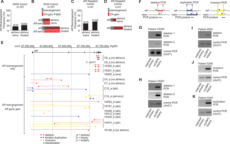
Purpose: Prostate cancer is the second leading cause of male cancer deaths. Castration-resistant prostate cancer (CRPC) is a lethal stage of the disease that emerges when endocrine therapies are no longer effective at suppressing activity of the androgen receptor (AR) transcription factor. The purpose of this study was to identify genomic mechanisms that contribute to the development and progression of CRPC.

Experimental design: We used whole-genome and targeted DNA-sequencing approaches to identify mechanisms underlying CRPC in an aggregate cohort of 272 prostate cancer patients. We analyzed structural rearrangements at the genome-wide level and carried out a detailed structural rearrangement analysis of the AR locus. We used genome engineering to perform experimental modeling of AR gene rearrangements and long-read RNA sequencing to analyze effects on expression of AR and truncated AR variants (AR-V).

Results: AR was among the most frequently rearranged genes in CRPC tumors. AR gene rearrangements promoted expression of diverse AR-V species. AR gene rearrangements occurring in the context of AR amplification correlated with AR overexpression. Cell lines with experimentally derived AR gene rearrangements displayed high expression of tumor-specific AR-Vs and were resistant to endocrine therapies, including the AR antagonist enzalutamide.

Conclusions: AR gene rearrangements are an important mechanism of resistance to endocrine therapies in CRPC.


Conflict of interest statement

Conflict of Interest Disclosure Statement: The authors declare no potential conflicts of interest

Figures

Figure 1:
Figure 1:
Diverse AR gene rearrangements are frequent in CRPC and detectable by whole genome DNA-seq. A, Schematic of the AR gene and classes of somatic alterations that occur in prostate cancer genomes. SNV, single nucleotide variant; CN, copy number; enh, enhancer. B, Frequency of AR gene alterations detected by whole genome DNA-seq of 130 primary prostate cancers or 101 metastatic CRPC specimens. Bars represent tumors with 1, 2, or more than 2 (3+) concurrent alterations in AR. Oncoprints on the right illustrate the type of AR alterations observed in each tumor sample. Each column represents an individual tumor. C, Map of AR gene rearrangements and breakpoint locations (triangles) within the AR gene body. Genome coordinates are genome build hg38. Locations of AR exons 1–8 are shown as black boxes. D, Coverage plots of DNA-seq reads in 3 tumors where an AR gene rearrangement was the only AR alteration detected by whole genome DNA-seq.
Figure 2:
Figure 2:
AR gene rearrangements are enriched in prostate cancer patients treated with AR-targeted endocrine therapies. A, Frequency of AR gene rearrangements in the 101-patient whole genome DNA-seq (WGS) cohort based on prior exposure of patients to abiraterone (abi) or enzalutamide (enz). B, Oncoprint illustrating AR alterations occurring in AR gene rearrangement-positive patients based on prior abiraterone or enzalutamide exposure. C, Frequency of AR gene rearrangements in a 41-patient AR-targeted DNA-seq cohort based on prior exposure of patients to abi or enz. D, Oncoprint illustrating AR alterations occurring in AR gene rearrangement-positive patients based on prior abi or enz exposure. E, Map of AR gene rearrangements and breakpoint locations (triangles) within the AR gene body discovered in a 41-patient AR-targeted DNA-seq cohort. Genome coordinates are genome build hg38. Patients are labeled based on whether tumor samples were obtained by autopsy (_a), biopsy (_b), or surgery (_s). Locations of AR exons 1–8 are shown as black boxes. F, Schematic of primer designs and PCR products for detection of genomic breakpoints arising from deletions, duplications, and inversions. Ref., reference genome; rearr., rearrangement. G-K, PCR products from patient-matched longitudinal samples of primary prostate cancer and metastatic CRPC (mCRPC).
Figure 3:
Figure 3:
Cells with engineered AR gene rearrangements have a clonal growth advantage under conditions of AR-targeted therapy. A, Schematic of experimental strategy for inducing and tracking clonal evolution of AR gene rearrangements using CRISPR/Cas9. Transfected cells were selected with puromycin (puro), and then cultured in medium supplemented with androgen-replete fetal bovine serum (FBS), charcoal-stripped, androgen-depleted FBS (CSS), enzalutamide (ENZ), or vehicle control (DMSO). B, AR gene rearrangements modeled using CRISPR/Cas9. C, Translocation-spanning PCR of DNA isolated from R1-AD1 cells that were transfected with gRNA and Cas9 plasmids as indicated. Quantitative PCR was used to track clonal enrichment or de-enrichment of translocations relative to the overall population of cells grown in D, 2-dimensional conditions or E, 3-dimensional (soft agar) conditions F, LNCaP cells were transfected as indicated and assayed as in (D). G-H, Deletion 1 from patient V5300_b, I-J, Deletion 2 from patient V5300_b, K-L, Deletion 1 from patient V5301_b, and M-N, Deletion 2 from patient V5301_b were modeled in R1-AD1 cells and monitored for clonal enrichment by breakpoint-spanning PCR as described for panels C and E. Gray bars represent mean +/− 95% confidence interval. Individual data points from biological replicate experiments are shown as black filled circles. P-values were determined using unpaired 2-sided t-tests.
Figure 4:
Figure 4:
Tumors harboring AR gene rearrangements as the only detectable AR gene alteration display high expression of AR variant mRNAs. A, Schematic of the translocation between AR and chromosome 11 in patient C9_a. B, Exon composition and quantification of AR transcripts isolated from a translocation-positive metastasis from patient C9_a cells using 3’ rapid amplification of cDNA ends (RACE) using a forward primer anchored in AR exon 1. Individual pixels represent discrete exons contained in individual AR transcripts. Pixel colors indicate whether that exon was spliced via annotated splice sites at the 5’ and/or 3’ ends of known exons. Read counts represent the number of single molecule transcripts that matched the indicated splicing pattern. AR transcripts were inspected manually for predicted translation, and annotated based on a previous nomenclature system. AR transcripts that had not been identified previously were classified as novel. C, Schematic of deletions in patient V5300_b and D, quantification of transcripts expressed in this metastasis as described in (B). E, Schematic of deletions in patient V5301_b and F, quantification of transcripts expressed in this metastasis as described in (B). G, Western blot of lysates from R1-AD1 cells transfected with gRNA and Cas9 plasmids as indicated and cultured in medium supplemented with the AR antagonist enzalutamide (ENZ), or vehicle control (DMSO) for 7 or 14 days as indicated. AR expression was determined using a pan-AR antibody that recognizes the AR N-terminal domain. Actin is a loading control. H, Schematic of deletions in patient V4002_b and I, quantification of transcripts expressed in this metastasis as described in (B). J, Comparison of AR-V7 mRNA 3’ terminus and AR-V7 protein C-terminus with AR mRNA variants discovered in prostate cancer metastases harboring AR gene rearrangements.
Figure 5:
Figure 5:
An AR-chromosome 11 translocation causes expression of a tumor-specific AR variant. A, CRISPR/Cas9 engineering strategy to generate an AR-chr11 translocation that models patient C9_a. Locations of PCR primers used for screening single cell clones are indicated. B, PCR-based characterization of parental R1-AD1 cells and a single cell clone (R1-X-11) derived by CRISPR/Cas9 engineering. C, Sanger sequencing of the PCR product from (B). D, Western blot of lysates from indicated cell lines. AR expression was determined using a pan-AR antibody that recognizes the AR N-terminal domain. Actin is a loading control. E, Schematic of the translocation between AR and chromosome 11, and location of the novel fusion exon expressed in patient C9_a. F, Exon composition and quantification of AR transcripts isolated from R1-X-11 cells using 3’ rapid amplification of cDNA ends (RACE) with a forward primer anchored in AR exon 1. Individual pixels represent discrete exons contained in individual AR transcripts. Pixel colors indicate whether that exon was spliced via annotated splice sites at the 5’ and/or 3’ ends of known exons. Read counts represent the number of single molecule transcripts that matched the indicated splicing pattern. AR transcripts were inspected manually for predicted translation, and annotated based on a previous nomenclature system. AR transcripts that had not been identified previously were classified as novel. G, Schematic of primers and siRNAs designed to study the C9a-AR-Vfusion1 transcript. H, RT-PCR with RNA isolated from indicated tumor and cell lines using primers illustrated in (G). I, Indicated cell lines were transfected with control (CTRL) siRNA or siRNAs illustrated in (G). Lysates were analyzed by Western blot as in (D).
Figure 6:
Figure 6:
A tumor-specific AR variant caused by AR-chromosome 11 translocation drives enzalutamide resistance. A, Growth of R1-AD1 and R1-X-11 cells was analyzed in culture medium containing enzalutamide (30μM) or DMSO as vehicle control. Bold black and red lines are mean +/− 95% CI from 3 independent experiments performed in biological quadruplicate (n=12). Light gray and red lines are the individual replicates. Significance was tested using unpaired 2-sided t-tests. B, Expression of AR target genes (FASN, FKBP5, and ABCC4) was tested by RT-PCR in R1-AD1 and R1-X-11 cells grown in culture medium containing enzalutamide (ENZ) or vehicle (DMSO) for 24h as in (A). Data are mean +/− 95% CI from 3 independent experiments performed in technical triplicate (n=9). Individual data points are shown. P-values were determined using unpaired 2-sided t-tests. C, Western blot of insoluble nuclear (chromatin) extracts from indicated cell lines. AR expression was determined using a pan-AR antibody that recognizes the AR N-terminal domain. Histone H3 is a loading control. D, R1-X-11 cells were transfected with control (CTRL) siRNA or siRNAs targeting exon f89 of the C9-a AR-Vfusion1 transcript and expression of AR target genes was tested by RT-PCR as in (B). Data are mean +/− 95% CI from 3 independent experiments performed in technical duplicate (n=6). Significance was tested using unpaired 2-sided t-tests. E, R1-AD1 and R1-X-11 cell lines were transfected with siRNAs as in (D) and growth was analyzed as in (A). Data are mean +/− 95% CI from 3 independent experiments performed in biological quadruplicate (n=12). P-values were determined using unpaired 2-sided t-tests.

References

    1. Attard G, Richards J, de Bono JS. New strategies in metastatic prostate cancer: targeting the androgen receptor signaling pathway. Clin Cancer Res 2011;17:1649–57. - PMC - PubMed
    1. Ryan CJ, Tindall DJ. Androgen receptor rediscovered: the new biology and targeting the androgen receptor therapeutically. J Clin Oncol 2011;29:3651–8. - PubMed
    1. Logothetis CJ, Gallick GE, Maity SN, Kim J, Aparicio A, Efstathiou E, et al. Molecular classification of prostate cancer progression: foundation for marker-driven treatment of prostate cancer. Cancer discovery 2013;3:849–61. - PMC - PubMed
    1. Watson PA, Arora VK, Sawyers CL. Emerging mechanisms of resistance to androgen receptor inhibitors in prostate cancer. Nat Rev Cancer 2015;15:701–11. - PMC - PubMed
    1. Guo Z, Yang X, Sun F, Jiang R, Linn DE, Chen H, et al. A novel androgen receptor splice variant is up-regulated during prostate cancer progression and promotes androgen depletion-resistant growth. Cancer Res 2009;69:2305–13. - PMC - PubMed

Publication types

MeSH terms