. 2020 Jan 13;11(1):23.
doi: 10.1038/s41419-019-2217-6.

Critical roles of conventional dendritic cells in autoimmune hepatitis via autophagy regulation

Affiliations

Critical roles of conventional dendritic cells in autoimmune hepatitis via autophagy regulation

Xiaoli Fan et al. Cell Death Dis. .

Abstract

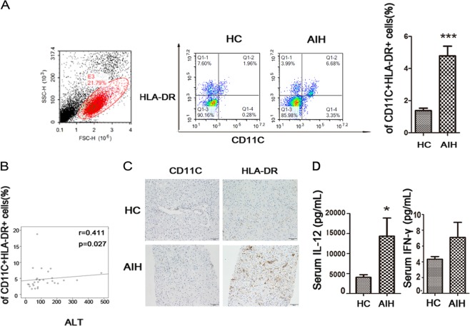
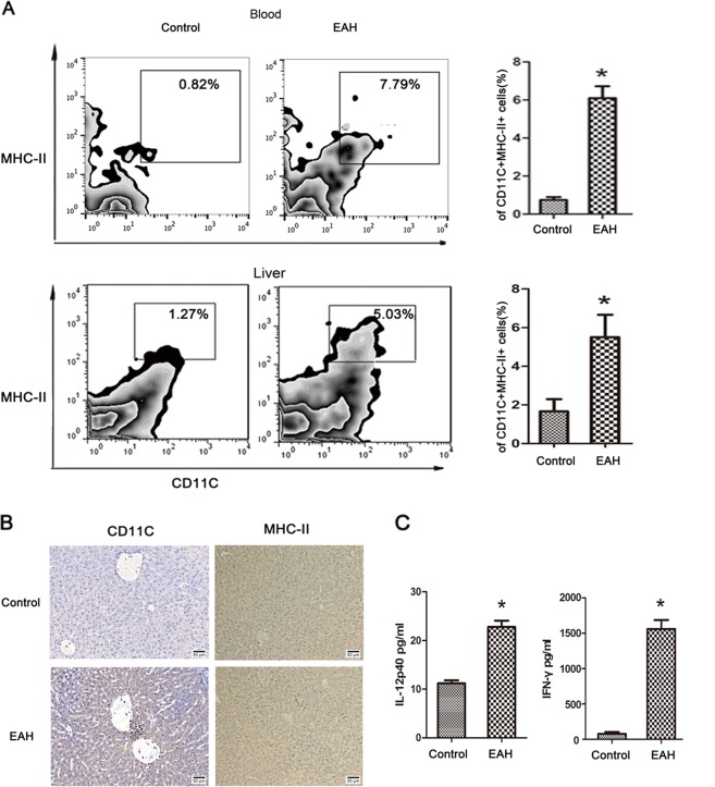
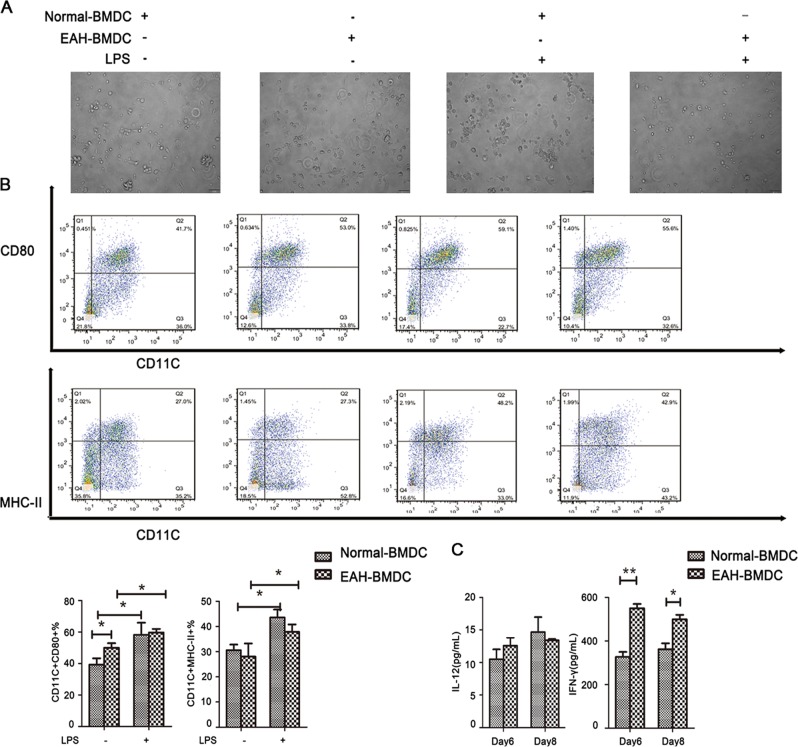
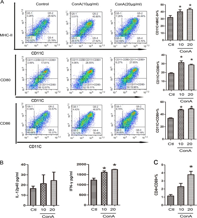
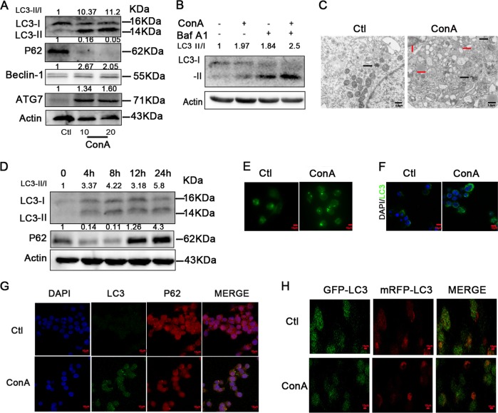
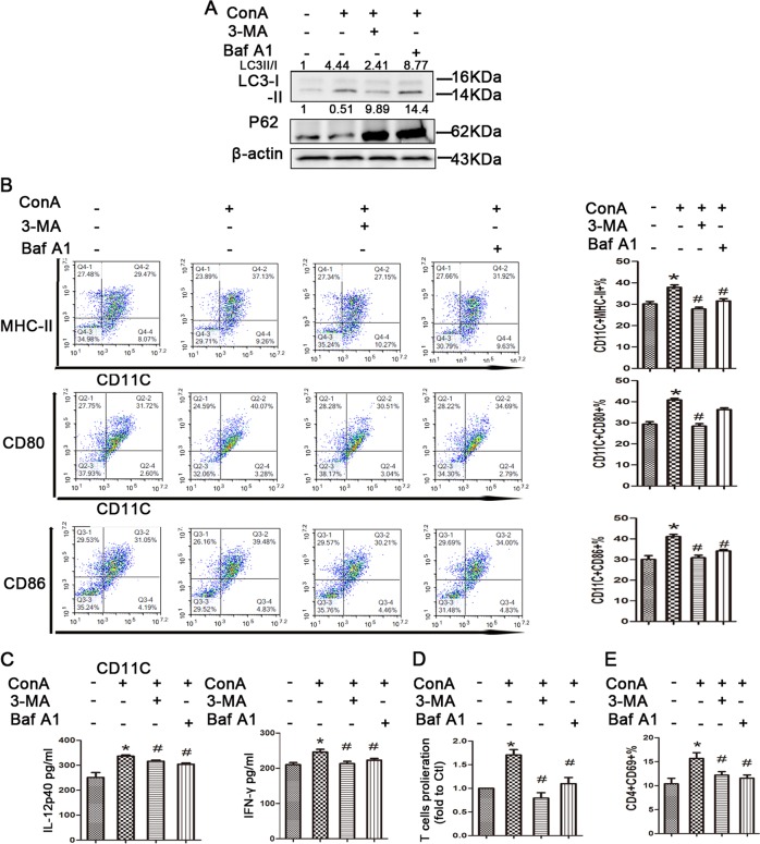
Autoimmune hepatitis (AIH) is a necroinflammatory disease associated with interactive cell populations of the innate and adaptive immune systems. The contribution of conventional dendritic cells (cDCs) to AIH and the underlying mechanism remain poorly understood. The frequency of peripheral mature cDCs increased in AIH patients and was positively correlated with disease severity. In experimental autoimmune hepatitis (EAH), hepatic accumulation of mature cDCs was observed, along with an increase in the periphery. Sequentially, bone marrow-derived dendritic cells (BMDC) from EAH mice exhibit more proinflammatory function than those from control mice. In vitro, ConA treatment promotes the maturation of BMDCs, which are characterized by higher expression of MHC-II, costimulatory molecules and cytokine secretion. ConA also induced the expression of autophagy-related protein and the formation of autophagosomes in DCs. To further investigate whether ConA-induced DC activation is associated with autophagy, we utilized 3-MA and bafilomycin A1 to block autophagy flux and accessed the maturation and function of DCs induced by ConA. 3-MA and bafilomycin A1 inhibited the mature status and proinflammatory cytokine secretion and diminished the proliferation and differentiation of CD4+ T cells when ConA-induced BMDCs cocultured CD4+ T cells. We demonstrated that cDCs contribute to the pathogenesis of AIH through excessive maturation. Aberrant autophagy flux plays a vital role in the immunogenic maturation of cDCs in AIH, and tolerogenic cDCs by inhibition of autophagy flux can be exploited as a new therapeutic approach for AIH.


Conflict of interest statement

The authors declare that they have no conflict of interest.

Figures

Fig. 1
Fig. 1. Circulating mature cDCs was increased in AIH patients.
a Analysis of CD11C + HLA-DR + DCs in the peripheral blood of HCs and AIH patients. b Frequency of circulating mature cDCs is positively correlated with hepatic inflammation marker in AIH. c Representative immunohistochemistry staining of CD11C and HLA-DR in the livers of HC and AIH patients. d ELISA analysis of cytokines in the peripheral blood of HCs and AIH patients. Original magnification: x200. Bars show the mean ± SD; *p < 0.05 and ***p < 0.001.
Fig. 2
Fig. 2. Circulating and intrahepatic cDCs accumulation in EAH model.
a Frequency of mature cDCs in blood and liver tissues in EAH, respectively; b IHC staining of CD11C and MHC-II in the hepatic tissues between the two groups. Original magnification: x200; c Serum IL-12 and IFN-γ. Bars show the mean ± SD; *p < 0.05 versus control.
Fig. 3
Fig. 3. Pro-inflammatory function of BMDCs from EAH mice.
a Morphology and phenotypic characteristics of BMDCs at day 8 treated with LPS. BMDCs were treated with or without LPS for 24 h. b FACS analysis of mature status when culture of BMDCs from normal and AIH mice before and after LPS challenged. c ELISA analysis of cytokines released of induced BMDCs on day 6 and day 8 from normal and AIH mice. *p < 0.05 and **p < 0.01.
Fig. 4
Fig. 4. ConA trigger phenotypic characteristics of BMDCs in vitro.
a Impact of ConA treatment on MHC-II and surface costimulatory molecules on BMDCs in each group. b ELISA analysis of cytokines released by BMDCs after ConA stimulation. c CD69 expression in T cells surface in each group. *p < 0.05 versus control.
Fig. 5
Fig. 5. ConA stimulation induces autophagy in BMDCs and DC2.4 cell line.
a Western blotting analysis of LC3-II,P62, Beclin-1 and ATG7 in BMDCs in each group; b Autophagic flux was calculated by dividing the levels of LC3-II in the presence of bafilomycin A1 by that without bafilomycin A1; c Transmission electron microscopy examination of autophagosomes and autolysosomes in the BMDCs. Autophagosomes were indicated by black arrows, and autolysosomes were indicated by red arrows; d Western blotting analysis of LC3-II and P62 in DC2.4 cell line in each group; e Acidic vacuoles in DC2.4 cells by ConA treatment, were stained with MDC and then observed under a fluorescence microscope; f LC3B-positive puncta in DC2.4 were examined for identifying autophagosome formation by fluorescence staining; g Immunofluorescence of LC3-II and p62 upon ConA treatment; h ConA-induced autophagy was evaluated by GFP-mRFP-LC3 plasmid transfection.
Fig. 6
Fig. 6. Effect of autophagy inhibitors on ConA‑induced BMDCs.
a Western blotting analysis of LC3-II and P62 levels in BMDCs in each group; b impact of autophagy inhibitors treatment on MHC-II and surface costimulatory molecules on BMDCs in each group. c ELISA analysis of cytokines released by BMDCs after autophagy inhibitors stimulation. d BMDCs-induced proliferation of T cells and CD69 expression in T cells surface in each group. *p < 0.05 versus control; #p < 0.05 versus ConA treatment.

References

    1. Manns MP, Lohse AW, Vergani D. Autoimmune hepatitis–Update 2015. J. Hepatol. 2015;62:S100–S111. doi: 10.1016/j.jhep.2015.03.005. - DOI - PubMed
    1. Webb GJ, Hirschfield GM, Krawitt EL, Gershwin ME. Cellular and molecular mechanisms of autoimmune hepatitis. Annu. Rev. Pathol. 2017 doi: 10.1146/annurev-pathol-020117-043534. - DOI - PubMed
    1. Floreani A, et al. Etiopathogenesis of autoimmune hepatitis. J. Autoimmun. 2018 doi: 10.1016/j.jaut.2018.10.020. - DOI - PubMed
    1. Sozzani S, Del Prete A, Bosisio D. Dendritic cell recruitment and activation in autoimmunity. J. Autoimmun. 2017;85:126–140. doi: 10.1016/j.jaut.2017.07.012. - DOI - PubMed
    1. Shortman K, Liu YJ. Mouse and human dendritic cell subtypes. Nat. Rev. Immunol. 2002;2:151–161. doi: 10.1038/nri746. - DOI - PubMed

Publication types