. 2020 Jan 14;12(1):4.
doi: 10.1038/s41368-019-0071-0.

The dimension and morphology of alveolar bone at maxillary anterior teeth in periodontitis: a retrospective analysis-using CBCT

Affiliations

The dimension and morphology of alveolar bone at maxillary anterior teeth in periodontitis: a retrospective analysis-using CBCT

Xue Zhang et al. Int J Oral Sci. .

Abstract

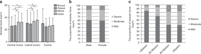
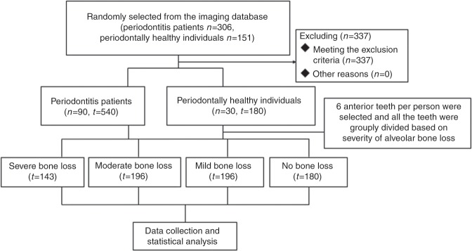
The morphology of the alveolar bone at the maxillary anterior teeth in periodontitis patients was evaluated by cone-beam computed tomography (CBCT) to investigate the distribution of alveolar defects and provide guidance for clinical practice. Ninety periodontitis patients and 30 periodontally healthy individuals were selected to determine the morphology of the alveolar bone at the maxillary anterior teeth according to the degree of bone loss, tooth type, sex and age. The differences in the dimensions between periodontitis patients and healthy individuals were compared, and the distribution of alveolar bone defects was analyzed. A classification system was established regarding the sagittal positions and angulations of the teeth. The buccal residual bone was thicker and the lingual bone was thinner in the periodontitis patients than in the periodontally healthy individuals, and there were differences between the different tooth types, sexes and age subgroups. The buccal undercut was close to the alveolar ridge, while fenestration was reduced and the apical bone height was higher in periodontitis patients than in periodontally healthy individuals. The apical bone height increased with the aggravation of bone loss and age. The proportions of different sagittal positions changed with the aggravation of bone loss. Moreover, the teeth moved more buccally regarding the positions of the maxillary anterior teeth. The morphology of the alveolar bone at the maxillary anterior teeth differed between periodontitis patients and healthy individuals, and the differences were related to the degree of bone loss, tooth type, sex and age.


Conflict of interest statement

The authors declare no competing interests.

Figures

Fig. 1
Fig. 1. Differences in alveolar bone morphology by tooth type, sex and age in patients with periodontitis.
a Distribution of alveolar bone loss at different teeth sites. Asterisk indicates Kruskal–Wallis test among different age group, P < 0.05. b Comparison in the proportion of alveolar bone loss between two sexes. c Comparison in the proportion of alveolar bone loss among different age groups.
Fig. 2
Fig. 2. Sagittal root position in relation to the anterior maxillary alveolar process in this study.
a Type P1. b Type B2. c Type M2. d Type P2. e Type N2. f Type B3. g Type M3. h Type P3. i Type N3.
Fig. 3
Fig. 3. Diagrams of the alveolar morphology measurements.
a Axial views at the maxillary arch level were perpendicular to the alveolar ridge. b Coronal views were parallel to the alveolar ridge. c Points C1, C2, C0, and A represent the buccal CEJ point, palatal CEJ point, midpoint of line C1C2 and apical point. Line C0A was the long axis of teeth. d Buccal line (line l) and palatal line (line l′) were marked by a line of best fit to the buccal and palatal alveolar surfaces, respectively. The long axis of the alveolar process (line l2) was marked by bisecting the line l and l′. e Lines perpendicular to the long axis of the teeth through points R and A were drawn. Bone loss was the vertical distance from C to R (buccal, palatal, mesial, and distal sites were h1, h2, h3, and h4, respectively), while the residual bone height was measured from R to A (buccal, palatal, mesial, and distal sites were h1′, h2′, h3′ and h4′, respectively). f Buccal and palatal bone thicknesses were obtained by measuring the distance perpendicular to the long axis of the teeth at the mid-root level (t1, t2), apical level (t3, t4) and 1 mm apical to the alveolar crest (t5, t6). g A line parallel to the long axis of the alveolar process and tangent to the buccal cortical bone was drawn. The distance from the deepest point D of the undercut to the line l2′ was defined as the buccal undercut depth. A line m perpendicular to the long axis of the alveolar process at point R was drawn. The distance from the buccal undercut convex-concave junction point P to line m was defined as the buccal undercut location. h The angulation between l1 and l2 was the required angle. The apical bone height h6 was measured along the long axis of the alveolar process from the root apex to the nasopalatine duct.
Fig. 4
Fig. 4. Classification of sagittal root position according to position and angulation.
a Type B. b Type M. c Type P. d Type N. e Type 1. f Type 2. g Type 3.
Fig. 5
Fig. 5. Patient flowchart of the study.
N represents number of the individuals and T represents number of the teeth.

References

    1. Papapanou P. N. et al. Periodontitis: consensus report of workgroup 2 of the 2017 World Workshop on the Classification of Periodontal and Peri-Implant Diseases and Conditions. J. Periodontol. 89, S173–S182 (2018). - PubMed
    1. Tonetti Maurizio S., Greenwell Henry, Kornman Kenneth S. Staging and grading of periodontitis: Framework and proposal of a new classification and case definition. Journal of Periodontology. 2018;89:S159–S172. doi: 10.1002/JPER.18-0006. - DOI - PubMed
    1. Gracco A, et al. Upper incisor position and bony support in untreated patients as seen on CBCT. Angle Orthod. 2009;79:692–702. doi: 10.2319/081908-437.1. - DOI - PubMed
    1. Cook DR, et al. Relationship between clinical periodontal biotype and labial plate thickness: an in vivo study. Int J. Periodontics Restor. Dent. 2011;31:345–354. - PubMed
    1. Rocca APL, et al. Anterior maxillary and mandibular biotype: relationship between gingival thickness and width with respect to underlying bone thickness. Implant Dent. 2012;21:507–515. doi: 10.1097/ID.0b013e318271d487. - DOI - PubMed

Publication types