. 2020 Jan 13;10(1):195.
doi: 10.1038/s41598-019-56853-6.

A butanolic fraction from the standardized stem extract of Cassia occidentalis L delivered by a self-emulsifying drug delivery system protects rats from glucocorticoid-induced osteopenia and muscle atrophy

Affiliations

A butanolic fraction from the standardized stem extract of Cassia occidentalis L delivered by a self-emulsifying drug delivery system protects rats from glucocorticoid-induced osteopenia and muscle atrophy

Subhashis Pal et al. Sci Rep. .

Abstract

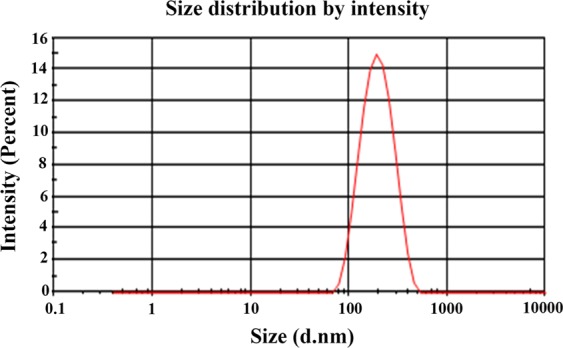
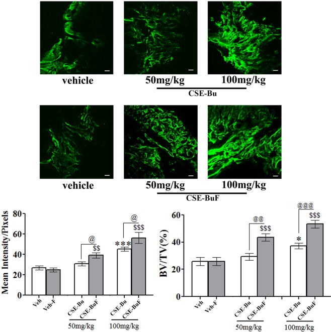
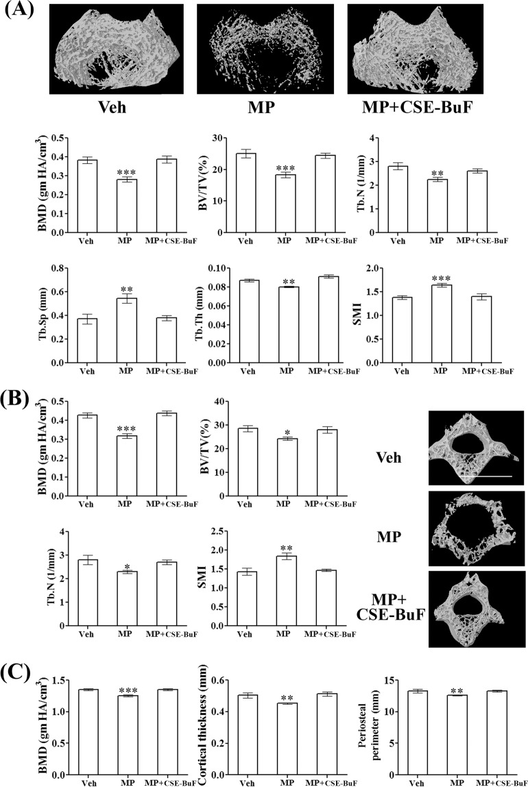
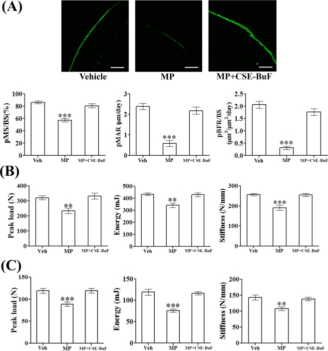
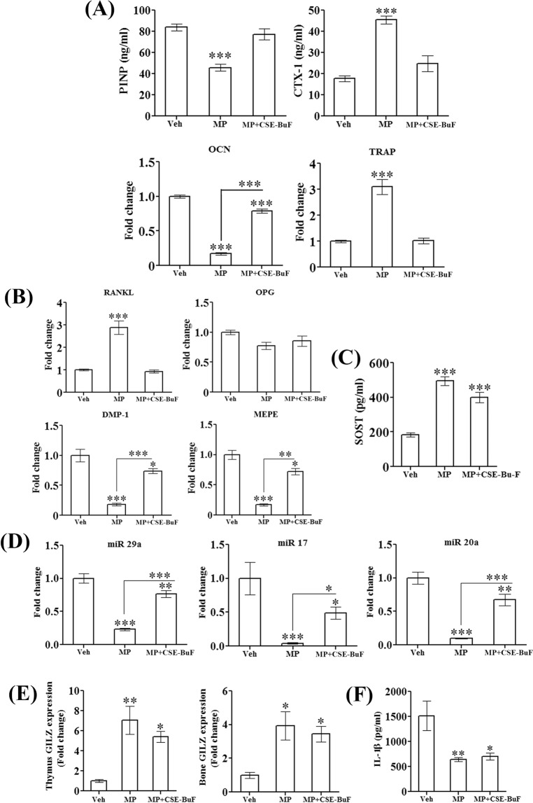
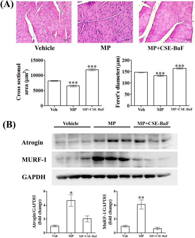
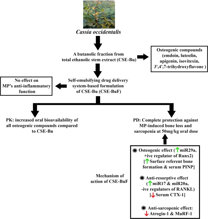
We recently reported that a butanol soluble fraction from the stem of Cassia occidentalis (CSE-Bu) consisting of osteogenic compounds mitigated methylprednisone (MP)-induced osteopenia in rats, albeit failed to afford complete protection thus leaving a substantial scope for further improvement. To this aim, we prepared an oral formulation that was a lipid-based self-nano emulsifying drug delivery system (CSE-BuF). The globule size of CSE-BuF was in the range of 100-180 nm of diluted emulsion and the zeta potential was -28 mV. CSE-BuF enhanced the circulating levels of five osteogenic compounds compared to CSE-Bu. CSE-BuF (50 mg/kg) promoted bone regeneration at the osteotomy site and completely prevented MP-induced loss of bone mass and strength by concomitant osteogenic and anti-resorptive mechanisms. The MP-induced downregulations of miR29a (the positive regulator of the osteoblast transcription factor, Runx2) and miR17 and miR20a (the negative regulators of the osteoclastogenic cytokine RANKL) in bone was prevented by CSE-BuF. In addition, CSE-BuF protected rats from the MP-induced sarcopenia and/or muscle atrophy by downregulating the skeletal muscle atrogenes, adverse changes in body weight and composition. CSE-BuF did not impact the anti-inflammatory effect of MP. Our preclinical study established CSE-BuF as a prophylactic agent against MP-induced osteopenia and muscle atrophy.


Conflict of interest statement

Although not applicable to this study, Naibedya Chattopadhyay has received (a) consultancy fees from Glenmark Pharmaceuticals, Navi Mumbai, India, (b) consultancy fees from Glaxo Smith Kline-Consumer Healthcare, Gurgaon, India, and (c) served as an Advisory Board Member of Alkem Laboratories Ltd, India. Subhashis Pal, Naresh Mittapelly, Athar Husain, Sapana Kushwaha, Sourav Chattopadhyay, Padam Kumar, Eppalapally Ramakrishna, Sudhir Kumar, Rakesh Maurya, Sabyasachi Sanyal, Jiaur R. Gayen and Prabhat Ranjan Mishra declare that they have no financial and non-financial competing interests.

Figures

Figure 1
Figure 1
Globule size and size distribution of SEDDS after dilution. The average globule size and PDI of formulation after dilution 1:100 in distilled water was found to be was 166.7 d.nm and 0.186 respectively.
Figure 2
Figure 2
CSE-BuF enhanced the bone regenerative effect of the butanolic fraction of the stem extract of Cassia occidentalis (CSE-Bu). Representative micrographs of calcein labeled callus from various groups (upper panel) and quantified data of calcein labeling and µCT analysis of callus at fracture site (lower panel). Scale bar, 50 µm. Water was used as vehicle (veh) for CSE-Bu group and blank SEDDS without extract as vehicle (veh-F). Data are expressed as mean ± SEM (n = 8–12/group); *p < 0.05 and ***p < 0.001 versus veh; $$p < 0.01 and $$$p < 0.001 versus veh-F; @p < 0.05, @@p < 0.01 and @@@p < 0.001 were used to compare between the formulated and unformulated groups.
Figure 3
Figure 3
CSE-BuF prevented the osteopenic effect of MP. (A) Upper panel showing 3-D representative images of different groups. Volumetric BMD (vBMD) and various trabecular parameters including BV/TV, Tb.N, Tb.Th, Tb.Sp and SMI of femur metaphysis are shown in the lower panels. (B) Left panel showing various trabecular parameters of L5 vertebra and right panel showing the 3-D representative images of indicated groups. (C) Assessment of cortical parameters at femur diaphysis was done by vBMD, cortical thickness and periosteal perimeter. Data are expressed as mean ± SEM (n = 8–10/group); *P < 0.05, **P < 0.01 and ***P < 0.001 compared to vehicle (Veh).
Figure 4
Figure 4
CSE-BuF promoted bone formation and bone strength in MP-treated rats. (A) Upper panel showing representative calcein labelling (20X) at femur diaphysis in the indicated groups and lower panel showing the quantified data including periosteal (p)-mineralizing surface/bone surface (p-MS/BS), mineral apposition rate (pMAR), and bone formation rate/bone surface (pBFR/BS). Scale bar, 50 µm. (B) L5 compression test and (C) femur 3-point bending of indicated groups are shown. Data are expressed as mean ± SEM (n = 6/group) **P < 0.01 and ***P < 0.001 compared to vehicle (Veh).
Figure 5
Figure 5
CSE-BuF promoted osteoblast activity and suppressed resorption without altering anti-inflammatory property of methylprednisolone. (A) Assessment of serum turnover markers (PINP and CTX-1), bone-specific osteogenic (OCN) and resorptive (TRAP) gene expression in the indicated groups. (B) Assessment of osteocyte markers (DMP-1, MEPE), RANKL and OPG in bones of indicated groups by qPCR. (C) Assessment of serum sclerostin (SOST) levels in the indicated groups by ELISA. (D) Assessment of expression of various corticosteroid-regulated miRNAs (miR 29a, miR17 and miR20a) involved in bone remodeling in the indicated groups. (E) Assessment of expression of GLIZ mRNA in thymus and proximal femur of indicated groups by qPCR. (F) Assessment of serum IL-1β levels in the indicated groups by ELISA. Data are expressed as mean ± SEM (n = 6/group) *P < 0.05, **P < 0.01 and ***P < 0.001 compared to vehicle (veh).
Figure 6
Figure 6
CSE-BuF prevented MP-induced muscle atrophy. (A) Upper panel showing representative H&E stained sections of gastrocnemius muscle (40X) and the lower panel showing quantified cross sectional area and feret’s diameter of muscle fibers in the indicated groups. Scale bar, 50 µm. (B) Western blot images and densitometric quantification of bands of atrogin-1 and MuRF-1 expression in gastrocnemius muscle from rats of the indicated groups are shown. Data are expressed as mean ± SEM (n = 6/group) *P < 0.05, **P < 0.01 and ***P < 0.001 compared to vehicle (veh).
Figure 7
Figure 7
A schematic diagram illustrating the effcts of CSE-Bu or CSE-BUF on rat bones and skeletal muscle given concurrently with MP treatment. Picture used in this figure was taken by Subhashis Pal (author of this manuscript). PK, pharmacokinetics and PD: pharmacodynamics.

References

    1. Sokka T, et al. QUEST-RA: quantitative clinical assessment of patients with rheumatoid arthritis seen in standard rheumatology care in 15 countries. Ann. Rheum. Dis. 2007;66:1491–1496. doi: 10.1136/ard.2006.069252. - DOI - PMC - PubMed
    1. Caplan A, Fett N, Rosenbach M, Werth VP, Micheletti RG. Prevention and management of glucocorticoid-induced side effects: A comprehensive review: A review of glucocorticoid pharmacology and bone health. J. Am. Acad. Dermatol. 2017;76:1–9. doi: 10.1016/j.jaad.2016.01.062. - DOI - PubMed
    1. Whittier X, Saag KG. Glucocorticoid-induced Osteoporosis. Rheum. Dis. Clin. North. Am. 2016;42:177–189, x. doi: 10.1016/j.rdc.2015.08.005. - DOI - PubMed
    1. Arazzi M, et al. [Therapy of glucocorticoid induced osteoporosis]. G. Ital. Nefrol.34, (2017). - PubMed
    1. Lane NE, et al. Parathyroid hormone treatment can reverse corticosteroid-induced osteoporosis. Results of a randomized controlled clinical trial. J. Clin. Invest. 1998;102:1627–1633. doi: 10.1172/JCI3914. - DOI - PMC - PubMed

MeSH terms