. 2020 Jan 13;11(1):236.
doi: 10.1038/s41467-019-13878-9.

Distinct DNA repair pathways cause genomic instability at alternative DNA structures

Affiliations

Distinct DNA repair pathways cause genomic instability at alternative DNA structures

Jennifer A McKinney et al. Nat Commun. .

Abstract

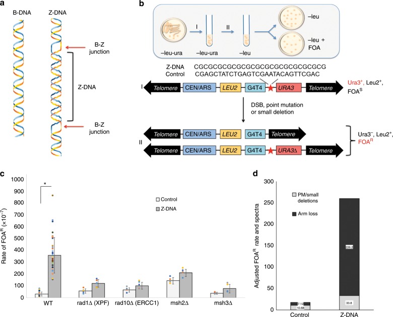
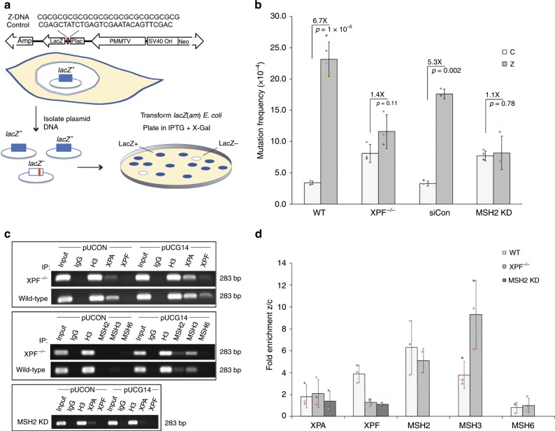
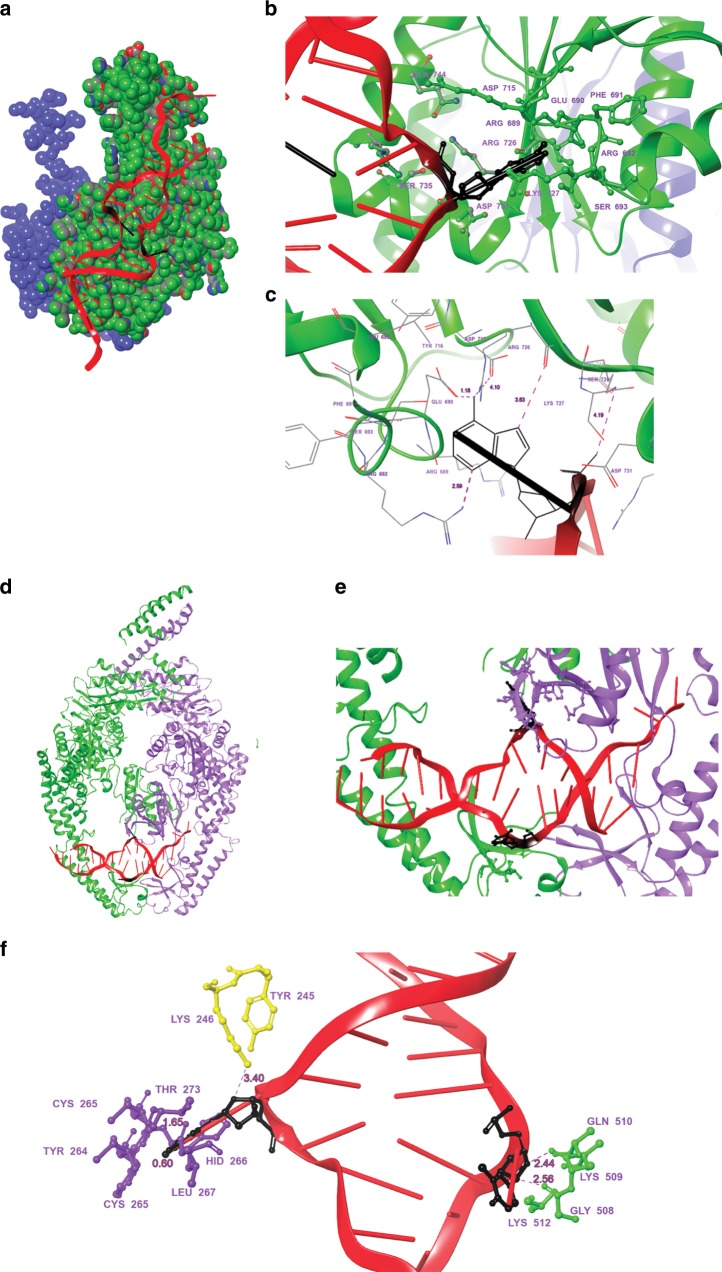
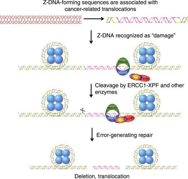
Alternative DNA structure-forming sequences can stimulate mutagenesis and are enriched at mutation hotspots in human cancer genomes, implicating them in disease etiology. However, the mechanisms involved are not well characterized. Here, we discover that Z-DNA is mutagenic in yeast as well as human cells, and that the nucleotide excision repair complex, Rad10-Rad1(ERCC1-XPF), and the mismatch repair complex, Msh2-Msh3, are required for Z-DNA-induced genetic instability in yeast and human cells. Both ERCC1-XPF and MSH2-MSH3 bind to Z-DNA-forming sequences, though ERCC1-XPF recruitment to Z-DNA is dependent on MSH2-MSH3. Moreover, ERCC1-XPF-dependent DNA strand-breaks occur near the Z-DNA-forming region in human cell extracts, and we model these interactions at the sub-molecular level. We propose a relationship in which these complexes recognize and process Z-DNA in eukaryotes, representing a mechanism of Z-DNA-induced genomic instability.


Conflict of interest statement

The authors declare no competing interests.

Figures

Fig. 1
Fig. 1. Rad10-Rad1 and Msh2-Msh3 are required for Z-DNA-induced genetic instability in S. cerevisiae.
a B-DNA (left panel) and left-handed Z-DNA flanked by two regions of B-DNA (right panel) B-Z junctions are labeled (red arrows). b Schematic of the YAC fragility assay. c Mutation rates calculated as rate of FOAR for WT, rad1Δ, rad10Δ, msh2Δ, and msh3Δ strains containing control B-DNA or Z-DNA-forming YACs [Student’s t-test was used to calculate a P value. *P < 0.001]. Data are presented as means ± SEM of triplicate experiments, and values of each repeat are shown as dot plots on the bars. d Mutation spectra of FOAR clones from control B-DNA or Z-DNA-forming YACs in the WT strain were determined by sequencing and PCR, and mutants were categorized as either point mutation (PM)/small deletion within the URA3 gene or complete loss of the right arm of the chromosome as a result of a DSB. “Adjusted FOAR rate and spectra” was calculated by multiplying the percentage of each category (calculated from 30 mutants) by the total FOAR ratio. See also Supplementary Figs. 1 and 2, and Supplementary Tables 1 and 2.
Fig. 2
Fig. 2. ERCC1-XPF and MSH2-MSH3 are required for Z-DNA-induced mutagenesis in human cells.
a Schematic of blue-white mutation assay (adapted from Vasquez et al.). b Mutation frequencies for WT, XPF-deficient, siRNA control, and siRNA-depleted MSH2 human cell lines. Student’s t-test was used to calculate statistical values. c Representative ChIP analysis performed on (pUCON and pUCG14) plasmids in WT and XPF-deficient human cells using antibodies against NER (top panel) and MMR (middle panel) proteins, and in MSH2-depleted cells (bottom panel). IgG and H3 served as negative and positive controls, respectively. d Quantification of ChIP analysis comparing the Z-DNA-forming sequence to the control B-DNA region (Z/C), normalized to input. Data in b, d are presented as means ± SEM of triplicate experiments, and values of each repeat are shown as dot plots on the bars. See also Supplementary Figs. 3–7, and Supplementary Table 1.
Fig. 3
Fig. 3. ERCC1-XPF cleaves near the Z-DNA-forming region.
a S1 nuclease assay schematic. b Top panel: SYBR Gold-stained agarose gel demonstrating S1 cleavage at single-stranded regions corresponding to the B-Z junctions on the Z-DNA-forming plasmid (Z), resulting in ~780 bp fragment (red arrow) that is absent in the control B-DNA plasmid (C). Bottom panel: higher exposure of S1 cleavage product. [NOC, nicked open circular; L, linear; SC, supercoiled species]. c PCR primer extension assay schematic using plasmid DNA in human XPF-proficient or XPF-deficient WCE. Purified DNA was used as a template for a primer extension assay using either the left or right primer (green arrows). d PCR products were separated on a 1.5% agarose gel revealing extra cleavage products (red arrows) from the Z-DNA plasmid in XPF-proficient WCE. Plasmid DNA only (P) served as a negative control, and EcoRI (E), restriction at the Z-DNA-forming insert served as a positive control, resulting in ~160 or ~180 bp products (black arrow). Lane 1: template only; lane 2: left primer; and lane 3: right primer. See also Supplementary Table 1. Extension products of >1000 bp resulted from primer “run off” from the template. Shorter extension products resulting from cleavage on plasmids in WCE are indicated by red arrows.
Fig. 4
Fig. 4. Models of the ERCC1-XPF and MSH2–3 complexes docked to Z-DNA.
a Corey-Pauling-Koltun (CPK) sphere representation of the ERCC1-XPF complex (ERCC1 complex in blue and XPF in green) docked to Z-DNA (red, B-Z junction in black) at the site with maximum interaction with the protein. b Interacting interface residues between the B-Z junction nucleotides with the ERCC1-XPF complex. c Distances between the major interacting and active residues with the B-Z junction. d Model showing different positions of Z-DNA (red, PDB: 2ACJ) bound to the MSH2-MSH3 complex (MSH2: green, MSH3: cyan, PDB: 3THY) in the increasing order of their interactions with the binding site residues (from left to right). e Highlighted interaction (from “d”) between the B-Z junction and the binding residues within 3 Å of the nucleotides. f Distances and labels of the interacting residues with the B-Z junction are highlighted.
Fig. 5
Fig. 5. Proposed mechanism for Z-DNA-induced genetic instability in eukaryotes.
During DNA metabolic processes, the CG repeat sequence is unwrapped from histones (blue circles) and negative supercoiling is generated, which stimulates Z-DNA formation. The structure of Z-DNA is recognized as “damage” by the MSH2-MSH3 complex, signaling repair. The ERCC1-XPF complex is recruited to the site for cleavage near the Z-DNA-forming region (processing of the B-Z junction on the right side of Z-DNA is shown in the figure; however, similar processing could also occur at the other B-Z junction on the left, marked as a scissor), resulting in DSBs in an attempt to repair the “damage”. The breaks may be processed in an error-free fashion, or the breaks may be processed in an error-generating fashion resulting in genomic instability in the form of large deletions and translocations, which may contribute to disease etiology.

Similar articles

Cited by

References

    1. Wang G, Christensen LA, Vasquez KM. Z-DNA-forming sequences generate large-scale deletions in mammalian cells. Proc. Natl Acad. Sci. USA. 2006;103:2677–2682. doi: 10.1073/pnas.0511084103. - DOI - PMC - PubMed
    1. Ditlevson JV, et al. Inhibitory effect of a short Z-DNA forming sequence on transcription elongation by T7 RNA polymerase. Nucleic Acids Res. 2008;36:3163–3170. doi: 10.1093/nar/gkn136. - DOI - PMC - PubMed
    1. Wang G, Carbajal S, Vijg J, DiGiovanni J, Vasquez KM. DNA structure-induced genomic instability in vivo. J. Natl Cancer Inst. 2008;100:1815–1817. doi: 10.1093/jnci/djn385. - DOI - PMC - PubMed
    1. Bacolla Albino, Tainer John A., Vasquez Karen M., Cooper David N. Translocation and deletion breakpoints in cancer genomes are associated with potential non-B DNA-forming sequences. Nucleic Acids Research. 2016;44(12):5673–5688. doi: 10.1093/nar/gkw261. - DOI - PMC - PubMed
    1. Eichman BF, Schroth GP, Basham BE, Ho PS. The intrinsic structure and stability of out-of-alternation base pairs in Z-DNA. Nucleic Acids Res. 1999;27:543–550. doi: 10.1093/nar/27.2.543. - DOI - PMC - PubMed

Publication types