. 2020 Feb;21(2):145-157.
doi: 10.1038/s41590-019-0568-x. Epub 2020 Jan 13.

Influenza-induced monocyte-derived alveolar macrophages confer prolonged antibacterial protection

Affiliations

Influenza-induced monocyte-derived alveolar macrophages confer prolonged antibacterial protection

Helena Aegerter et al. Nat Immunol. 2020 Feb.

Abstract

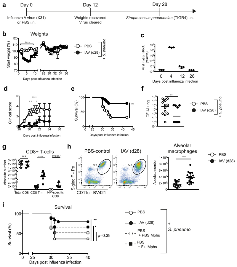
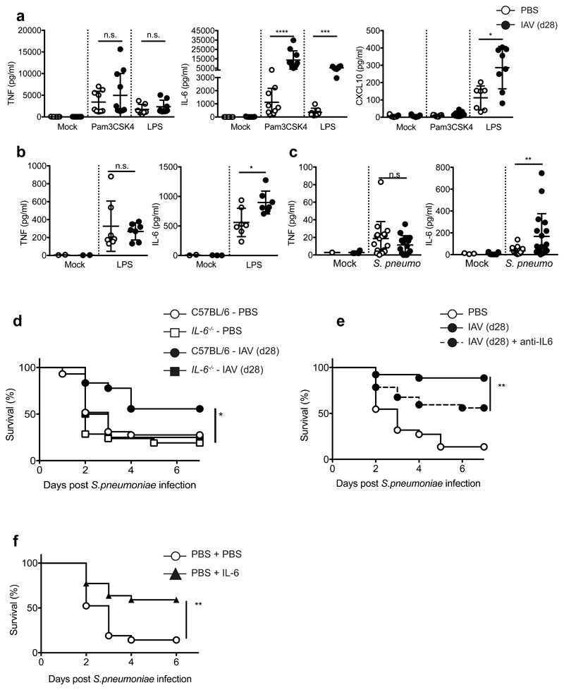
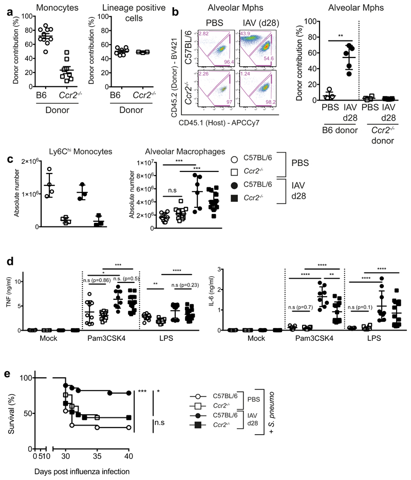
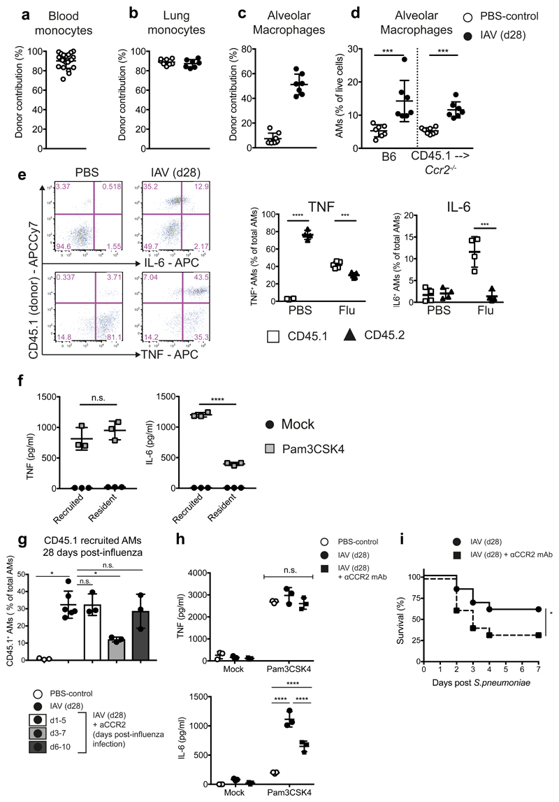
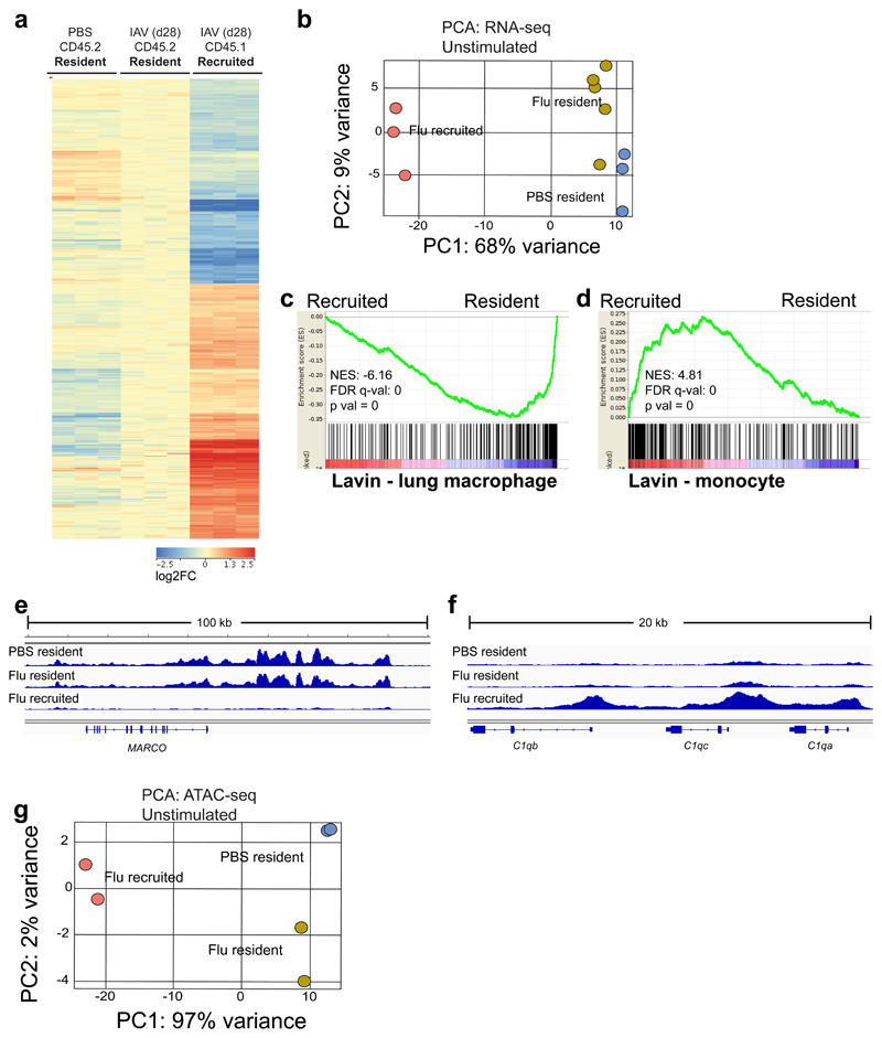
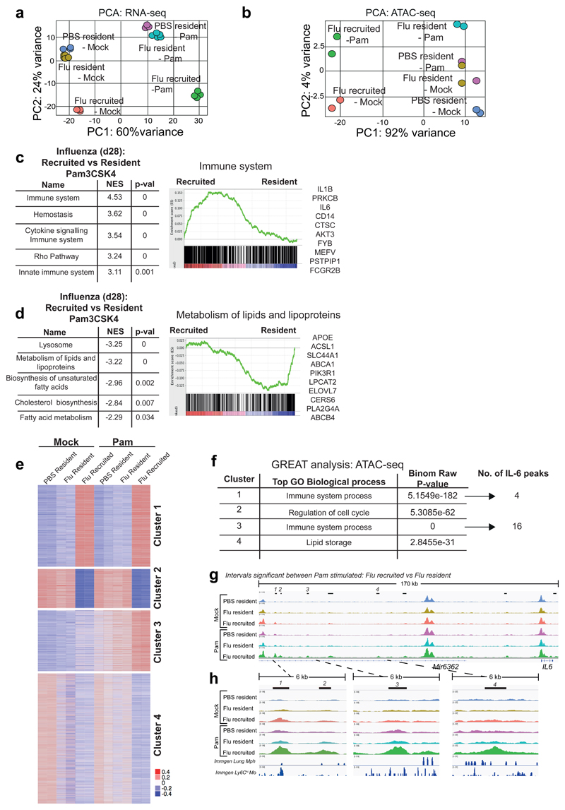
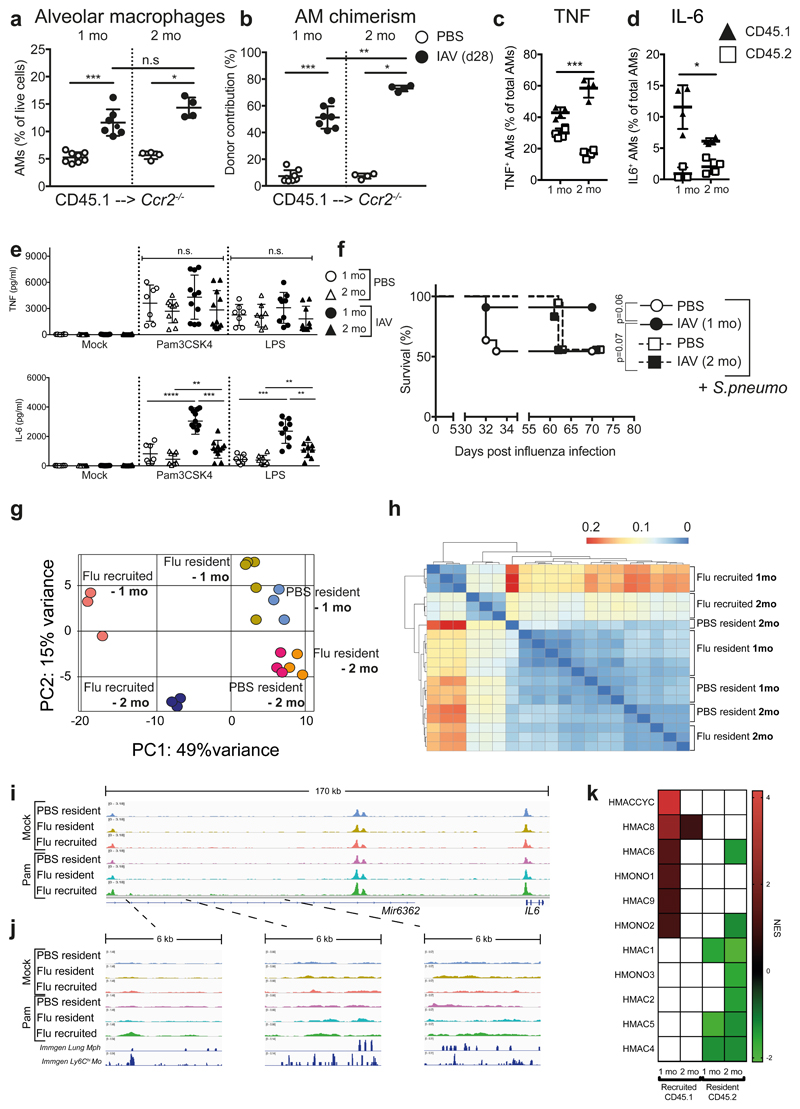
Despite the prevalence and clinical importance of influenza, its long-term effect on lung immunity is unclear. Here we describe that following viral clearance and clinical recovery, at 1 month after infection with influenza, mice are better protected from Streptococcus pneumoniae infection due to a population of monocyte-derived alveolar macrophages (AMs) that produce increased interleukin-6. Influenza-induced monocyte-derived AMs have a surface phenotype similar to resident AMs but display a unique functional, transcriptional and epigenetic profile that is distinct from resident AMs. In contrast, influenza-experienced resident AMs remain largely similar to naive AMs. Thus, influenza changes the composition of the AM population to provide prolonged antibacterial protection. Monocyte-derived AMs persist over time but lose their protective profile. Our results help to understand how transient respiratory infections, a common occurrence in human life, can constantly alter lung immunity by contributing monocyte-derived, recruited cells to the AM population.


Conflict of interest statement

Competing Interests statement

E.M.H. and S.B. were employees of GSK at the time of this study. The other authors declare that they have no conflict of interest.

Figures

Figure 1
Figure 1. At one month post-influenza, mice are more protected against S. pneumoniae infection, and have an increased population of alveolar macrophages which confers resistance.
a, Schematic of infection schedule. b,d-f, Female C57BL/6 mice were inoculated with 8000 TCID50 X31 (H3N2)/30μl i.n. (influenza - black circles) or PBS (white circles). 28 days later, all mice were infected with 5 x 10⁵ CFU TIGR4/30μl i.n. (S. pneumoniae) and monitored for (b) weight loss, (d) clinical scores and (e) survival (p=0.0002). n=32 mice (PBS)/ n=30 mice (X31) from 4 independent experiments. c, qRT-PCR of influenza matrix protein of whole lung at indicated time points post-influenza infection. f, Lung colony forming units (CFUs) from whole lung at 48 hours post-S. pneumoniae. (p=0.0047). n=14 mice (PBS)/ n=11 mice (X31) from one representative experiment. g-h, Quantification of cells identified in lung by flow cytometry. n= 9 mice (PBS)/n=17 mice (X31) from 3 independent experiments. (g) n.s.=0.0789 i, Alveolar macrophages were isolated from naive or post-influenza mice and transferred (106 AMs/20μl i.t.) into naive mice. 24 hours later, mice were infected with S. pneumoniae and survival was monitored. n=25 mice (X31/PBS + PBS Mphs)/n=30 mice (PBS), n= 35 mice (PBS+ X31 Mphs) from 3 independent experiments (** = 0.0012, *=0.0128, n.s. = 0.386). Data shown as arithmetic means ±SEM (weights) or ±SD for cell quantification and statistical significance assessed by a two-tailed test using 2-way ANOVA (weights and clinical scores), Log Rank (Mantel-Cox) test for survival or Mann-Whitney U for cell quantification and lung CFUs. Data in (c) and (f) are shown as geometric means. * p=≤0.05, ** p=≤0.01, *** p=≤0.001, **** p=≤0.0001.
Figure 2
Figure 2. Alveolar macrophages from post-influenza mice produce increased IL-6 that confers protection from S. pneumoniae infection.
a, Alveolar macrophages isolated from the lungs of naive and post-influenza mice were stimulated ex vivo for 16 hours with 100ng/ml Pam3CSK4 or LPS. Cytokines were quantified in the supernatants by 36plex. n= 7 mice (PBS)/ n=11 mice (X31) from 2-3 independent experiments. (a, TNF– Pam3CSK4 n.s. = 0.37/LPS n.s. = 0.15) b-c, Cytokine quantification by ELISA in the broncho-alveolar lavage (BAL) of mice (b) 16 hours after in vivo administration of 1.5μg/50μl LPS i.n. (* = 0.038, n.s. = 0.8) or (c) 6 hours post infection with 5 x 10⁵ CFU TIGR4/30μl i.n. (** = 0.0071, n.s. = 0.1915) d, Male and female C57BL/6 (PBS n=36 mice /X31 =25 mice) and IL-6-/- (PBS n = 21 mice, X31 n=13 mice) mice were infected with 5 x 10⁵ CFU TIGR4/30μl i.n. (*=0.0171) in 5 independent experiments e, Mice were treated with 90μg anti-IL6 (MPF-20F3) in 50μl i.n. at -2 hours prior to S. pneumoniae infection and survival was monitored. n=26 mice (X31/X31 +anti-IL-6)/n=24 mice (PBS) from 3 independent experiments (**=0.0088) f, Naive mice were treated with 500ng IL6 in 50μl i.n. at -3hrs and +16hrs post-S. pneumoniae infection and survival was monitored. n=21 mice (PBS)/ n=22 mice (PBS + IL-6) from 4 independent experiments (**=0.0027). Data shown as arithmetic means ±SD and statistical significance assessed by a two-tailed test using Mann-Whitney U (a-c) or Log Rank (Mantel-Cox) test for survival (d-f). * p=≤0.05, *** p=≤0.01, *** p=≤0.001, **** p=≤0.0001.
Figure 3
Figure 3. CCR2-dependent monocyte recruitment contributes to the population of AMs and their altered functional profile at one month post-influenza.
a,b, CD45.1 mice were treated with Busulfan and reconstituted with CD45.2 B6 or Ccr2-/- BM. a, Frequency and chimerism of blood monocytes (n=10 mice C57BL/6, n=8 mice Ccr2-/-) and lineage (CD3/NK1.1/CD19) positive cells (n=7 mice C57BL/6, n=3 mice Ccr2-/-) 40 days post-reconstitution, prior to influenza infection. 2 independent experiments. b, Chimerism of alveolar macrophages in the lung one month post-influenza (n=5 mice from two independent experiments **=0.0079). c, Quantification of cells identified in naive and post-influenza lungs of B6 (n=10 mice (PBS)/n=6 mice (X31)) and Ccr2-/- (n=13 mice (PBS)/n=12 mice (X31) mice by flow cytometry. 3 independent experiments (n.s.=0.1010, ***=0.0005). d, Alveolar macrophages were isolated from the lungs of naive and post-influenza C57BL/6 and Ccr2-/-mice and stimulated ex vivo for 16 hours with 100ng/ml Pam3CSK4 or LPS. IL-6 and TNF were quantified in the supernatants by ELISA. n=5-15 mice from 2-4 independent experiments. e, 8-10 week old female C57BL/6 (n=30 mice (PBS)/ n=28 mice (X31)) and Ccr2-/- (n=25 mice (PBS/X31) mice were infected with 8000 TCID50 X31/30μl i.n. 28 days later, all mice were infected with 5 x 10⁵ CFU TIGR4/30μl i.n. (S. pneumoniae) and monitored for survival. 3 independent experiments (*= 0.0167, n.s = 0.08). Data shown as arithmetic means ±SD and statistical significance assessed using two-tailed tests; Mann-Whitney U or Log Rank (Mantel-Cox) test for survival (e). * p=≤0.05, *** p=≤0.01, *** p=≤0.001, **** p=≤0.0001.
Figure 4
Figure 4. Quantifying the contribution of CCR2-dependent monocytes to alveolar macrophages and their cytokine production at one month post-influenza.
Ccr2-/- mice were treated with Busulfan and reconstituted with CD45.1 WT BM. a, Chimerism of blood monocytes 30 days post-reconstitution and prior to influenza infection (n=22 mice). b,c, Chimerism of (b) lung monocytes and (c) alveolar macrophages in naïve (n=8 mice) and influenza-experienced (n=7 mice) mice. d, Frequency of alveolar macrophages in naive and influenza experienced WT and chimeric mice. (b-d) Represents two independent experiments. e, Naive and IAV (d28) alveolar macrophages were isolated from the lungs of chimeric mice and stimulated ex vivo with 100ng/ml Pam3CSK4. ICCS for IL6 (top panels) and TNF (bottom panels) n=4 mice from 1 experiment. f, Donor (CD45.1) and host (CD45.2) alveolar macrophage populations from post-influenza mice were FACS-sorted and stimulated for 16hr with 100ng/ml Pam3CSK4. IL-6 and TNF were quantified by ELISA in the supernatants. n=3 mice from one experiment. (n.s.= 0.74) g, Chimeras as above were treated with anti-CCR2 mAb (MC21) at the indicated time points post-influenza and the BM-derived (CD45.1) cell contribution to AMs measured at d28 post-influenza. n=3 (all groups except for n=6 mice (X31)) from 1 experiment. n.s. = 0.9, *=0.02 h,i, Female C57BL/6 mice were influenza-infected and administered anti-CCR2 mAb or PBS at d3-7. h, Alveolar macrophages were isolated on d28 from whole lung and stimulated ex vivo for 16 hours with 100ng/ml Pam3CSK4. Cytokines were quantified in the supernatants by ELISA. n=3 mice from 1 experiment. i, At d28 post-influenza, mice were infected with 5 x 10⁵ CFU TIGR4/30μl i.n. (S. pneumoniae) and monitored for survival. n=25 mice (X31)/n=24 mice (X31 + anti-CCR2) from three independent experiments *=0.0479. Data shown is arithmetic means ±SD and statistical significance assessed by a two-tailed test using Mann-Whitney U or Log Rank (Mantel-Cox) test for survival (i). * p=≤0.05, *** p=≤0.001, **** p=≤0.0001.
Figure 5
Figure 5. Recruited macrophages are distinct in their expression and chromatin profile, relative to resident macrophages at one month post-influenza.
RNA-seq and ATAC-seq of AMs FACS-sorted from the lungs of naïve and post-influenza Busulfan chimeras. a, Heat map of genes differentially expressed (FC>1.5, 1-way ANOVA with Benjamini-Hochberg correction, p<0.05) between CD45.2 (Ccr2-/-) resident and CD45.1 recruited AMs. b, Principal component analysis (PCA) plot of RNA-seq data from resident and recruited macrophages from post-influenza and naive lungs of Ccr2-/- Busulfan chimeras. c,d, Gene set enrichment analysis (GSEA) of Flu resident (CD45.2) vs Flu recruited (CD45.1) macrophages using clusters from Lavin et al. representing (c) lung macrophages and (d) blood monocytes. e,f, IGV tracks from ATAC-seq data; depicting chromatin in unstimulated resident and recruited macrophages from naïve and post-influenza lungs of Ccr2-/- Busulfan chimeras. All tracks were group-autoscaled to enable comparison. g, PCA plot of ATAC-seq data from unstimulated resident and recruited macrophages from naïve and post-influenza lungs.
Figure 6
Figure 6. Following TLR stimulation, origin determines gene expression, chromatin profile and IL-6 gene accessibility.
AMs were FACS-sorted based on congenic markers from the lungs of naïve and post-influenza Busulfan chimeras, and stimulated ex vivo with Pam3CSK4. PCA plot of (a) RNA-seq data and (b) ATAC-seq data from unstimulated and Pam3CSK4-stimulated CD45.2 (Ccr2-/-) resident and CD45.1 recruited AMs. c,d, Top 5 pathways (FDR q-val <0.01) from GSEA of Pam3CSK4 stimulated AMs from post-influenza lungs. NES = Normalized Enrichment Score. Representative GSEA plots are shown, with the top 10 highest ranked genes from each GSEA listed. e, ATAC-seq data as in (b) shown as heatmap of mean centred k-means clustering of rlog values generated by DESeq2 for differentially accessible intervals found to be significant (FDR ≤ 0.01) in at least one pairwise comparison. f, GREAT analysis of ATAC-seq-derived intervals from each cluster in (e). g,h, IGV tracks of ATAC-seq data for AMs, showing the IL-6 locus and upstream peaks. Significantly differentially accessible intervals in the comparison Flu_resident_Pam vs Flu_recruited_Pam are marked using bars. ImmGen chromatin tracks from lung macrophages and blood Ly6Chi monocytes are shown underneath (h). All tracks were group-autoscaled to enable comparison.
Figure 7
Figure 7. Monocyte-derived alveolar macrophages persist for 2 months post-influenza, but do not produce increased IL-6 and afford bacterial protection.
a,b, Frequency (a) and chimerism (b) of alveolar macrophages in the lungs of CD45.1 --> Ccr2-/- Busulfan chimeras at one (n=8 mice PBS/n=7 mice X31) and two months (n=4 mice) post-influenza infection. From 1-2 separate experiments. c,d, Alveolar macrophages were isolated from the lungs of CD45.1 --> Ccr2-/- Busulfan chimeras at one and two months post-influenza infection and stimulated ex vivo with Pam3CSK4. TNF (c) and IL-6 (d) were assessed by intracellular cytokine staining. n=3-5 mice from 1 experiment. e, Alveolar macrophages were isolated from the lungs of naive and 2-mo post-influenza C57BL/6 mice and stimulated ex vivo for 16 hours with 100ng/ml Pam3CSK4 or LPS. IL-6 and TNF were quantified by ELISA in the supernatants. n=7-16 mice from 4 independent experiments. f, At one or two months post-influenza infection, mice were infected with 5 x 10⁵ CFU TIGR4/30μl i.n. (S. pneumoniae) and monitored for survival. n=11 mice (PBS 1 mo/X31 1mo) n=12 mice (X31 2 mo) n=18 mice (PBS 2 mo) from 2-3 independent experiments. n.s.= 0.0734 g, PCA plot of RNA-seq data from unstimulated resident (CD45.2) and recruited (CD45.1) AMs from the lungs of post-influenza and naive Ccr2-/- Busulfan chimeras at one and two months post-infection. h, Hierarchical clustering of RNA-seq data using Pearson dissimilarity (1-correlation) of the samples’ rlog values generated by DESeq2. i,j, IGV tracks from ATAC-seq of unstimulated and Pam3CSK4-stimulated resident (CD45.2) and recruited (CD45.1) AMs from the lungs of post-influenza and naive Ccr2-/-Busulfan chimeras at two months post-infection. The intervals chosen are identical to those in Fig. 6g,h. The scale on corresponding tracks are identical to Fig. 6g,h. to to enable comparison. k, Heatmap showing the NES of GSEA performed using human macrophage, monocyte and dendritic cell datasets obtained from Zilionis et al.. Data shown as arithmetic means ±SD and statistical significance assessed by two-tailed tests using 2-way ANOVA or Log Rank (Mantel-Cox) test for survival. * p=≤0.05, ** p=≤0.01, *** p=≤0.001.

References

    1. Dahl ME, Dabbagh K, Liggitt D, Kim S, Lewis DB. Viral-induced T helper type 1 responses enhance allergic disease by effects on lung dendritic cells. Nat Immunol. 2004;5:337–343. doi: 10.1038/ni1041. - DOI - PubMed
    1. Didierlaurent A, et al. Sustained desensitization to bacterial Toll-like receptor ligands after resolution of respiratory influenza infection. J Exp Med. 2008;205:323–329. doi: 10.1084/jem.20070891. - DOI - PMC - PubMed
    1. Machiels B, et al. A gammaherpesvirus provides protection against allergic asthma by inducing the replacement of resident alveolar macrophages with regulatory monocytes. Nat Immunol. 2017;18:1310–1320. doi: 10.1038/ni.3857. - DOI - PubMed
    1. Beura LK, et al. Normalizing the environment recapitulates adult human immune traits in laboratory mice. Nature. 2016;532:512–516. doi: 10.1038/nature17655. - DOI - PMC - PubMed
    1. Reese TA, et al. Sequential Infection with Common Pathogens Promotes Human-like Immune Gene Expression and Altered Vaccine Response. Cell Host Microbe. 2016;19:713–719. doi: 10.1016/j.chom.2016.04.003. - DOI - PMC - PubMed

Publication types

MeSH terms