. 2020 Dec;14(6):2569-2586.
doi: 10.1007/s11682-019-00209-1.

Altered brain structures in the dorsal and ventral language pathways in individuals with and without developmental language disorder (DLD)

Affiliations

Altered brain structures in the dorsal and ventral language pathways in individuals with and without developmental language disorder (DLD)

Joanna C Lee et al. Brain Imaging Behav. 2020 Dec.

Abstract

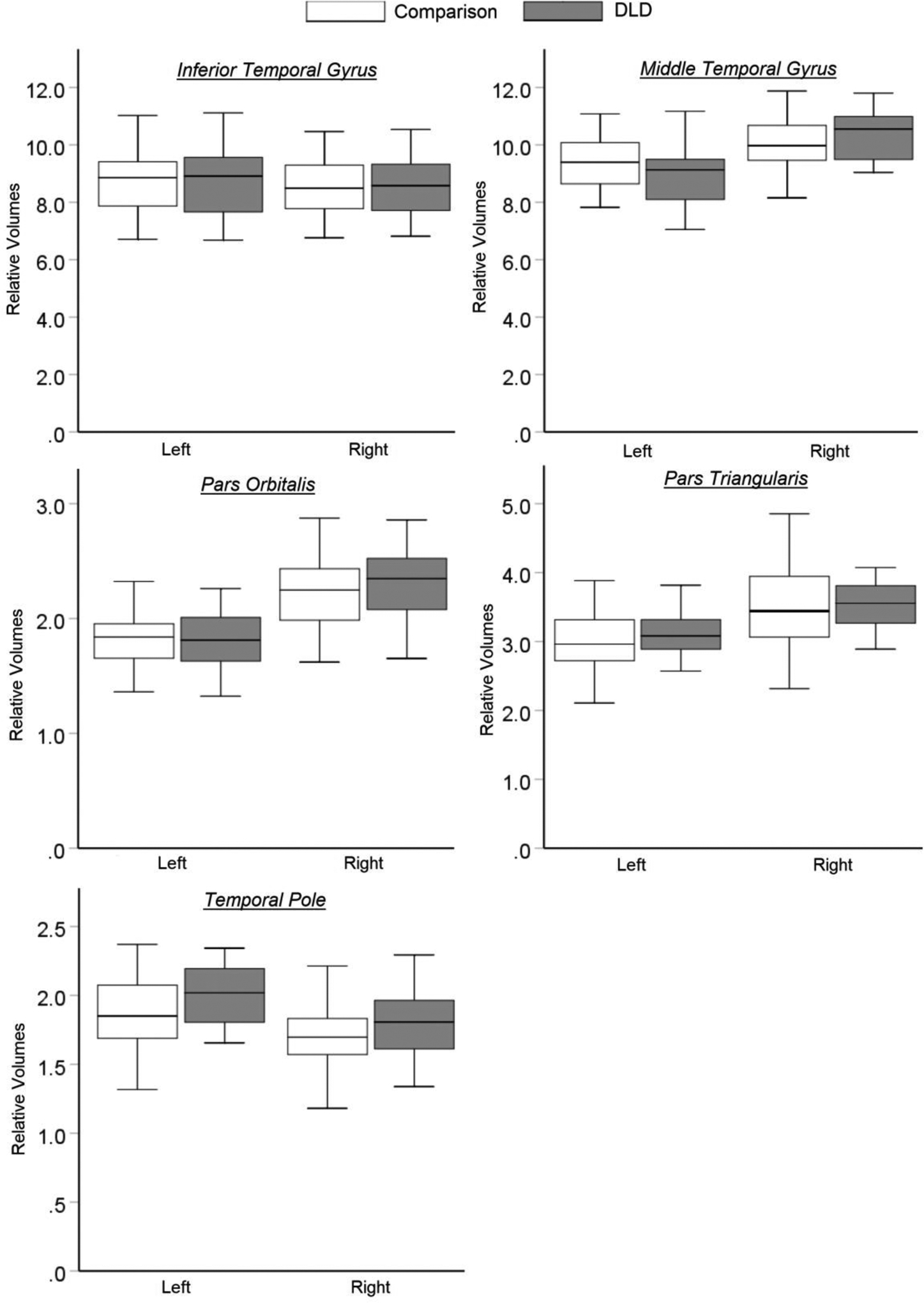
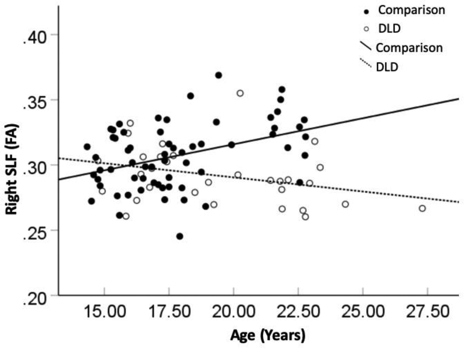
Developmental Language Disorder (DLD) is a neurodevelopmental disorder characterized by difficulty learning and using language, and this difficulty cannot be attributed to other developmental conditions. The aim of the current study was to examine structural differences in dorsal and ventral language pathways between adolescents and young adults with and without DLD (age range: 14-27 years) using anatomical magnetic resonance imaging (MRI) and diffusion tensor imaging (DTI). Results showed age-related structural brain differences in both dorsal and ventral pathways in individuals with DLD. These findings provide evidence for neuroanatomical correlates of persistent language deficits in adolescents/young adults with DLD, and further suggest that this brain-language relationship in DLD is better characterized by taking account the dynamic course of the disorder along development.

Keywords: Developmental language disorder; Dorsal pathway; Structural brain imaging; Ventral pathway.


Figures

Figure 1.
Figure 1.
FreeSurfer map of the regions of interest in the left hemisphere. The transverse temporal gyrus (TTG) is buried within the lateral sulcus. ITG: Inferior temporal gyrus; MTG: Middle temporal gyrus; SMG: Supramarginal gyrus; STG: Superior temporal gyrus; TP: Temporal Pole; TTG: Transverse temporal gyrus.
Figure 2.
Figure 2.
JHU white matter tractography atlas of the regions of interest. IFOF: Inferior fronto-occipital fasciculus; ILF: Inferior longitudinal fasciculus; SLF: Superior longitudinal fasciculus; UF: Uncinate fasciculus.
Figure 3a.
Figure 3a.
Relative gray matter volumes of the regions of interest in the dorsal pathway. C: the comparison group; DLD: the Developmental Language Disorder group.
Figure 3b.
Figure 3b.
Relative gray matter volumes of the regions of interest in the ventral pathway. C: the comparison group; DLD: the Developmental Language Disorder group.
Figure 4.
Figure 4.
Significant age-by-group interaction effect on fractional anisotropy (FA) measure in the right superior longitudinal fasciculus (SLF) in the dorsal pathway.
Figure 5.
Figure 5.
Significant age-by-group interaction effect on fractional anisotropy (FA) measure in the right uncinate fasciculus (UF) in the ventral pathway.
Figure 6.
Figure 6.
Significant age-by-group interaction effect on fractional anisotropy (FA) measure in the left inferior longitudinal fasciculus (ILF) in the ventral pathway.

References

    1. Alexander AL, Lee JE, Lazar M, & Field AS (2007). Diffusion tensor imaging of the brain. Neurotherapeutics, 4, 316–329. - PMC - PubMed
    1. Anthony JL, Davis C, Williams JM, & Anthony TI (2014). Preschoolers’ oral language ability: A multilevel examination of dimensionality. Learning and Individual Differences, 35, 56–61.
    1. Aron AR, Robbins TW, & Poldrack RA (2004). Inhibition and the right inferior frontal cortex. Trends in Cognitive Sciences, 8(4), 170–177. - PubMed
    1. Asato MR, Terwilliger R, Woo J, & Luna B (2010). White matter development in adolescence: A DTI study. Cerebral Cortex, 20(9), 2122–2131. - PMC - PubMed
    1. Badcock NA, Bishop DVM, Hardiman MJ, Barry JG, & Watkins KE (2012). Co-localisation of abnormal brain structure and function in specific language impairment. Brain and Language, 120(3), 310–320. - PMC - PubMed

LinkOut - more resources