

Cellular and genomic approaches for exploring structural chromosomal rearrangements
- PMID: 31933061
- PMCID: PMC7131874
- DOI: 10.1007/s10577-020-09626-1
Cellular and genomic approaches for exploring structural chromosomal rearrangements
Abstract
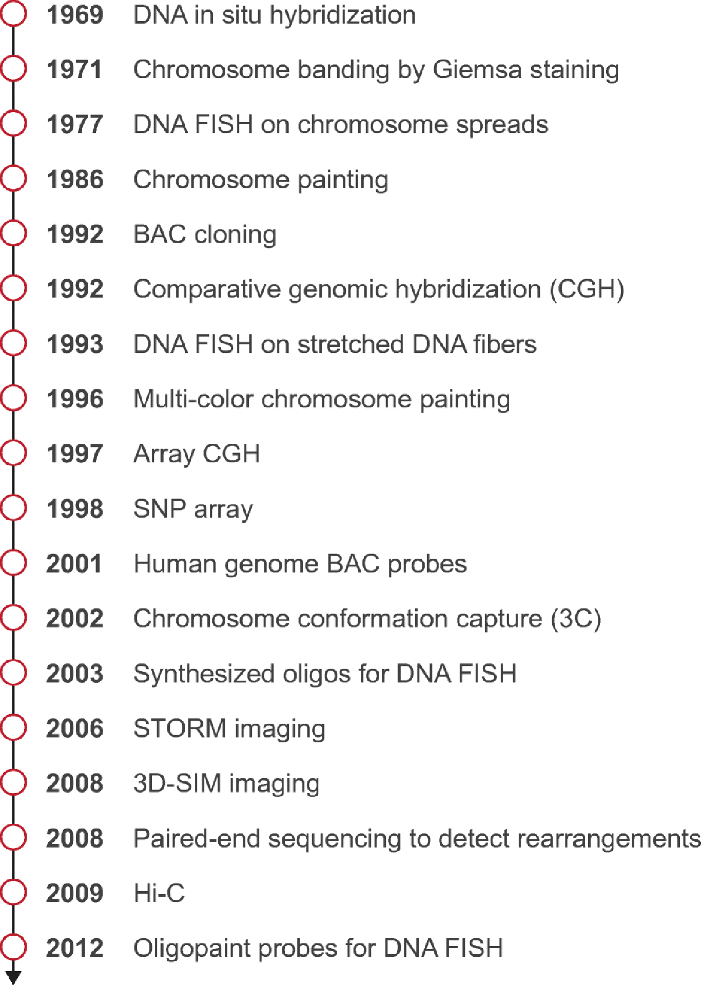
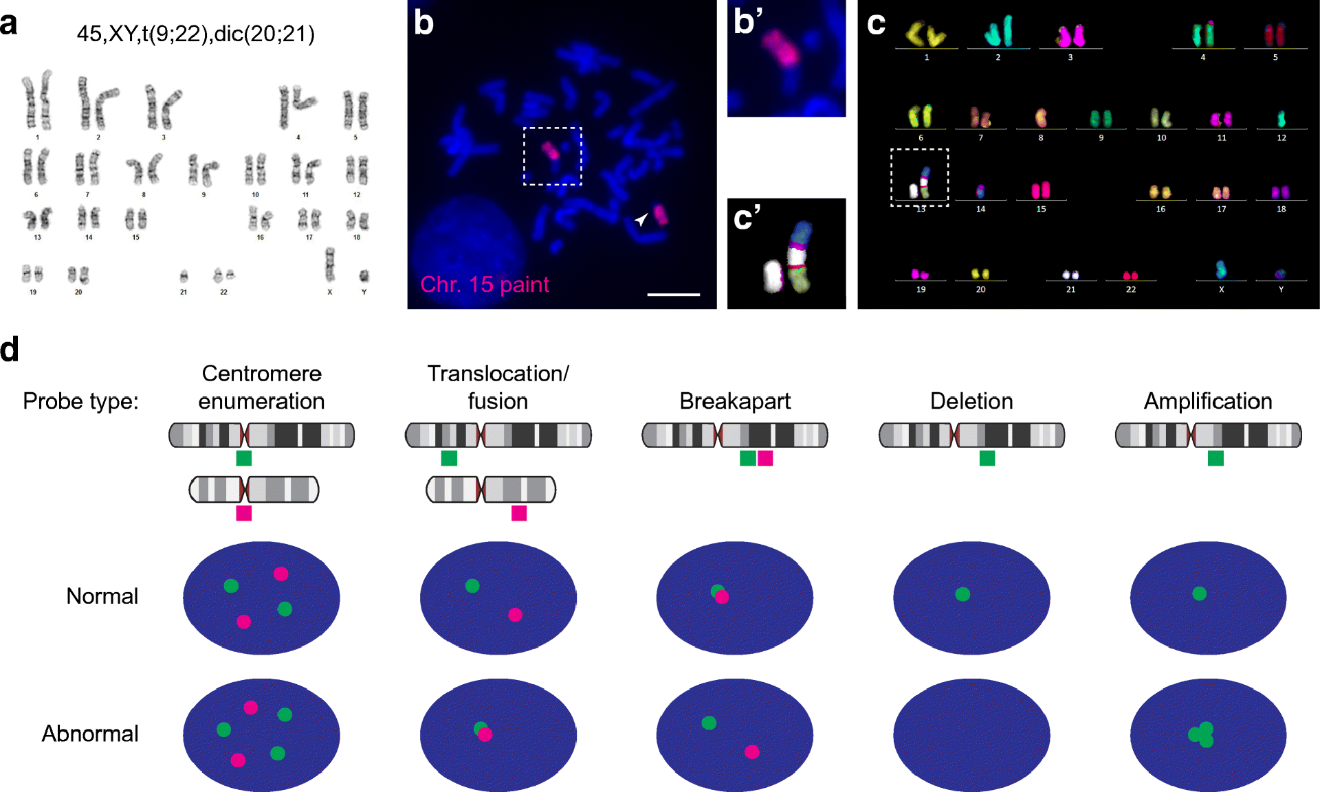
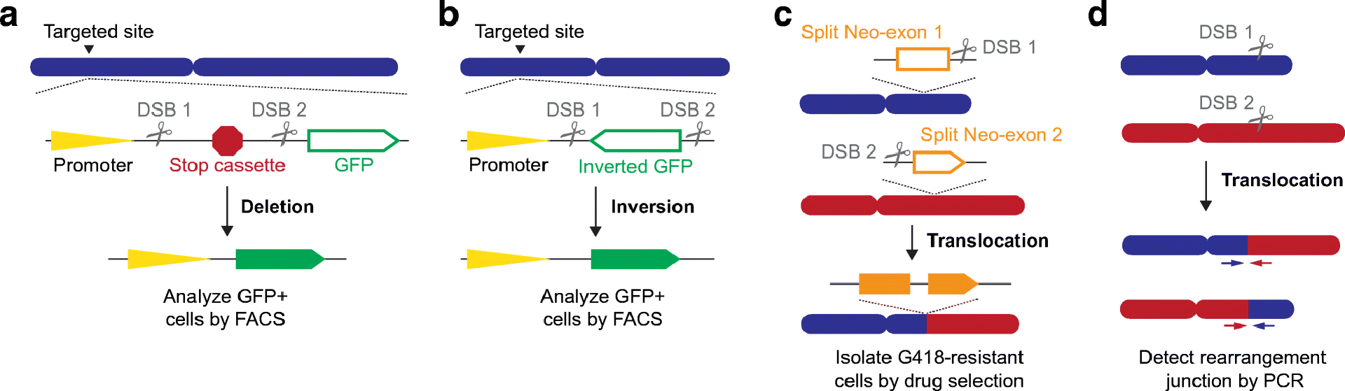
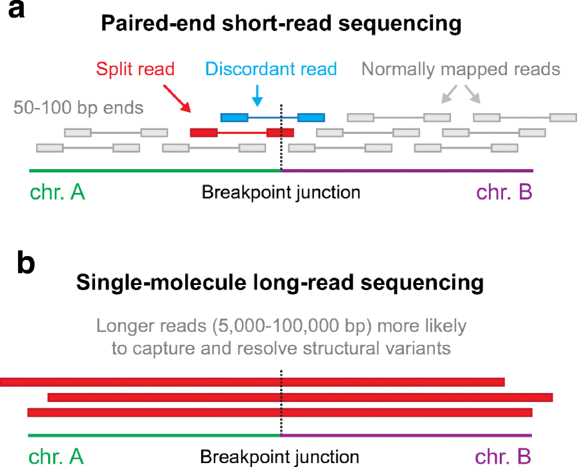
Human chromosomes are arranged in a linear and conserved sequence order that undergoes further spatial folding within the three-dimensional space of the nucleus. Although structural variations in this organization are an important source of natural genetic diversity, cytogenetic aberrations can also underlie a number of human diseases and disorders. Approaches for studying chromosome structure began half a century ago with karyotyping of Giemsa-banded chromosomes and has now evolved to encompass high-resolution fluorescence microscopy, reporter-based assays, and next-generation DNA sequencing technologies. Here, we provide a general overview of experimental methods at different resolution and sensitivity scales and discuss how they can be complemented to provide synergistic insight into the study of human chromosome structural rearrangements. These approaches range from kilobase-level resolution DNA fluorescence in situ hybridization (FISH)-based imaging approaches of individual cells to genome-wide sequencing strategies that can capture nucleotide-level information from diverse sample types. Technological advances coupled to the combinatorial use of multiple methods have resulted in the discovery of new rearrangement classes along with mechanistic insights into the processes that drive structural alterations in the human genome.
Keywords: Chromosome rearrangements; Cytogenetics; FISH; Genome instability; Karyotype; Structural variants.
Figures
References
-
- Lupski JR, Genomic rearrangements and sporadic disease. Nat Genet 39, S43–47 (2007). - PubMed
-
- Rowley JD, A new consistent chromosomal abnormality in chronic myelogenous leukaemia identified by quinacrine fluorescence and Giemsa staining. Nature 243, 290–293 (1973). - PubMed
-
- Lupski JR, Genomic disorders: structural features of the genome can lead to DNA rearrangements and human disease traits. Trends Genet 14, 417–422 (1998). - PubMed
-
- Trask BJ, Human cytogenetics: 46 chromosomes, 46 years and counting. Nat Rev Genet 3, 769–778 (2002). - PubMed
Publication types
MeSH terms
Grants and funding
LinkOut - more resources
Full Text Sources
Other Literature Sources
