. 2019 Apr 1;12(4):1224-1232.
eCollection 2019.

DLC-3 suppresses cellular proliferation, migration, and invasion in triple-negative breast cancer by the Wnt/β-catenin pathway

Affiliations

DLC-3 suppresses cellular proliferation, migration, and invasion in triple-negative breast cancer by the Wnt/β-catenin pathway

Bin Kong et al. Int J Clin Exp Pathol. .

Erratum in

Abstract

Triple-negative breast cancer (TNBC) is the most aggressive breast cancer subtype. Our study investigated the functional role of DLC-3 in TNBC. The expression of DLC-3 was assessed by immunohistochemistry in TNBC to evaluate the clinicopathologic significance of DLC-3. Recombinant lentiviral vectors encoding the DLC-3 gene were constructed for transfection into MDA-MB-231. Real-time qPCR and western blot analysis were employed to evaluate the expression of DLC-3, β-catenin, GSK-3β and c-myc in DLC-3-transfected cells. Moreover, cell proliferation assays, cell colony formation assays, and cell migration and invasion assays were performed to elucidate the role of DLC-3 in TNBC development and progression. Our data revealed that DLC-3 was downregulated in TNBC, and its expression level was associated with lymph node status and differentiation grade in breast cancer. Both real-time qPCR and western blot analyses showed that the DLC-3 gene and protein were overexpressed in the DLC-3-transfected MDA-MB-231 cells. In addition, the expression of GSK-3β was upregulated and the expression of β-catenin and c-myc gene was downregulated in the DLC-3-transfected cells. Furthermore, DLC-3 overexpression inhibited cell proliferation, colony formation, migration, and invasion in vitro. DLC-3, functioning as a tumor-suppressor gene, inhibits cell growth and invasion in TNBC, possibly through regulation of the Wnt/β-catenin signaling pathway.

Keywords: DLC-3; Triple-negative breast cancer; invasion; proliferation.


Conflict of interest statement

None.

Figures

Figure 1
Figure 1
DLC-3 expression was frequently down-regulated in TNBC. (A) IHC results of DLC-3 expression in adjacent noncancerous and breast cancer tissue samples. DLC-3 was expressed in the nucleus of normal breast epithelial cells (Adjacent noncancerous) (a). Loss of expression of DLC-3 in poorly differentiated TNBC (b) and weak staining in well-differentiated TNBC (c). (B) Compared with normal breast tissue, DLC-3 expression was significantly decreased in TNBC (*P < 0.05). (C) IHC results of Ki-67 expression in breast cancer samples. (a) represents the low expression level group, (b) represents the high expression level group (× 200).
Figure 2
Figure 2
Effects of pcDNA3.1(+)-DLC-3 on DLC-3 expression in MDA-MB-231. A. The expression of DLC-3 in HCC-1937, MDA-MB-468, MDA-MB-231 and MCF-10A was detected by western blotting and normalized to GAPDH expression. B. DLC-3 protein expression in the pcDNA3.1(+)-DLC-3 cells was higher than that in the pcDNA3.1(+) group and the untransfected MDA-MB-231 group (*P < 0.05). No difference was observed between the pcDNA3.1(+) group and the untransfected MDA-MB-231 group.
Figure 3
Figure 3
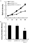
DLC-3 overexpression inhibits proliferation and colony formation of MDA-MB-231 cells. A. Cell proliferation of the pcDNA3.1(+)-DLC-3 cells was decreased compared with the pcDNA3.1(+) group and the untransfected MDA-MB-231 group. B. The colony formation of pcDNA3.1(+)-DLC-3 cells was decreased compared with the pcDNA3.1(+) group and the untransfected MDA-MB-231 group (*P < 0.05).
Figure 4
Figure 4
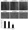
Upregulation of DLC-3 suppresses breast cancer migration and invasion in vitro. A. Compared to the control group, upregulation of DLC-3 significantly inhibited MDA-MB-231 cells migration by wound healing assay. B and C. The number of pcDNA3.1(+)-DLC-3 cells that passed through the membrane was lower than the number in the pcDNA3.1(+) group and the untransfected MDA-MB-231 group (*P < 0.05). No difference was observed between the pcDNA3.1(+) group and the untransfected MDA-MB-231 group.
Figure 5
Figure 5
DLC-3 overexpression regulates the Wnt/β-catenin signaling pathway. A. GSK-3β mRNA expression was higher in the pcDNA3.1(+)-DLC-3 group, while β-catenin and c-myc mRNA expression was lower when compared to these levels in the pcDNA3.1(+) group and the untransfected MDA-MB-231 cells. B, C. GSK-3β protein expression was higher in the pcDNA3.1(+)-DLC-3 group, while β-catenin and c-myc protein expression were lower, when compared to these levels in the pcDNA3.1(+) group and the untransfected MDA-MB-231 cells (*P < 0.05).

References

    1. Safonov A, Jiang T, Bianchini G, Győrffy B, Karn T, Hatzis C, Pusztai L. Immune gene expression is associated with genomic aberrations in breast cancer. Cancer Res. 2017;12:3317–3324. - PubMed
    1. Lv ZD, Yang ZC, Liu XP, Jin LY, Dong Q, Qu HL, Li FN, Kong B, Sun J, Zhao JJ, Wang HB. Silencing of Prrx1b suppresses cellular proliferation, migration, invasion and epithelial-mesenchymal transition in triple-negative breast cancer. J Cell Mol Med. 2016;20:1640–1650. - PMC - PubMed
    1. Huang GH, Sun ZL, Li HJ, Feng DF. Rho GTPase-activating proteins: regulators of Rho GTPase activity in neuronal development and CNS diseases. Mol Cell Neurosci. 2017;80:18–31. - PubMed
    1. Gobbi G, Mirandola P, Carubbi C, Masselli E, Sykes SM, Ferraro F, Nouvenne A, Thon JN, Italiano JE, Vitale M. Proplatelet generation in the mouse requires PKCε-dependent RhoA inhibition. Blood. 2013;7:1305–1311. - PMC - PubMed
    1. Winge MC, Ohyama B, Dey CN, Boxer LM, Li W, Ehsani-Chimeh N, Truong AK, Wu D, Armstrong AW, Makino T, Davidson M. RAC1 activation drives pathologic interactions between the epidermis and immune cells. J Clin Invest. 2016;7:2661–2677. - PMC - PubMed

LinkOut - more resources