

Spindle cell lipoma: clinicopathologic characterization of 40 cases
- PMID: 31934089
- PMCID: PMC6949558
Spindle cell lipoma: clinicopathologic characterization of 40 cases
Abstract
Objective: In view of the existence of multifarious pathologic subtypes of spindle cell lipoma (SCL), which is easily misdiagnosed as other benign and malignant soft tissue tumors, we performed this study and aimed to better define the category of SCL.
Methods: We collected and analyzed 40 cases of SCL with complete clinical and pathologic information from January 2010 to December 2018. Clinical and histopathologic analyses of SCL were performed, as well as immunohistochemical staining and fluorescence in situ hybridization (FISH) using probes for RB1 and MDM2, and the related literature was reviewed.
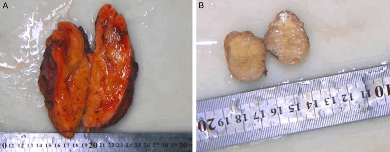
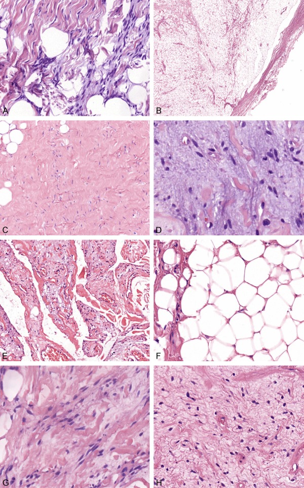
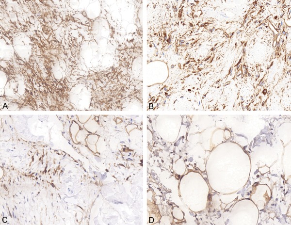
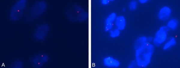
Results: In 40 cases, the male to female ratio was 3.4:1, and the mean age was 54 years old. SCL of our study included six pathologic subtypes: classic (25/40), fibrous (4/40), myxoid (4/40), low-fat (3/40), pseudoangiomatous (2/40), and fat-rich (2/40) changes. Microscopically, SCL showed distinctive morphology, with uniform spindle cells and a variably adipocytic component. The spindle cells were bland in morphology, without prominent atypia or pleomorphism, set in a myxoid or fibrous matrix. Immunohistochemically, CD34 and vimentin were positive in spindle cells, and spindle cells of 6 cases also expressed S-100 protein. FISH analysis of 10 cases revealed that heterozygous deletion of RB1 was in six samples with chromosome 13 aberrations and MDM2 gene amplification was not detected in any cases. Surgical resection is considered as the primary treatment for SCL, as there was no any recurrence or metastasis in our cases after 2-105 months of follow-up.
Conclusions: SCL is a rare benign lipoma, and the proportion of spindle cells and adipocytic component varies, which may form various pathologic changes. The diagnosis needs to be combined with clinicopathologic features, immunophenotypes, and genetics. It has to be differentiated from mammary-type myofibroblastoma, cellular angiolipoma, solitary fibrous tumor, and myxoid liposarcoma.
Keywords: MDM2; RB1; S-100; Spindle cell lipoma (SCL); differential diagnosis.
IJCEP Copyright © 2019.
Conflict of interest statement
None.
Figures
Similar articles
-
Atypical Spindle Cell Lipomatous Tumor: Clinicopathologic Characterization of 232 Cases Demonstrating a Morphologic Spectrum.Am J Surg Pathol. 2017 Feb;41(2):234-244. doi: 10.1097/PAS.0000000000000770. Am J Surg Pathol. 2017. PMID: 27879515
-
Dysplastic Lipoma: A Distinctive Atypical Lipomatous Neoplasm With Anisocytosis, Focal Nuclear Atypia, p53 Overexpression, and a Lack of MDM2 Gene Amplification by FISH; A Report of 66 Cases Demonstrating Occasional Multifocality and a Rare Association With Retinoblastoma.Am J Surg Pathol. 2018 Nov;42(11):1530-1540. doi: 10.1097/PAS.0000000000001129. Am J Surg Pathol. 2018. PMID: 30001242
-
[Spindle cell lipoma and pleomorphic lipoma: a clinicopathologic analysis of 65 cases].Zhonghua Bing Li Xue Za Zhi. 2018 Apr 8;47(4):263-268. doi: 10.3760/cma.j.issn.0529-5807.2018.04.007. Zhonghua Bing Li Xue Za Zhi. 2018. PMID: 29690665 Chinese.
-
Anisometric cell lipoma: Insight from a case series and review of the literature on adipocytic neoplasms in survivors of retinoblastoma suggest a role for RB1 loss and possible relationship to fat-predominant ("fat-only") spindle cell lipoma.Ann Diagn Pathol. 2017 Aug;29:52-56. doi: 10.1016/j.anndiagpath.2017.04.012. Epub 2017 Apr 29. Ann Diagn Pathol. 2017. PMID: 28807343 Review.
-
Spindle Cell Lipoma and Pleomorphic Lipoma: An Update and Review.Cancer Diagn Progn. 2023 May 3;3(3):282-290. doi: 10.21873/cdp.10213. eCollection 2023 May-Jun. Cancer Diagn Progn. 2023. PMID: 37168965 Free PMC article. Review.
Cited by
-
Spindle cell lipoma: An uncommon variant of lipoma affecting the foot sole.Clin Case Rep. 2022 Feb 13;10(2):e05455. doi: 10.1002/ccr3.5455. eCollection 2022 Feb. Clin Case Rep. 2022. PMID: 35198205 Free PMC article.
-
Histological and Ultrastructural Description of Benign Adipocytic Tumors in Farmed Striped Sea Bream (Lythognathus mormyrus).Animals (Basel). 2021 Nov 30;11(12):3413. doi: 10.3390/ani11123413. Animals (Basel). 2021. PMID: 34944190 Free PMC article.
-
Intradermal Low-Fat Spindle Cell Lipoma: A Case Report.Ann Dermatol. 2023 May;35(Suppl 1):S10-S13. doi: 10.5021/ad.21.051. Ann Dermatol. 2023. PMID: 37853856 Free PMC article.
-
Well-differentiated Spindle Cell Liposarcoma of the Larynx: A Rare Case Report and Review of Literature.In Vivo. 2021 Sep-Oct;35(5):2779-2783. doi: 10.21873/invivo.12563. In Vivo. 2021. PMID: 34410968 Free PMC article. Review.
-
Case report: Atypical spindle cell/pleomorphic lipomatous tumor masquerading as a myxoid liposarcoma or intramuscular myxoma.Front Oncol. 2022 Nov 10;12:1033114. doi: 10.3389/fonc.2022.1033114. eCollection 2022. Front Oncol. 2022. PMID: 36439417 Free PMC article.
References
-
- Enzinger FM, Harvey DA. Spindle cell lipoma. Cancer. 1975;36:1852–1859. - PubMed
-
- Ko JS, Daniels B, Emanuel PO, Elson P, Khachaturov V, McKenney JK, Goldblum JR, Billings SD. Spindle cell lipomas in women: a report of 53 cases. Am J Surg Pathol. 2017;41:1267–1274. - PubMed
-
- Bhat A, Vijaya C, Rao SB. Pseudoangiomatous variant of spindle cell lipoma: report of a rare case. Indian J Pathol Microbiol. 2016;59:376–378. - PubMed
-
- Billings SD, Folpe AL. Diagnostically challenging spindle cell lipomas: a report of 34 “low-fat” and “fat-free” variants. Am J Dermatopathol. 2007;29:437–442. - PubMed
-
- Cheah A, Billings S, Goldblum J, Hornick J, Uddin N, Rubin B. Spindle cell/pleomorphic lipomas of the face: an under-recognized diagnosis. Histopathology. 2015;66:430–437. - PubMed
LinkOut - more resources
Full Text Sources
Research Materials
Miscellaneous