Review
. 2019 Dec 24:2019:7286737.
doi: 10.1155/2019/7286737. eCollection 2019.

Pro- and Antioxidant Effects of Vitamin C in Cancer in correspondence to Its Dietary and Pharmacological Concentrations

Affiliations
Review

Pro- and Antioxidant Effects of Vitamin C in Cancer in correspondence to Its Dietary and Pharmacological Concentrations

Elzbieta Pawlowska et al. Oxid Med Cell Longev. .

Abstract

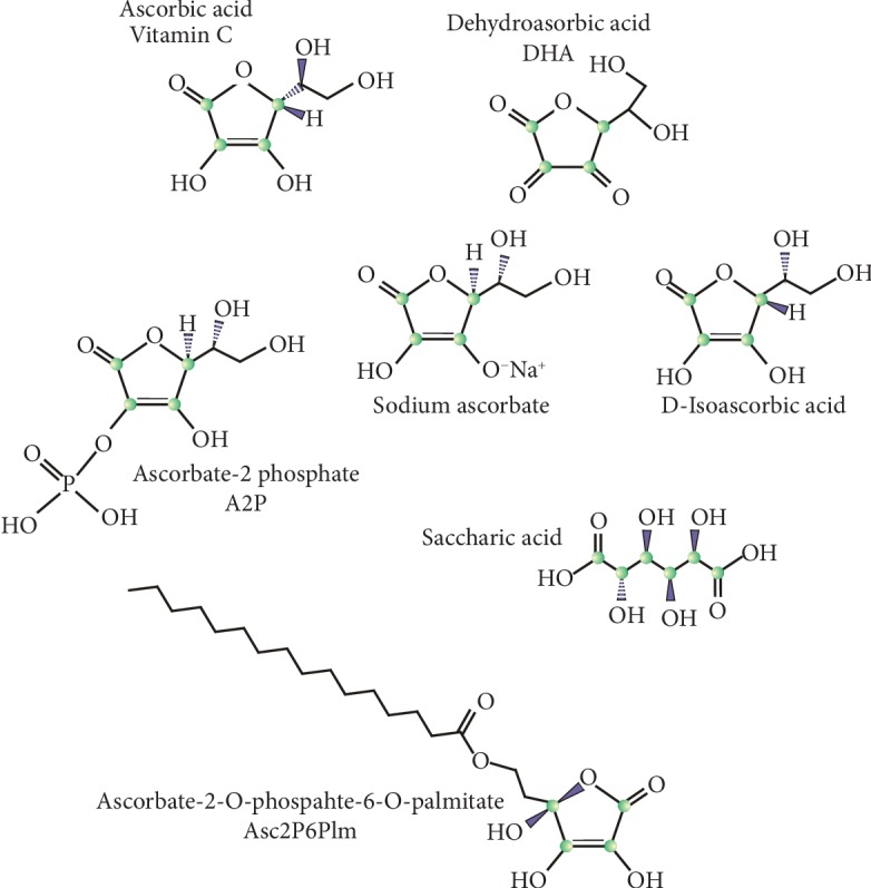
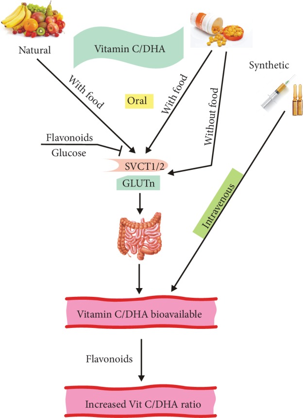
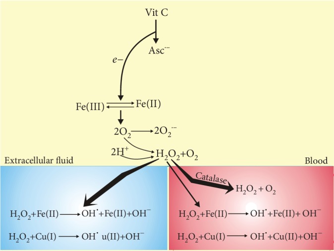
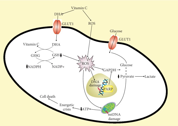
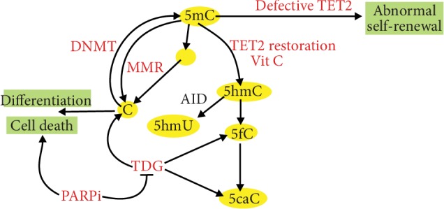
Vitamin C is an antioxidant that may scavenge reactive oxygen species preventing DNA damage and other effects important in cancer transformation. Dietary vitamin C from natural sources is taken with other compounds affecting its bioavailability and biological effects. High pharmacological doses of vitamin C may induce prooxidant effects, detrimental for cancer cells. An oxidized form of vitamin C, dehydroascorbate, is transported through glucose transporters, and cancer cells switch from oxidative phosphorylation to glycolysis in energy production so an excess of vitamin C may limit glucose transport and ATP production resulting in energetic crisis and cell death. Vitamin C may change the metabolomic and epigenetic profiles of cancer cells, and activation of ten-eleven translocation (TET) proteins and downregulation of pluripotency factors by the vitamin may eradicate cancer stem cells. Metastasis, the main reason of cancer-related deaths, requires breakage of anatomical barriers containing collagen, whose synthesis is promoted by vitamin C. Vitamin C induces degradation of hypoxia-inducible factor, HIF-1, essential for the survival of tumor cells in hypoxic conditions. Dietary vitamin C may stimulate the immune system through activation of NK and T cells and monocytes. Pharmacological doses of vitamin C may inhibit cancer transformation in several pathways, but further studies are needed to address both mechanistic and clinical aspects of this effect.


Conflict of interest statement

The authors declare no conflict of interest.

Figures

Figure 1
Figure 1
Molecular structure of vitamin C and its derivatives displaying anticancer properties that are discussed in this work.
Figure 2
Figure 2
Absorption and bioavailability of natural and synthetic vitamin C. Vitamin C that is partly oxidized to dehydroascorbate (DHA) in an oxygen environment is transported by two sodium-dependent transporters SVCT1 and SVCT2, while DHA is taken up by the glucose transporter GLUTn, where n is 1-3 or 8. Vitamin C/DHA can be taken as either natural or synthetic ascorbic acid, and the latter can be given orally (with or without food) or intravenously. The final concentration of vitamin C in circulation depends not only on the route of ingestion but also on its excretion (not presented here) and the action of other dietary compounds, including glucose and flavonoids. Flavonoids can block the absorption of vitamin C, but they can also reduce some oxidants leading to an increase in the vitamin C/DHA ratio.
Figure 3
Figure 3
Vitamin C may differently produce reactive oxygen species (ROS) in blood and the extracellular space. After oral or intravenous administration, vitamin C reaches the same concentration in blood and extracellular fluid and loses one electron (e) to form ascorbate radical Asc·− and reduces a protein-centered metal ion, such as Fe(III). Reduced metal donates an electron to oxygen forming ROS, including superoxide (O2·−) that can be dismutated to hydrogen peroxide. These reactions in blood are inhibited by plasma and red cell membrane proteins, and hydrogen peroxide in blood is neutralized by antioxidant enzymes hardly present in extracellular fluid. Unless H2O2 is decomposed, it may produce hydroxyl radicals in the Fe(II)- or Cu(I)-catalyzed Fenton-like reaction yielding hydroxyl peroxide (HO·).
Figure 4
Figure 4
Vitamin C is oxidized in the extracellular space to dehydroascorbate (DHA) that is taken up by cancer cells via glucose transporters such as GLUT1. Inside the cell, DHA is reduced back to vitamin C by reduced glutathione (GSH) that is oxidized to glutathione disulfide (GSSG) and converted back to GSH by reduced nicotinamide adenine dinucleotide phosphate (NADPH). Depletion of GSH and NADPH results in ROS overproduction that may damage biomolecules and kill cancer cells. ROS-damaged DNA activates poly(ADP-ribose) polymerase (PARP) that requires NAD+. ROS can also inhibit glyceraldehyde 3-phosphate dehydrogenase (GAPDH) resulting in decreased production of pyruvate and ATP by mitochondria and finally energetic crisis and cell death [–84].
Figure 5
Figure 5
Vitamin C induces the ten-eleven translocation 2 (TET2) proteins to kill leukemic blasts. TETs are involved in active DNA demethylation that is achieved through TET2-mediated oxidation of 5-methylcytosine (5mC) to 5-hydroxymethylcytosine (5hmC), 5-formylcytosine (5fC), and 5-carboxylcytosine (5caC). Oxidized 5mC is progressively lost in subsequent cellular divisions or converted to nonmethylated C by thymine DNA glycosylase (TDG). 5mC can undergo spontaneous or activation-induced deaminase- (AID-) mediated deamination converting it into thymine (T) that can be replaced by C by TDG or in mismatch repair (MMR). AID can convert 5hmC to 5-hydroxymethyluracil (5hmU) or T. If TET2 is deficient in leukemic stem cells, their self-renewal is disturbed leading to increased blast production and progression of the disease. Vitamin C exerts similar effects as restoration of TET2 that leads to increased differentiation and less aggressive disease. Vitamin C-induced oxidation of 5mC results in an increased sensitivity of the cells to inhibitors of poly(ADP-ribose) polymerase (PARPi) that can induce cell death and inhibit disease progression.

References

    1. Nishikimi M., Fukuyama R., Minoshima S., Shimizu N., Yagi K. Cloning and chromosomal mapping of the human nonfunctional gene for L-gulono-gamma-lactone oxidase, the enzyme for L-ascorbic acid biosynthesis missing in man. The Journal of biological chemistry. 1994;269(18):13685–13688. - PubMed
    1. Granger M., Eck P. Dietary vitamin C in human health. Advances in food and nutrition research. 2018;83:281–310. doi: 10.1016/bs.afnr.2017.11.006. - DOI - PubMed
    1. Ang A., Pullar J. M., Currie M. J., Vissers M. C. M. Vitamin C and immune cell function in inflammation and cancer. Biochemical Society transactions. 2018;46(5):1147–1159. doi: 10.1042/bst20180169. - DOI - PMC - PubMed
    1. Vissers M. C. M., Das A. B. Potential mechanisms of action for vitamin C in cancer: reviewing the evidence. Frontiers in physiology. 2018;9:p. 809. doi: 10.3389/fphys.2018.00809. - DOI - PMC - PubMed
    1. van Gorkom G. N. Y., Lookermans E. L., Van Elssen C., Bos G. M. J. The effect of vitamin C (ascorbic acid) in the treatment of patients with cancer: a systematic review. Nutrients. 2019;11(5):p. 977. doi: 10.3390/nu11050977. - DOI - PMC - PubMed