. 2020 Feb 4;92(3):2794-2801.
doi: 10.1021/acs.analchem.9b05099. Epub 2020 Jan 14.

Injection Molded Microfluidics for Establishing High-Density Single Cell Arrays in an Open Hydrogel Format

Affiliations

Injection Molded Microfluidics for Establishing High-Density Single Cell Arrays in an Open Hydrogel Format

Ying Li et al. Anal Chem. .

Abstract

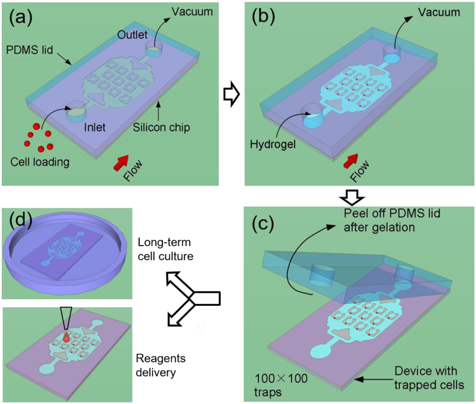
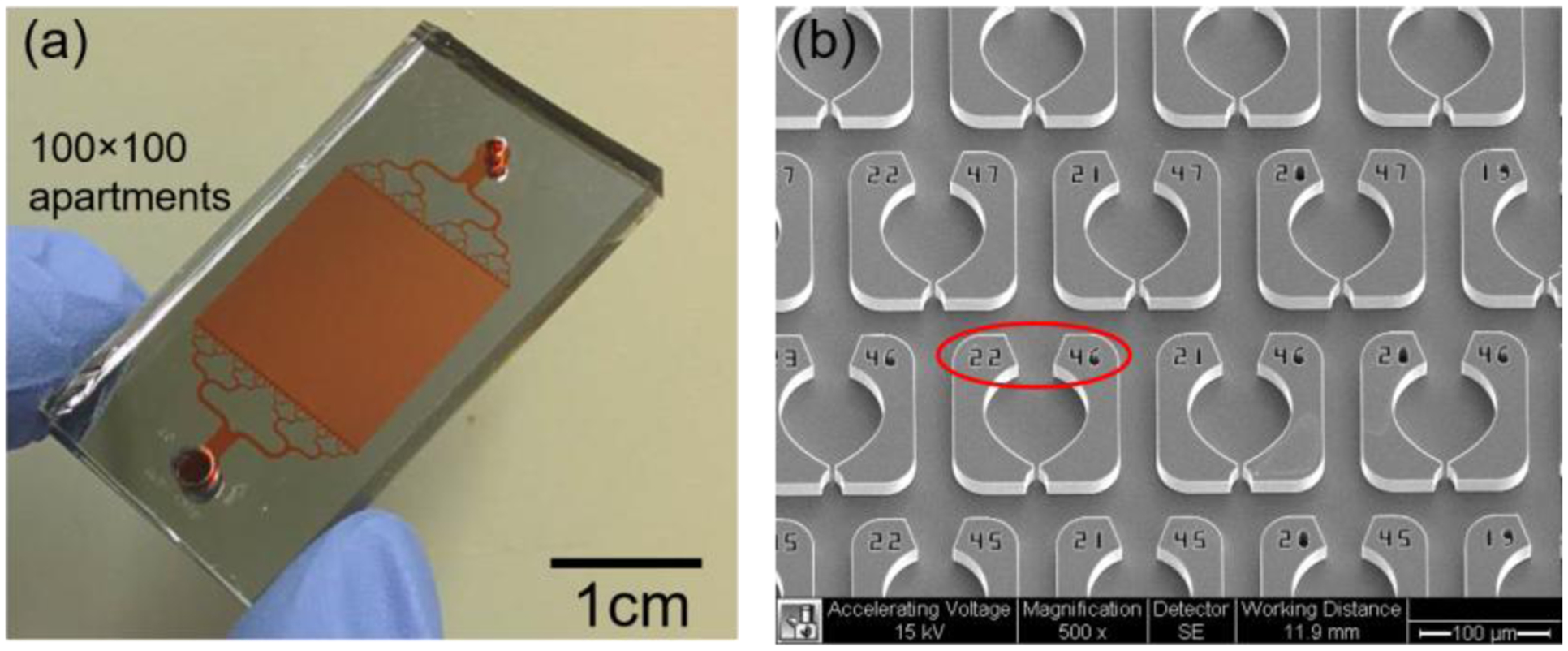
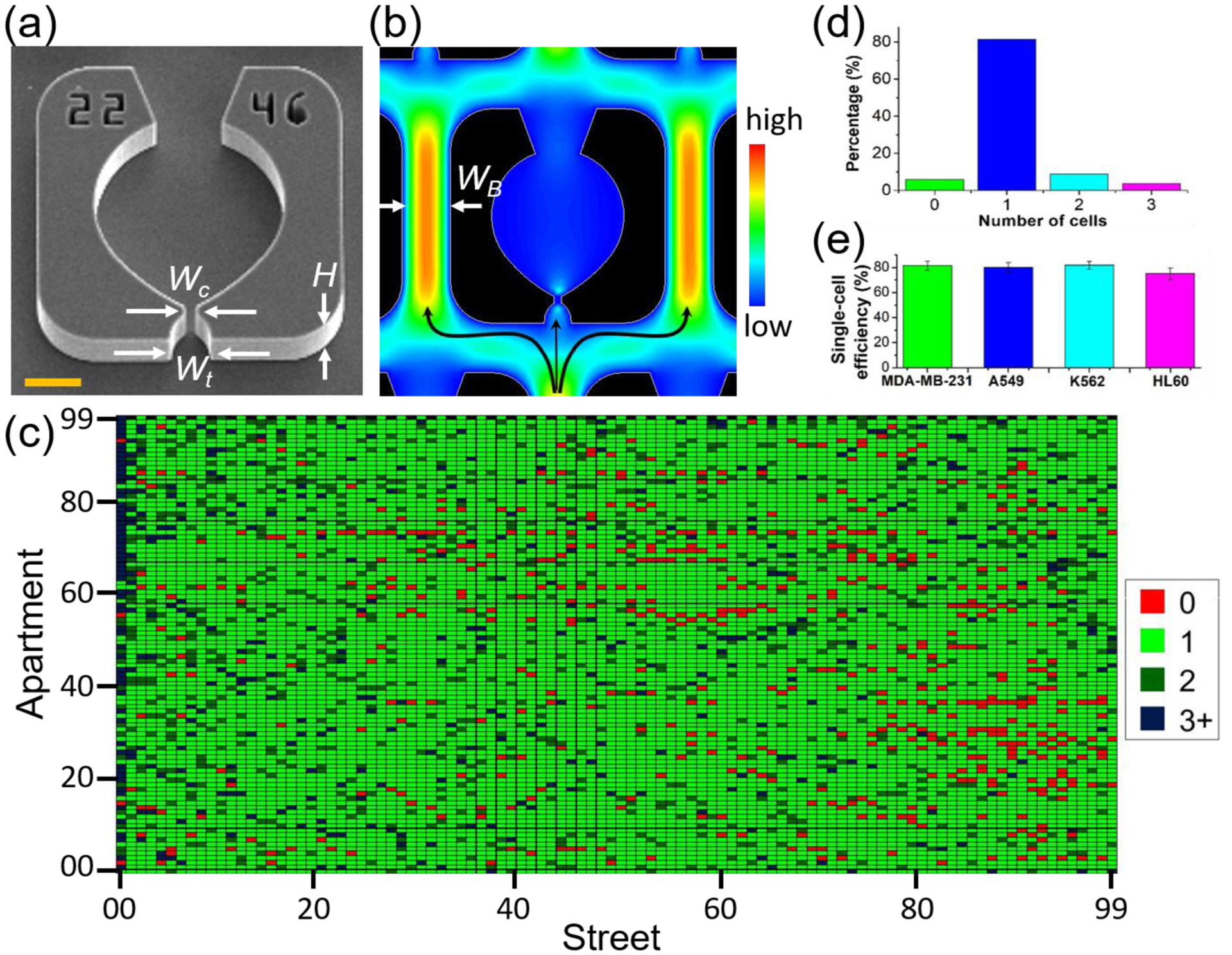
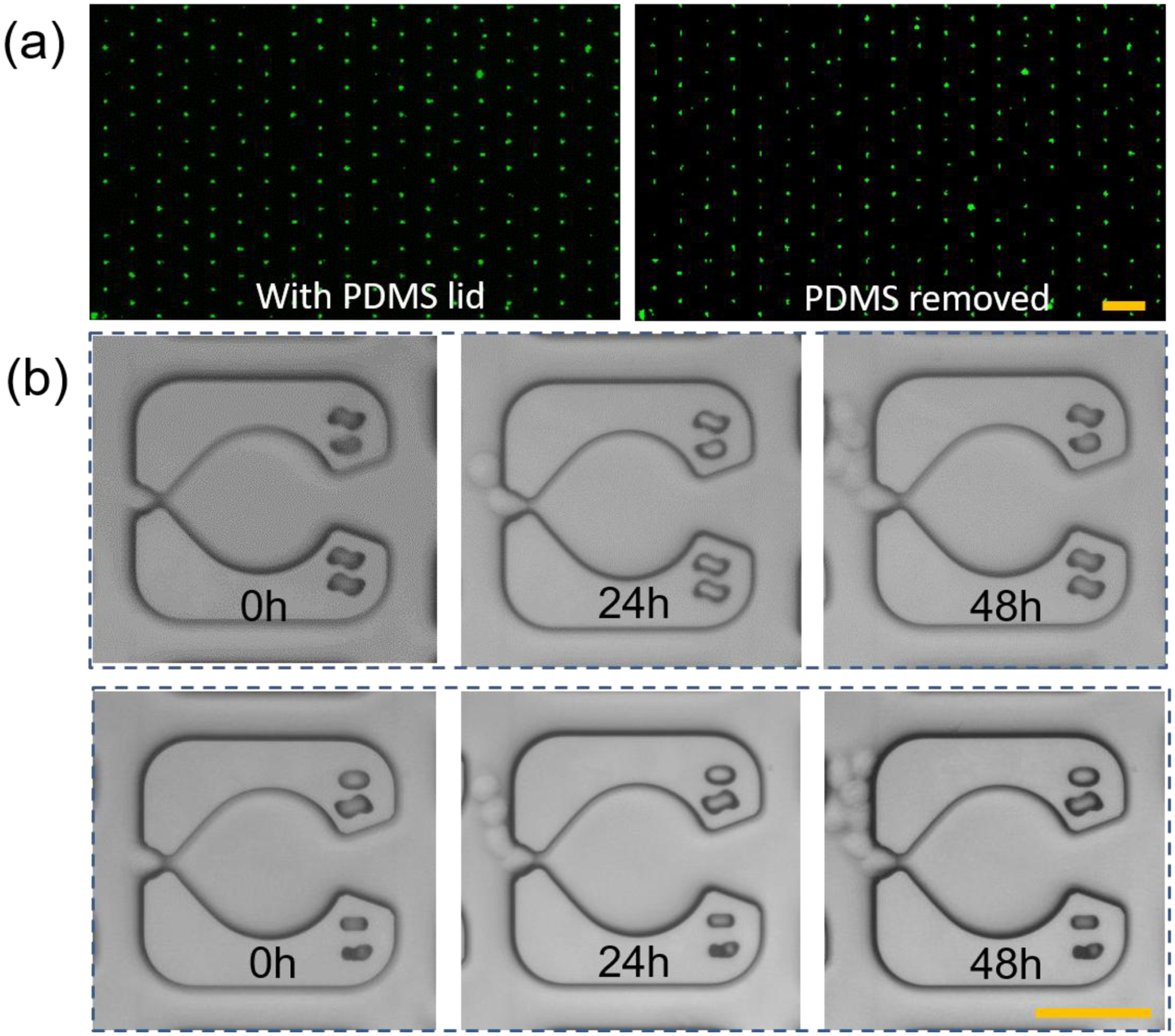
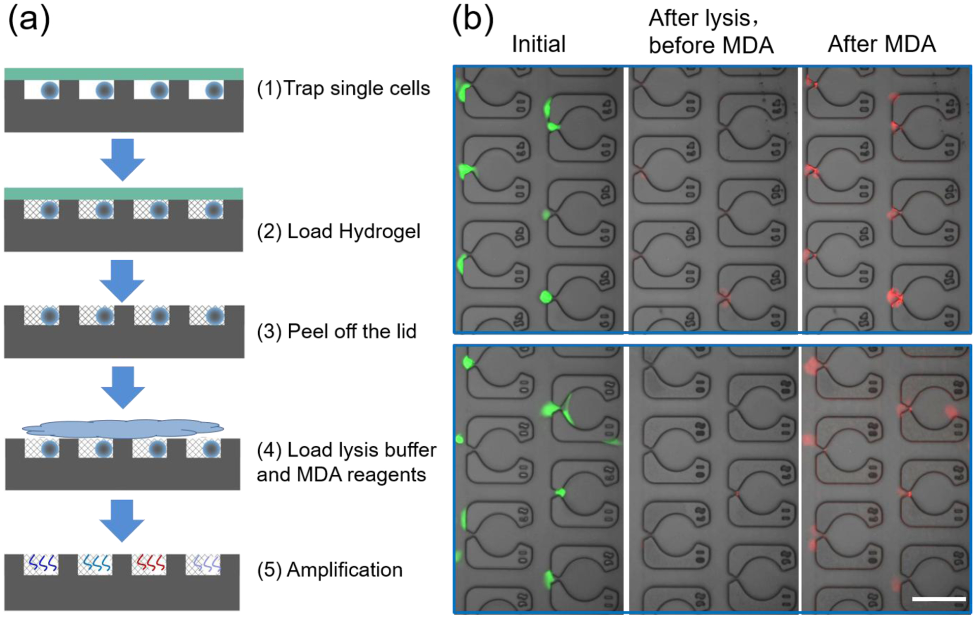
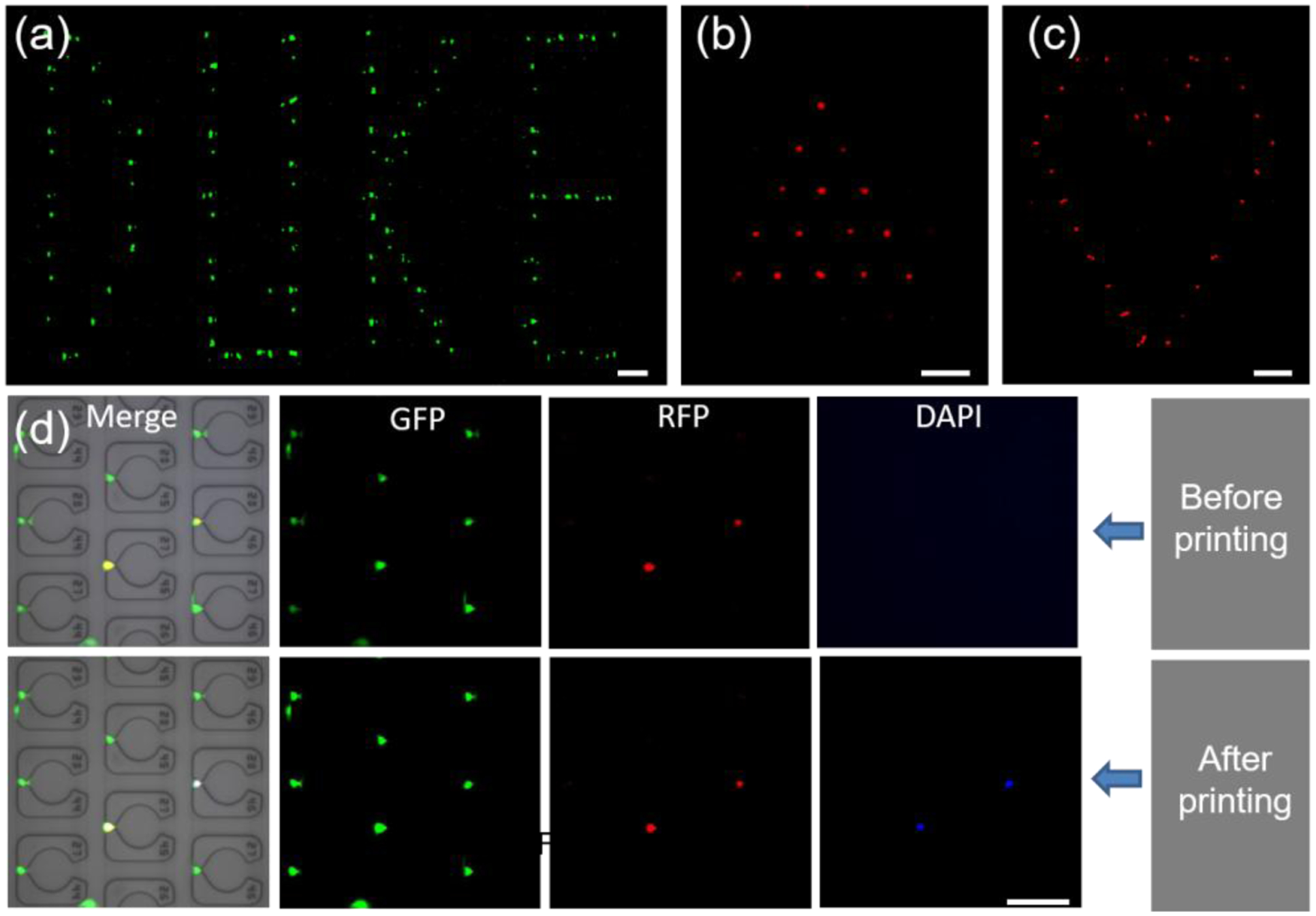
Here, we develop an injection molded microfluidic approach for single cell analysis by making use of (1) rapidly curing injectable hydrogels, (2) a high density microfluidic weir trap array, and (3) reversibly bonded PDMS lids that are strong enough to withstand the injection molding process, but which can be peeled off after the hydrogel sets. This approach allows for single cell patterns to be created with densities exceeding 40 cells per mm2, is amenable to high speed imaging, and creates microfluidic devices that enable efficient nutrient and gas exchange and the delivery of specific biological and chemical reagents to individual cells. We show that it is possible to organize up to 10 000 single cells in a few minutes on the device, and we developed an image analysis program to automatically analyze the single-cell capture efficiency. The results show single cell trapping rates were better than 80%. We also demonstrate that the genomic DNA of the single cells trapped in the hydrogel can be amplified via localized, multiple displacement amplification in a massively parallel format, which offers a promising strategy for analyzing single cell genomes. Finally, we show the ability to perform selective staining of individual cells with a commercial bioprinter, providing proof of concept of its ability to deliver tailored reagents to specific cells in an array for future downstream analysis. This injection molded microfluidic approach leverages the benefits of both closed and open microfluidics, allows multiday single cell cultures, direct access to the trapped cells for genotypic end point studies.


Conflict of interest statement

CONFLICTS OF INTEREST

Y.L., J.D.M., and B.B.Y. are named inventors on a pending patent application.

Figures

Figure 1.
Figure 1.. Working principle of the injection molded microfluidic approach.
(a) Cells are loaded into the device by applying vacuum at the outlet to form a single cell array, after which (b) the hydrogel solution is injected into the device by vacuum and allowed to cure. (c) After curing, the PDMS lid is peeled off and finally (d) the device is placed in a cell culture flask for long term incubation, or alternatively, reagents are delivered to specific locations of the device.
Figure 2.
Figure 2.. Images of the devices.
An optical photograph of the assembled device is shown in (a). The device contains a 100×100 trap array organized in an area of 1.5×1.5 cm2. (b) An SEM image of the device is shown. Each weir has an individual address (indicated by the red circle) that allows for the identification of the trapped cells.
Figure 3
Figure 3. Evaluation of the trapping efficiency.
(a) SEM image of a single apartment is shown, where the critical dimensions are labeled for the trap width (Wt = 16 μm) and the constriction width (Wc = 5 μm) and the channel height (H= 20 μm). Scale bar is 20 μm. (b) Simulated flow velocity profile in a single trap. In simulations and experiments, the width of the bypass WB is fixed at 35 μm. (c) The single-cell trapping efficiency is determined by a custom MATLAB code which uses fluorescent intensity analysis combined with circle tracking to determine the number of cells in each apartment. (d) Bar-plotting of the result in (c) to show the percentage of different numbers of cells trapped in each trapping area. (e) Average single-cell trapping efficiency for different cell lines on the 100×100 array (N=5 devices for each cell line).
Figure 4
Figure 4. Biocompatibility of the injection-molded single cell arrays.
(a) The trapped K562 cells on the device with (left) and without (right) the PDMS lid. Scale bar, 200 μm. This result showed that the crosslinked hydrogel was stable in the device and it could retain the cells after removing the PDMS lid. (b) Long-term culture of the single cells encapsulated in the hydrogel. Scale bar, 50 μm.
Figure 5
Figure 5. Massively parallel single cell MDA.
(a) Single cells are trapped and allowed to attach to the device, followed by hydrogel loading/crosslinking and lid removal. Then cells are lysed, and MDA is carried out to amplify the whole genome of single cells at 30 °C for 8 hours. (b) Merged images for the single cells at the initial state (left panel), after lysis but before MDA reaction (central panel) and after the MDA reaction (right panel). The red fluorescent signal increased significantly after MDA in comparison to that before MDA, indicating that amplification is occurring in a site selective manner. Scale bar, 100 μm.
Figure 6
Figure 6. Localized printing of biochemical reagents on the device and cell-array.
(a) A pattern of ‘DUKE’ is generated by printing FITC on a blank chip. (b-c) ‘Filled’ triangle and ‘unfilled’ heart cell-patterns obtained by printing Hoechst 33342 on an injection molded single-cell array. Red is used as a pseudo-color for Hoechst 33342 to show the pattern more clearly. See also supporting data in Figure S10–S12. (d) Precise printing of staining dye on the selected MDA-MB-231 cells in a single-cell array. Cells showed both green and red fluorescence were selected as target cells to print Hoechst 33342. After the printing, the target cells showed green, red and blue fluorescence. The scale bar is 200 μm for (a-c) and 100 μm for (d).

References

    1. Bhatia SN; Ingber DE Nat Biotechnol 2014, 32, 760–772. - PubMed
    1. Navin NE Genome Biol 2014, 15. - PMC - PubMed
    1. Zhang K; Chou CK; Xia X; Hung MC; Qin L Proc Natl Acad Sci U S A 2014, 111, 2948–2953. - PMC - PubMed
    1. Papalexi E; Satija R Nat Rev Immunol 2018, 18, 35–45. - PubMed
    1. Dhar M; Lam JN; Walser T; Dubinett SM; Rettig MB; Di Carlo D Proc Natl Acad Sci U S A 2018, 115, 9986–9991. - PMC - PubMed

Publication types