. 2020 Mar:77:105-113.
doi: 10.1016/j.neuro.2020.01.003. Epub 2020 Jan 11.

Chronic exposure to methylmercury induces puncta formation in cephalic dopaminergic neurons in Caenorhabditis elegans

Affiliations

Chronic exposure to methylmercury induces puncta formation in cephalic dopaminergic neurons in Caenorhabditis elegans

Tao Ke et al. Neurotoxicology. 2020 Mar.

Abstract

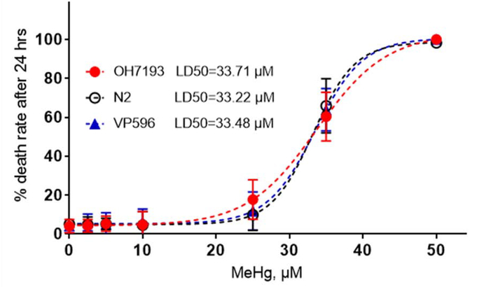
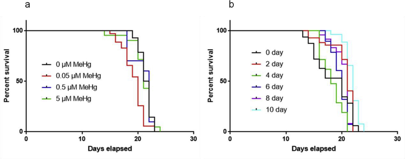
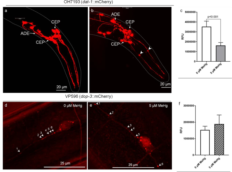
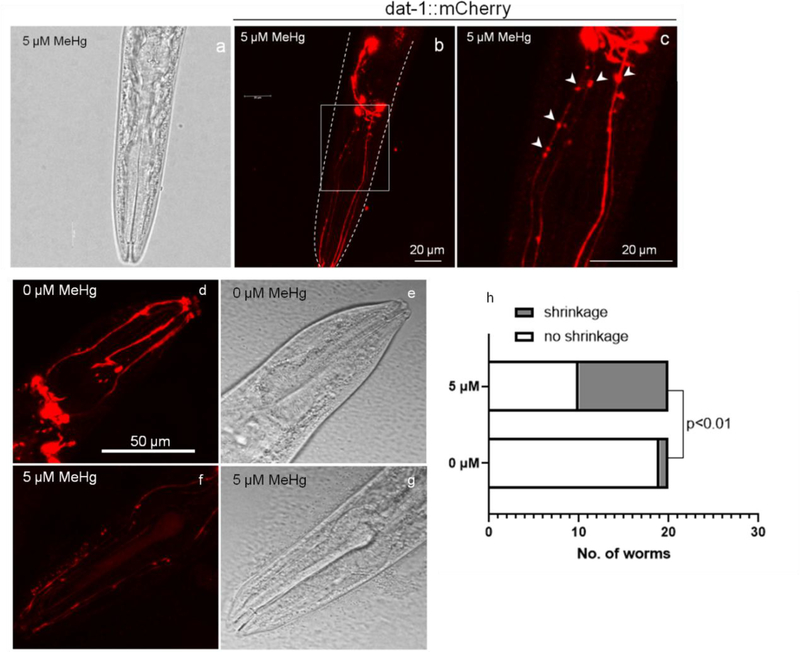
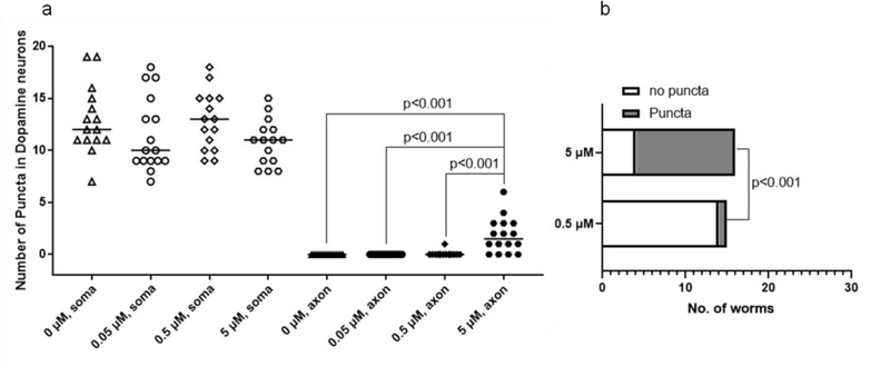
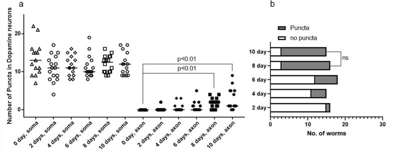
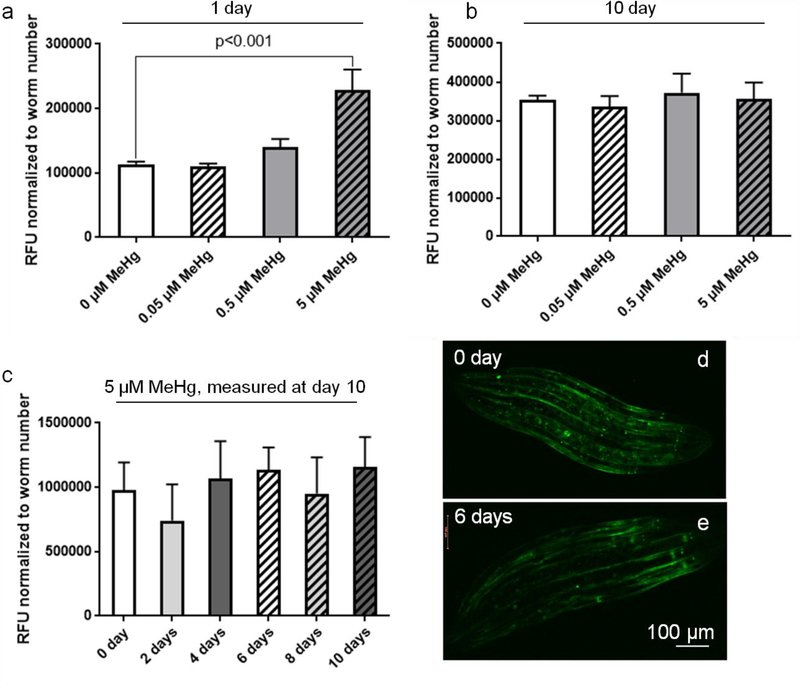
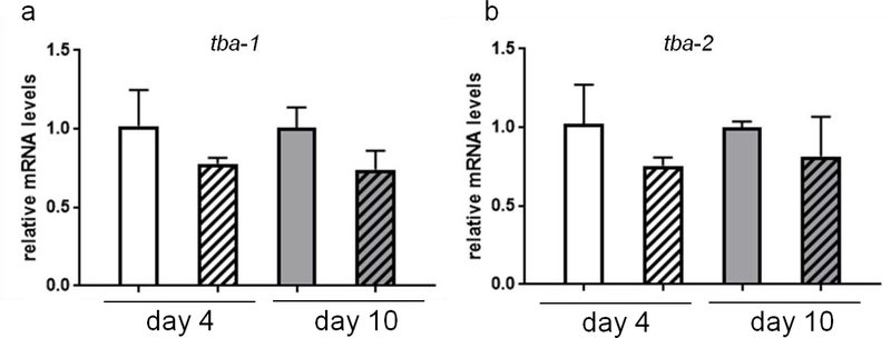
The neurotransmitter dopamine is a neuromodulator in the positive and negative regulation of brain circuits. Dopamine insufficiency or overload has been implicated in aberrant activities of neural circuits that play key roles in the pathogenesis of neurological and psychiatric diseases. Dopaminergic neurons are vulnerable to environmental insults. The neurotoxin methylmercury (MeHg) produces dopaminergic neuron damage in rodent as well as in Caenorhabditis elegans (C. elegans) models. Previous studies have demonstrated the utility of C. elegans as an alternative and complementary experimental model in dissecting out mechanism of MeHg-induced dopaminergic neurodegeneration. However, a sensitive pathological change that marks early events in neurodegeneration induced by environmental level of MeHg, is still lacking. By establishing a chronic exposure C. elegans model, for the first time, we have shown the propensity of MeHg (5 μM, 10 days) to induce bright puncta of dat-1::mCherry aggreagtes in the dendrites of cephalic (2 CEPs) dopaminergic neurons in a dose- and time-dependent manner, while these changes were not found in other dopaminergic neurons: anterior deirids (2 ADEs) and posterior deirids (2 PDEs), cholinergic neurons (2 AIYs) or glutamatergic neurons (2 PVDs). The bright puncta appear as an aggregation of mCherry proteins accumulating in dendrites. Further staining shows that the puncta were not inclusions in lysosome, or amyloid protein aggregates. In addition, features of the puncta including enlarged sphere shape (0.5-2 μm diameters), bright and accompanying with the shrinkage of the dendrite suggest that the puncta are likely composed of homologous mCherry molecules packaged at the dendritic site for exportation. Moreover, in the glutathione S-transferase 4 (gst-4) transcriptional reporter strain and RT-PCR assay, the expression levels of gst-4 and tubulins (tba-1 and tba-2) genes were not significantly modified under this chronic exposure paradigm, but gst-4 did show significant changes in an one day exposure paradigm. Collectively, these results suggest that CEP dopaminergic neurons are a sensitive target of MeHg, and the current exposure paradigm could be used as a model to investigate mechanism of dopaminergic neurotoxicity.

Keywords: C. elegansdopamine; Methylmercury; Toxicity; puncta formation.


Conflict of interest statement

Declaration of Competing Interest The authors report no declarations of interest.

Figures

Fig 1.
Fig 1.
OH7193, N2 and VP596 strains have comparable sensitivity to MeHg toxicity. 1000 worms were treated with 0–50 μM MeHg for 24 h, followed by manual counting of dead worms. Data are expressed as mean ± SD. Dose-response lethality curves and LD50 estimation were generated with a sigmoidal dose-response model with a top constraint at 100%.
Fig 2.
Fig 2.
MeHg exposure does not affect lifespan of OH7193 strain at 0–5 μM concentration. (a) OH7193 strain was exposed to 0–5 μM MeHg for 10 days, followed by lifespan assay. (b) OH7193 strain was exposed to 5 μM MeHg for 0–10 days, followed by lifespan assay. At least 50 worms per treatment were counted for lifespan assay. Comparisons of the survival curves were made using Log-rank (Mantel-Cox) test.
Fig 3.
Fig 3.
Chronic MeHg exposure induces mCherry puncta formation in dendrites of CEP neurons. (a,b) Representative images of dopaminergic CEP neurons in worms treated without (a) or with MeHg (b). Arrowheads show the puncta in the dendrites of CEP neurons following MeHg treatment. (c) Effect of MeHg on the mCherry fluorescence level of CEP dendrites. (d, e) Representative images of glutaminergic PVD neurons in worms treated without (d) or with MeHg (e). Arrowheads show the normal mCherry aggregates in PVD neurons. (f) Effect of MeHg on the mCherry fluorescence level of PVD dendrites. The fluorescence level in dendritic areas of CEP and PVD were assessed with ImageJ software. Data are expressed as mean ± SD (one-way ANOVA, n=3).
Fig. 4
Fig. 4
Characterization of CEP dendrites of worms treated with MeHg. (a-c) Representative images of puncta positive dendrites of CEP neurons in worms treated with MeHg. Arrowheads show the beading-shape puncta in the dendrites of CEP. scable bar = 20 μm. (d-g) Shrinkage of CEP dendrites in worms treated with MeHg. scable bar = 50 μm. (h) Number of worms with CEP dendritic shrinkage following MeHg treatment (Fisher’s exact test).
Fig. 5
Fig. 5
MeHg dose-dependently increases puncta in the dendrites of CEP neurons. (a) OH9173 strain was treated with 0–5 μM MeHg for 10 days, followed by counting the number of puncta in CEP soma and dendrites. The horizontal lines represent median values. (Kruskal-Wallis test followed by Dunn’s multiple comparisons test, n=3). (b) Reanalysis of data presented in (a) showing the number of worms with positive puncta (Fisher’s exact test).
Fig. 6
Fig. 6
MeHg time-dependently increases puncta in the dendrites of CEP neurons. (a) OH9173 strain was treated with 5 μM MeHg for 0–10 days, followed by counting the number of puncta in CEP soma and dendrites. The horizontal lines represent median values (Kruskal-Wallis test followed by Dunn’s multiple comparisons test, n=3). (b) Reanalysis of data presented in (a) showing the number of worms with positive puncta (Chi-square test).
Fig. 7
Fig. 7
Chronic exposure to MeHg doesn’t invoke overexpression of gst-4. (a) CL2166 strain was treated with 0–5 μM MeHg for 1 day, followed by measurement of fluorescence intensity with ImageJ software. (b) CL2166 strain was treated with 0–5 μM MeHg for 10 days, followed by measurement of fluorescence intensity with ImageJ software. (c) CL2166 strain was treated with 5 μM MeHg for 0–10 days, followed by measurement of fluorescence intensity with ImageJ software. (d,e) Representative images of worms treated without MeHg (d) or with MeHg for 6 days (e). Data are expressed as mean ± SD (one-way ANOVA followed by Tukey’s post hoc test, n=3).
Fig. 8
Fig. 8
Expression of tubulin genes is not associated with MeHg exposure. (a) N2 strain was treated with 5 μM MeHg for 4 or 10 days, followed by measurement of tba-1 mRNA with RT-PCR. (b) N2 strain was treated with 5 μM MeHg for 4 or 10 days, followed by measurement of tba-2 mRNA with RT-PCR. Data are expressed as mean ± SD (one-way ANOVA followed by Tukey’s post hoc test; n=3).

References

    1. Angelo G, Van Gilst MR, 2009. Starvation protects germline stem cells and extends reproductive longevity in C. elegans. Science 326(5955), 954–958. - PubMed
    1. Arvidson B, 1987. Retrograde axonal transport of mercury. Exp Neurol 98(1), 198–203. - PubMed
    1. Arvidson B, 1990. Accumulation of mercury in brainstem nuclei of mice after retrograde axonal transport. Acta Neurol Scand 82(4), 234–237. - PubMed
    1. Arvidson B, 1992. Accumulation of Inorganic Mercury in Lower Motoneurons of Mice. Neurotoxicology 13(1), 277–280. - PubMed
    1. Arvidson B, Arvidsson J, 1990. Retrograde axonal transport of mercury in primary sensory neurons innervating the tooth pulp in the rat. Neurosci Lett 115(1), 29–32. - PubMed

Publication types

MeSH terms