Review
. 2020 Apr:61:113-123.
doi: 10.1016/j.sbi.2019.11.011. Epub 2020 Jan 11.

Evolving understanding of HIV-1 reverse transcriptase structure, function, inhibition, and resistance

Affiliations
Review

Evolving understanding of HIV-1 reverse transcriptase structure, function, inhibition, and resistance

Francesc Xavier Ruiz et al. Curr Opin Struct Biol. 2020 Apr.

Abstract

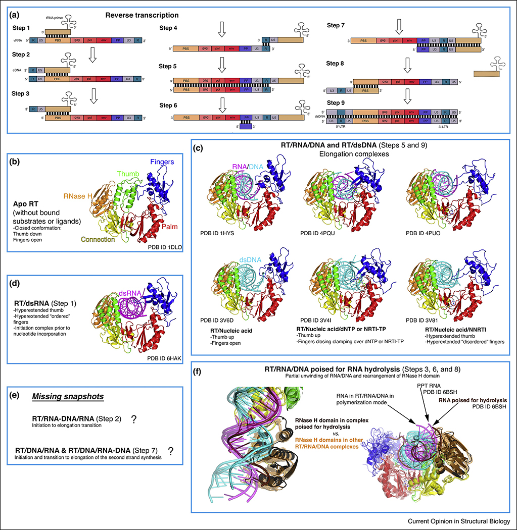
The essential role of reverse transcription in the HIV life cycle is illustrated by the fact that half of the ∼30 FDA-approved drugs for HIV treatment target HIV-1 reverse transcriptase (RT). Even though more than 160 structures of RT deposited in the Protein Data Bank (PDB) have revealed the molecular architecture of RT in great detail, some key states of RT function and inhibition remain still unknown. Recent structures of RT initiation complexes, RT poised for RNA hydrolysis, and RT with approved drugs and investigational compounds have provided a deeper understanding of RT function and inhibition, suggesting novel avenues for targeting this central enzyme of HIV.


Conflict of interest statement

Conflict of interest statement

Nothing declared.

Figures

Figure 1
Figure 1. Overview of HIV-1 reverse transcriptase (RT) structure and main binding sites for substrates and inhibitors.
Structure of RT in complex with dsDNA and incoming EFdA-TP (PDB ID 5J2M), with template-primer, with p66 polymerase fingers, palm, thumb, connection subdomains and RNase H domain, and with p51 indicated, with the RT/rilpivirine (brown spheres, PDB ID 4G1Q) and RT/11b (light pink spheres, PDB ID 6AOC) structures superposed. Zoom-ins of: i) the NNIBP (NNRTI binding pocket) of the RT/rilpivirine (RPV, circled) complex (PDB ID 4G1Q) superposed to the former structure, with residues, catalytic motif YMDD and primer grip domain location highlighted; ii) the polymerase active site in the presence of incoming EFdA-TP (circled, 2-fluorine (2-F), 4’-ethynyl and 3’-OH moieties indicated), residues, template-primer, catalytic metals, N-site, and P-site indicated; iii) the RNase H active site with 11b bound, catalytic metals and residues indicated.
Figure 2
Figure 2. Conformational states of RT during reverse transcription.
(a) Schematic representation of retroviral reverse transcription, displaying the genomic and host nucleic acids (modified from https://en.wikipedia.org/wiki/Reversetranscriptase#/media/File:Reversetranscription.svg, Creative Commons Attribution License CC BY 3.0 license). In brief, the different steps correspond to: 1- Initiation of first strand synthesis; 2- Initiation to elongation transition (RNA/DNA hybrid primer); 3- Hydrolysis of the 5’ region of the viral RNA (vRNA); 4- tRNA jump to the 3’ region of the vRNA; 5- Elongation (dsDNA); 6- vRNA degradation (except polypurine tract, PPT); 7- Initiation of the second strand synthesis; 8- PPT hydrolysis and second jump (tRNA exit); 9- Second strand elongation and addition of the long terminal repeats (LTRs). (b-f) The rest of the boxes (except from the one indicating the challenges remaining, that is, uncharacterized conformational states) contain all the described conformations of RT complexed with nucleic acids (and apo form) during reverse transcription, with color coding and annotations included for clarity. The notion of ‘ordered’ versus ‘disordered’ hyperextended fingers subdomain (boxes C and D) is based on B-factors (lower versus higher) and number of interactions with the nucleic acid strand (more versus less).
Figure 3
Figure 3. Comparison of the cryo-EM and X-ray crystallographic structures of the RT initiation complex (RTIC).
(a) On top, nucleotide sequences of i) dsRNA 23-mer-17-mer template-primer used in X-ray crystallography structure (a template G was cross-linked to the Q258C position of RT without primer extension and with the G and C positions of the sixth base pair switched compared with the naturally annealed PBS-tRNALys3 sequence), and ii) the annealed PBS region of vRNA-tRNALys3 in the RTIC for which the cryo-EM structure was reported (the vRNA-tRNALys3 was cross-linked to RT at ‘G’ and primer extended with catalytic incorporation of a ddCMP). Below, comparison of the crystal lattice dsRNA/dsRNA interaction reminiscent of vRNA-tRNALys3 RNA interactions in the RTIC complex and fitting of the above complex in the 8 Å cryo-EM density (EMBD-7032) by aligning the RT/dsRNA complex and the dsRNA (sym) positioned exactly on the density for the H1 helix and connecting loop of vRNA. (b) On top, superposition of the RT/dsRNA structure (blue protein, orange RNA) on the structure of the RTIC core (PDB ID 6B19; yellow protein and RNA) showing high structural resemblance. Detail of the relative locations of the primer 3′ ends: P′ of RT/dsRNA (blue/orange), (P+ 1)′′ of cryo-EM RTIC N complex (yellow), and P of RT/dsDNA (gray) structures. The incorporated ddCMP is at (P + 1)′′ and the nucleotide corresponding to P′ of RT/dsRNA is located at P′′ in the RTIC structure. The primer 3′ end has to reach the P-site for nucleotide incorporation by RT. Detail of the RT/dsRNA complex (blue/orange), closely resembling RT in the RT/dsDNA/nevirapine complex (white/yellow/cyan spheres, PDB ID 3V81). Close-up view of the polymerase site and NNRTI pocket shows that the primer 3′ ends in both structures are displaced away from the polymerase site, and also about 3.5 Å apart. Only minimal structural arrangements of the NNRTI pocket region of RT/dsRNA structure are needed to accommodate nevirapine; the major adjustment required is the repositioning of W229 as indicated by an arrow. The side chains of Y181 and Y188 have disordered electron density, and therefore are not included in the RT/dsRNA structure. Figure adapted from Ref. [••].
Chart 1
Chart 1
Molecular formulas of the compounds cited in the manuscript.

Similar articles

Cited by

References

    1. UNAIDS Fact Sheet July 2018. http://www.unaids.org/en/resources/fact-sheeton World Wide Web URL: http://www.unaids.org/en/resources/fact-sheet.
    1. Das K, Arnold E: HIV-1 reverse transcriptase and antiviral drug resistance. Part 1. Curr Opin Virol 2013, 3:111–118. - PMC - PubMed
    1. Das K, Arnold E: HIV-1 reverse transcriptase and antiviral drug resistance. Part 2. Curr Opin Virol 2013, 3:119–128. - PMC - PubMed
    1. Sarafianos SG, Marchand B, Das K, Himmel DM, Parniak MA, Hughes SH, Arnold E: Structure and function of HIV-1 reverse transcriptase: molecular mechanisms of polymerization and inhibition. J Mol Biol 2009, 385:693–713. - PMC - PubMed
    1. Isel C, Ehresmann C, Marquet R: Initiation of HIV reverse transcription. Viruses 2010, 2:213–243. - PMC - PubMed

Publication types

MeSH terms

Substances