

Electrical Field-Assisted Gene Delivery from Polyelectrolyte Multilayers
- PMID: 31935814
- PMCID: PMC7022892
- DOI: 10.3390/polym12010133
Electrical Field-Assisted Gene Delivery from Polyelectrolyte Multilayers
Abstract
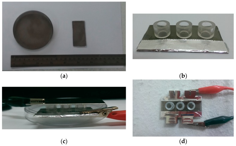
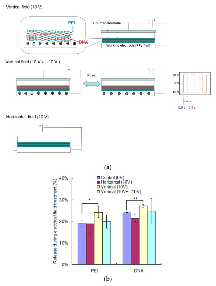
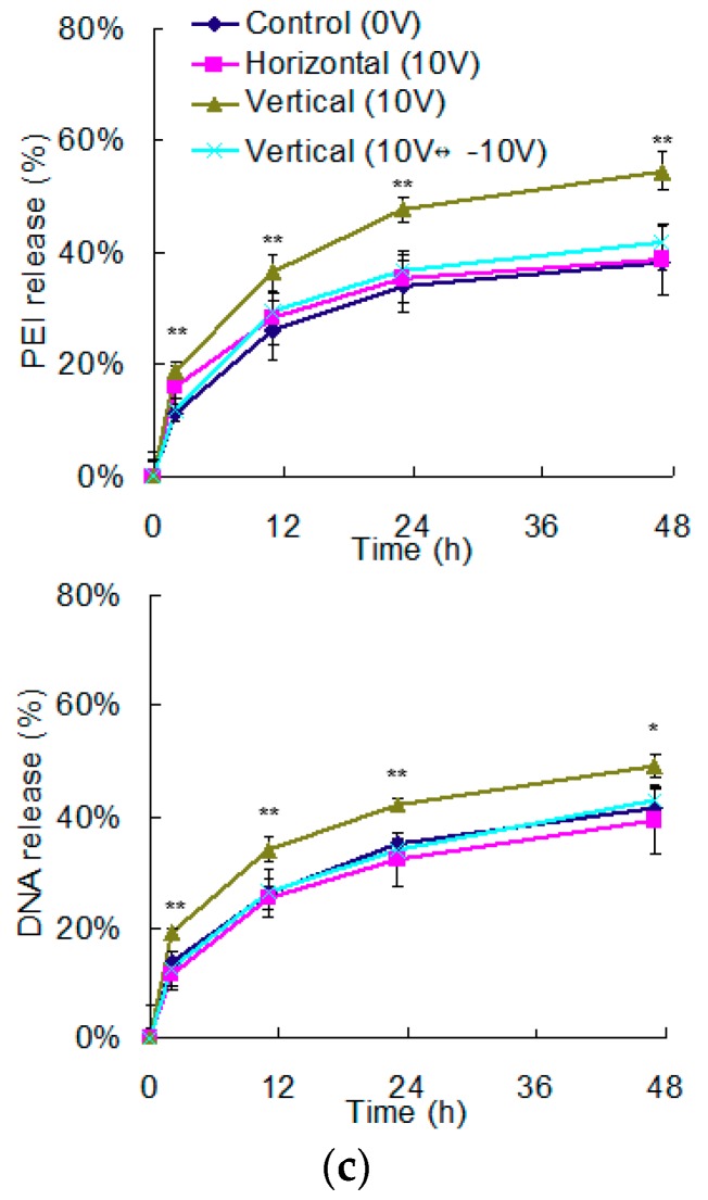
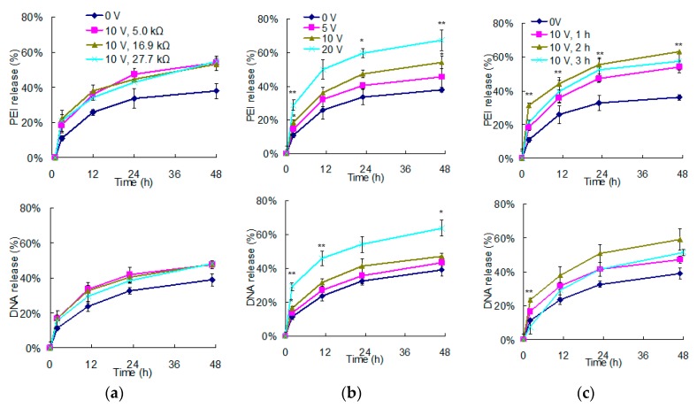
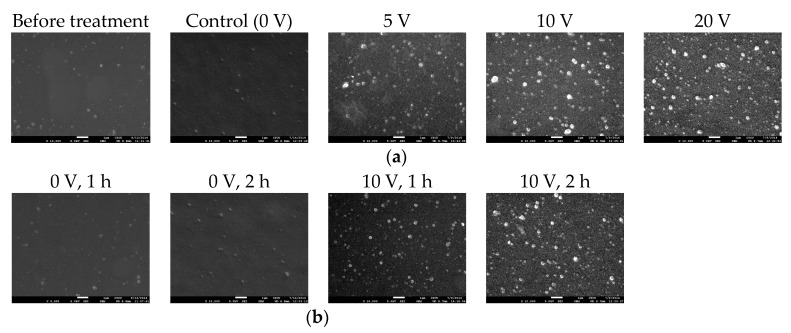
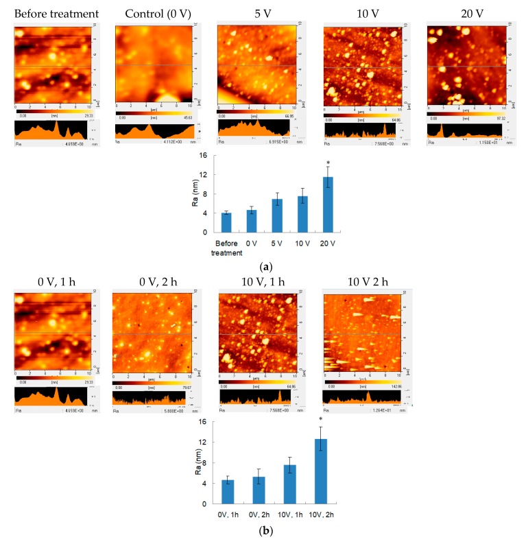
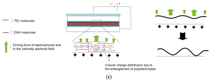
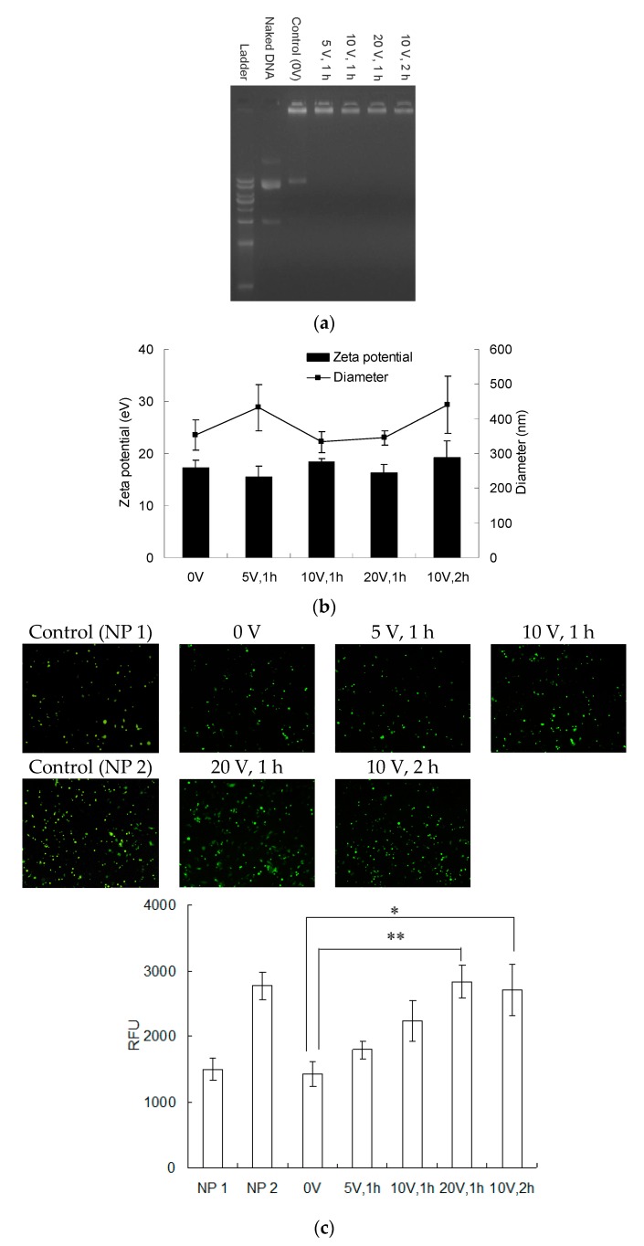
To sustain gene delivery and elongate transgene expression, plasmid DNA and cationic nonviral vectors can be deposited through layer-by-layer (LbL) assembly to form polyelectrolyte multilayers (PEMs). Although these macromolecules can be released for transfection purposes, their entanglement only allows partial delivery. Therefore, how to efficiently deliver immobilized genes from PEMs remains a challenge. In this study, we attempt to facilitate their delivery through the pretreatment of the external electrical field. Multilayers of polyethylenimine (PEI) and DNA were deposited onto conductive polypyrrole (PPy), which were placed in an aqueous environment to examine their release after electric field pretreatment. Only the electric field perpendicular to the substrate with constant voltage efficiently promoted the release of PEI and DNA from PEMs, and the higher potential resulted in the more releases which were enhanced with treatment time. The roughness of PEMs also increased after electric field treatment because the electrical field not only caused electrophoresis of polyelectrolytes and but also allowed electrochemical reaction on the PPy electrode. Finally, the released DNA and PEI were used for transfection. Polyplexes were successfully formed after electric field treatment, and the transfection efficiency was also improved, suggesting that this electric field pretreatment effectively assists gene delivery from PEMs and should be beneficial to regenerative medicine application.
Keywords: electrical field; gene delivery; layer-by-layer assembly; polyelectrolyte multilayer; polypyrrole.
Conflict of interest statement
The authors declare no conflict of interest.
Figures
Similar articles
-
Electrophoretic deposition to promote layer-by-layer assembly for in situ gene delivery application.Colloids Surf B Biointerfaces. 2015 Sep 1;133:171-8. doi: 10.1016/j.colsurfb.2015.05.046. Epub 2015 Jun 6. Colloids Surf B Biointerfaces. 2015. PMID: 26101817
-
The effect of alginate on DNA delivery from layer-by-layer assembled films.Carbohydr Polym. 2014 Jan 30;101:240-8. doi: 10.1016/j.carbpol.2013.09.025. Epub 2013 Sep 19. Carbohydr Polym. 2014. PMID: 24299770
-
From polyelectrolyte complexes to polyelectrolyte multilayers: Electrostatic assembly, nanostructure, dynamics, and functional properties.Adv Colloid Interface Sci. 2017 Jun;244:71-89. doi: 10.1016/j.cis.2016.12.004. Epub 2016 Dec 16. Adv Colloid Interface Sci. 2017. PMID: 28499602
-
Weak polyelectrolyte-based multilayers via layer-by-layer assembly: Approaches, properties, and applications.Adv Colloid Interface Sci. 2020 Aug;282:102200. doi: 10.1016/j.cis.2020.102200. Epub 2020 Jun 15. Adv Colloid Interface Sci. 2020. PMID: 32585489 Review.
-
A closer physico-chemical look to the Layer-by-Layer electrostatic self-assembly of polyelectrolyte multilayers.Adv Colloid Interface Sci. 2020 Aug;282:102197. doi: 10.1016/j.cis.2020.102197. Epub 2020 Jun 12. Adv Colloid Interface Sci. 2020. PMID: 32579951 Review.
Cited by
-
Chitosan-miRNA functionalized microporous titanium oxide surfaces via a layer-by-layer approach with a sustained release profile for enhanced osteogenic activity.J Nanobiotechnology. 2020 Sep 9;18(1):127. doi: 10.1186/s12951-020-00674-7. J Nanobiotechnology. 2020. PMID: 32907598 Free PMC article.
-
Electrical Stimuli-Responsive Decomposition of Layer-by-Layer Films Composed of Polycations and TEMPO-Modified Poly(acrylic acid).Polymers (Basel). 2022 Dec 7;14(24):5349. doi: 10.3390/polym14245349. Polymers (Basel). 2022. PMID: 36559714 Free PMC article.
References
-
- Decher G. Fuzzy nanoassemblies: Toward layered polymeric multicomposites. Science. 1997;277:1232–1237. doi: 10.1126/science.277.5330.1232. - DOI
-
- Xu X.Y., Chen Y.F., Tan Q.G., Chen Z.J., Li Y., Wu W.G., Wang X.F., Liu Y.B. Construction of multilayer films with bactericidal and long-term antitumor drug release properties as a non-vascular stent coating for therapy in obstruction. J. Mater. Chem. B. 2019;7:4963–4972. doi: 10.1039/C9TB01036J. - DOI - PubMed
-
- Chang H., Ren K.F., Zhang H., Wang J.L., Wang B.L., Ji J. The (prs/hgf-pdna) multilayer films for gene-eluting stent coating: Gene-protecting, anticoagulation, antibacterial properties, and in vivo antirestenosis evaluation. J. Biomed. Mater. Res. Part B. 2015;103:430–439. doi: 10.1002/jbm.b.33224. - DOI - PubMed
-
- de Avila E.D., Castro A.G.B., Tagit O., Krom B.P., Lowik D., van Well A.A., Bannenberg L.J., Vergani C.E., van den Beucken J. Anti-bacterial efficacy via drug-delivery system from layer-by-layer coating for percutaneous dental implant components. Appl. Surf. Sci. 2019;488:194–204. doi: 10.1016/j.apsusc.2019.05.154. - DOI
Grants and funding
LinkOut - more resources
Full Text Sources
