. 2020 Jan 6;25(1):236.
doi: 10.3390/molecules25010236.

Tracking Extracellular Matrix Remodeling in Lungs Induced by Breast Cancer Metastasis. Fourier Transform Infrared Spectroscopic Studies

Affiliations

Tracking Extracellular Matrix Remodeling in Lungs Induced by Breast Cancer Metastasis. Fourier Transform Infrared Spectroscopic Studies

Karolina Chrabaszcz et al. Molecules. .

Abstract

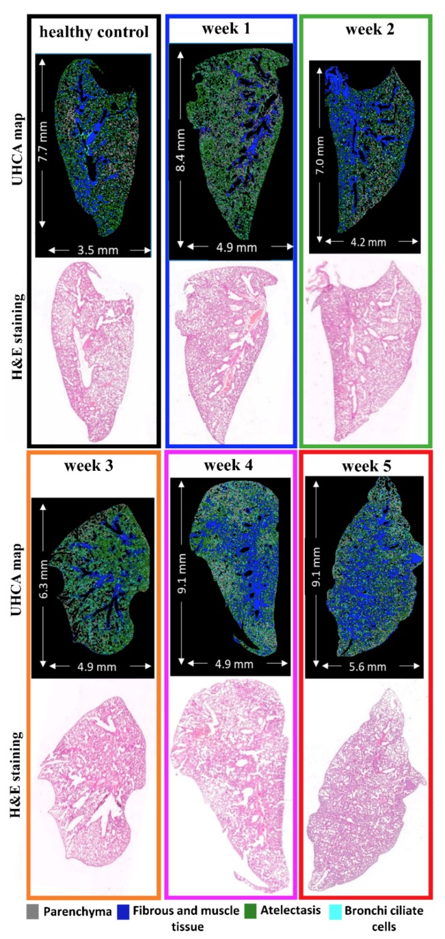
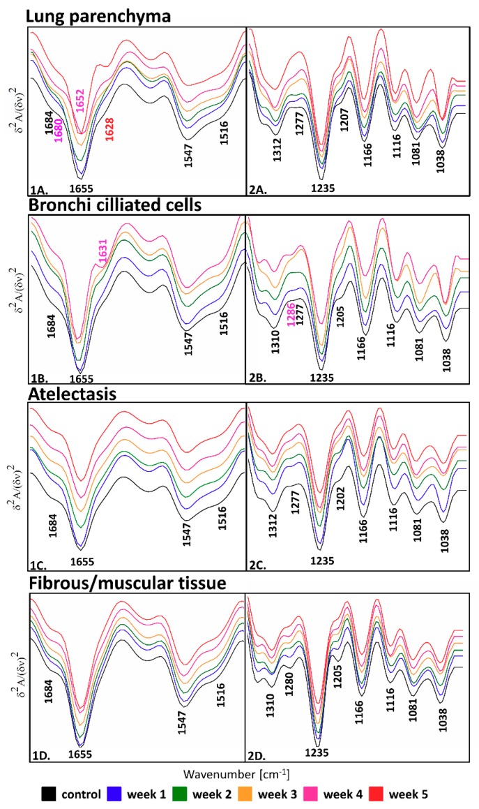
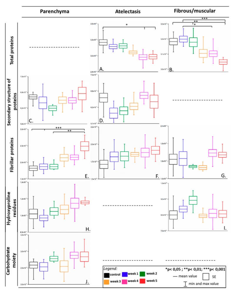
This work focused on a detailed assessment of lung tissue affected by metastasis of breast cancer. We used large-area chemical scanning implemented in Fourier transform infrared (FTIR) spectroscopic imaging supported with classical histological and morphological characterization. For the first time, we differentiated and defined biochemical changes due to metastasis observed in the lung parenchyma, atelectasis, fibrous, and muscle cells, as well as bronchi ciliate cells, in a qualitative and semi-quantitative manner based on spectral features. The results suggested that systematic extracellular matrix remodeling with the progress of the metastasis process evoked a decrease in the fraction of the total protein in atelectasis, fibrous, and muscle cells, as well as an increase of fibrillar proteins in the parenchyma. We also detected alterations in the secondary conformations of proteins in parenchyma and atelectasis and changes in the level of hydroxyproline residues and carbohydrate moieties in the parenchyma. The results indicate the usability of FTIR spectroscopy as a tool for the detection of extracellular matrix remodeling, thereby enabling the prediction of pre-metastatic niche formation.

Keywords: FTIR imaging; cancer metastases; extracellular matrix remodeling; fibrous proteins.


Conflict of interest statement

The authors declare no conflict of interest.

Figures

Figure 1
Figure 1
Comparison of false-color unsupervised hierarchical cluster analysis (UHCA) maps with hematoxylin and eosin (H&E) microphotographs collected from lung cross-sections. UHCA classification of the most abundant tissue types in lungs: parenchyma (grey), atelectasis (green), fibrous/muscular tissue (blue), and bronchi ciliated cells (aqua) corresponds to tissue structure observed in the H&E images. Magnified microphotographs after H&E staining with marked tissue types are shown in Figure S2 in Supplementary Materials.
Figure 2
Figure 2
The comparison of second derivative IR spectra extracted from UHCA analysis for lung parenchyma (averaged from n = 37 spectra) (A), bronchi ciliated cells (averaged from n = 23 spectra) (B), atelectasis (averaged from n = 43 spectra), (C) and fibrous/muscular tissue (averaged from n = 44 spectra) (D). Spectra are shown in the regions of 1700–1480 (1) and 1340–1000 cm−1 (2). On week 5, bronchi ciliated cells were not found since atelectasis covers the entire area of the lungs.
Figure 3
Figure 3
Box diagrams of integral intensities for the selected bands showing biochemical changes in parenchyma, atelectasis, and fibrous/muscular tissue in the metastasis progression in the lungs. Integration regions: total proteins [(1707–1608 cm−1) + (1589–1485 cm−1)], secondary structure of proteins [(1589–1485 cm−1)/(1707–1608 cm−1)], fibrillar proteins (1286–1193 cm−1), hydroxyproline residues (1187–1140 cm−1), carbohydrate moieties (1137–1015 cm−1). The bands’ assignment is given in Table 2.

References

    1. Klein C.A. Parallel progression of primary tumours and metastases. Nat. Rev. Cancer. 2009;9:302–312. doi: 10.1038/nrc2627. - DOI - PubMed
    1. Weigelt B., Peterse J.L., van ’t Veer L.J. Breast cancer metastasis: Markers and models. Nat. Rev. Cancer. 2005;5:591–602. doi: 10.1038/nrc1670. - DOI - PubMed
    1. Peinado H., Lavotshkin S., Lyden D. The secreted factors responsible for pre-metastatic niche formation: Old sayings and new thoughts. Semin. Cancer Biol. 2011;21:139–146. doi: 10.1016/j.semcancer.2011.01.002. - DOI - PubMed
    1. Alderton G.K. Metastasis. Exosomes drive premetastatic niche formation. Nat. Rev. Cancer. 2012;12:447. doi: 10.1038/nrc3304. - DOI - PubMed
    1. Murata T. Histological Studies on the Respiratory Portions of the Lungs of Cetacea. Sci. Rep. 1951;6:35–47.