Review
. 2020 Jan 7;9(1):140.
doi: 10.3390/cells9010140.

Sterculic Acid: The Mechanisms of Action beyond Stearoyl-CoA Desaturase Inhibition and Therapeutic Opportunities in Human Diseases

Affiliations
Review

Sterculic Acid: The Mechanisms of Action beyond Stearoyl-CoA Desaturase Inhibition and Therapeutic Opportunities in Human Diseases

Rafael Peláez et al. Cells. .

Abstract

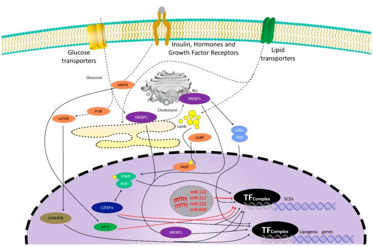
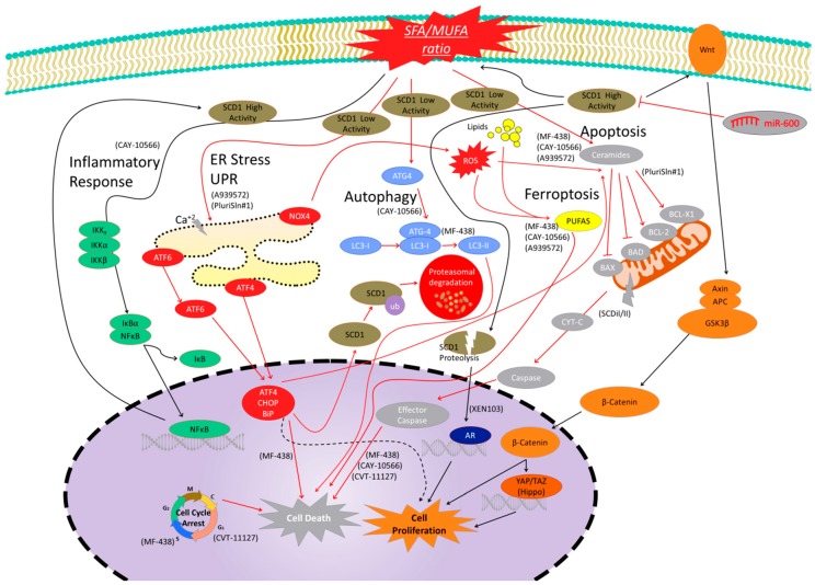
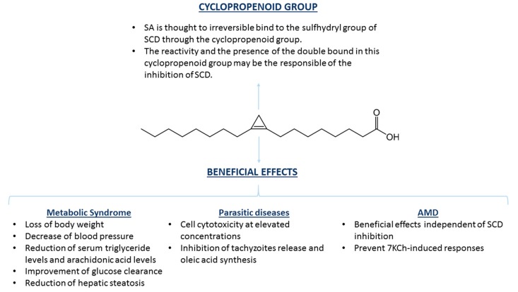
In many tissues, stearoyl-CoA desaturase 1 (SCD1) catalyzes the biosynthesis of monounsaturated fatty acids (MUFAS),(i.e., palmitoleate and oleate) from their saturated fatty acid (SFA) precursors (i.e., palmitate and stearate), influencing cellular membrane physiology and signaling, leading to broad effects on human physiology. In addition to its predominant role in lipid metabolism and body weight control, SCD1 has emerged recently as a potential new target for the treatment for various diseases, such as nonalcoholic steatohepatitis, Alzheimer's disease, cancer, and skin disorders. Sterculic acid (SA) is a cyclopropene fatty acid originally found in the seeds of the plant Sterculia foetida with numerous biological activities. On the one hand, its ability to inhibit stearoyl-CoA desaturase (SCD) allows its use as a coadjuvant of several pathologies where this enzyme has been associated. On the other hand, additional effects independently of its SCD inhibitory properties, involve anti-inflammatory and protective roles in retinal diseases such as age-related macular degeneration (AMD). This review aims to summarize the mechanisms by which SA exerts its actions and to highlight the emerging areas where this natural compound may be of help for the development of new therapies for human diseases.

Keywords: cell death; inflammation; macular degeneration; metabolism; sterculic acid.


Conflict of interest statement

I.M.L. holds a patent on “Methods of treatment using sterculic acid”. “The funders had no role in the design of the study; in the collection, analyses, or interpretation of data; in the writing of the manuscript, or in the decision to publish the results”.

Figures

Figure 1
Figure 1
Genetic control of the stearoyl-CoA desaturase (SCD) family and other lipogenic gene expression. Promoter regions of SCD genes present many transcription factor binding sites, such as sterol regulatory element binding protein 1 (SREBP1), carbohydrate response element binding protein (ChREBP), CCAAT/enhancer-binding protein (C/EBP), liver X receptor (LXR), or peroxisome proliferator-activated receptor (PPAR). Black arrows represent inductive signals while red lines represent repressive signals that negatively modulate SCD genes expression. Glucose uptake or insulin signaling, but also lipid uptake, hormones, or growth factors binding to their receptors, signaling pathways such as phosphatidylinositol 3-kinase (PI3K), mitogen-activated protein kinase (MAPK), mammalian target of rapamycin (mTOR), or endoplasmic reticulum (ER) stress, promote direct or indirect transcription factor binding to SCDs promoters to modulate SCD gene expression. Some microRNAs (mirRNA121, 221, 222, and 600) have been reported to downregulate SCDs expression. Activator protein 1 (AP-1) is a transcription factor that reduces SCD1 expression after leptin stimulation. C/EBP induction can also downregulate SCD1 expression after bacterial infection.
Figure 2
Figure 2
Signaling pathways modulated by SCD1 activity and levels. Black arrows are signals of cell proliferation or associated with high SCD1 expression or activity, while red arrows are signals linked to cell death or decreased SCD1 expression or activity. Inflammatory pathway is shown in green. SCD1 inhibition reduces the pro-inflammatory environment and nuclear factor kappa-B (NFκB) signaling to reduce cell proliferation, while NFκB signaling activation promotes transcription factors binding to SCD1 promotor and gene expression. The Wnt/β-catenin pathway is related to cell proliferation and it is shown in orange. High SCD1 levels are linked to increased β-catenin signaling and increased levels of wingless-related integration site (Wnt) ligands to induce cell proliferation. Hippo pathway is a β-catenin-related signal cascade, which is associated with cell proliferation, and SCD1 inhibition has been demonstrated to be associated with decreased levels of Hippo target genes. This pathway is shown in dark orange. The SCD1 peptides from proteolytic controlled degradation activate androgen receptor (AR) signaling to promote cell proliferation. This pathway is shown in dark blue. The autophagy cell death pathway is a protein cascade which is upregulated after SCD1 inhibition. Some central elements of this pathway are shown in light blue. The endoplasmic reticulum (ER) stress signal pathway is shown in red. This cascade is linked to misfolded and unfolded proteins to induced cell death. SCD1 inhibition has been linked to the upregulation of the elements of the ER pathway to promote cell death. SCD1 is also ubiquitinated by this pathway to promote protein degradation. However, light ER pathway activation has also been related to cell survival (dashed black arrows). Apoptosis is a programmed cell death shown in grey that is activated after SCD1 inhibitor treatments. Increased ceramides, mitochondrial effector pathway, caspases, and other pathway effectors have been detected after treatments. Ferroptosis is another cell death mechanism (yellow) that has been shown to be activated after SCD1 Inhibition. Finally, SCD1 inhibition has been demonstrated to induce cell cycle arrest in different checkpoints to promote cell death. Molecules in parentheses are SCD1 inhibitors used in the literature to elucidate the SCD1-related pathways.
Figure 3
Figure 3
Chemical structure of SA and beneficial effects exerted in several pathologies. The cyclopropene group of SA has been suggested to be responsible for both binding and inhibition of SCD as a consequence of the reactivity of the double bond between C9 and C10. Inhibition of SCD by SA has been described as potentially therapeutic for several diseases, such as those related with metabolic syndrome and parasitic diseases. Positive effects of SA have also been shown in age-related macular degeneration (AMD), although the effects seem to be independent of SCD inhibition.

References

    1. Smith U., Kahn B.B. Adipose tissue regulates insulin sensitivity: Role of adipogenesis, de novo lipogenesis and novel lipids. J. Intern. Med. 2016;280:465–475. doi: 10.1111/joim.12540. - DOI - PMC - PubMed
    1. Song Z., Xiaoli A.M., Yang F. Regulation and Metabolic Significance of De Novo Lipogenesis in Adipose Tissues. Nutrients. 2018;10:1383. doi: 10.3390/nu10101383. - DOI - PMC - PubMed
    1. Aarsland A., Chinkes D., Wolfe R.R. Hepatic and whole-body fat synthesis in humans during carbohydrate overfeeding. Am. J. Clin. Nutr. 1997;65:1774–1782. doi: 10.1093/ajcn/65.6.1774. - DOI - PubMed
    1. Poudyal H., Brown L. Stearoyl-CoA desaturase: A vital checkpoint in the development and progression of obesity. Endocr. Metab. Immune Disord Drug Targets. 2011;11:217–231. doi: 10.2174/187153011796429826. - DOI - PubMed
    1. Paton C.M., Ntambi J.M. Biochemical and physiological function of stearoyl-CoA desaturase. Am. J. Physiol. Endocrinol. Metab. 2009;297:E28–E37. doi: 10.1152/ajpendo.90897.2008. - DOI - PMC - PubMed

Publication types

MeSH terms

LinkOut - more resources