

Significance of 5- S-Cysteinyldopa as a Marker for Melanoma
- PMID: 31936623
- PMCID: PMC7013534
- DOI: 10.3390/ijms21020432
Significance of 5- S-Cysteinyldopa as a Marker for Melanoma
Abstract
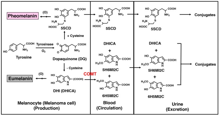
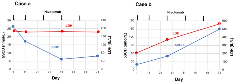
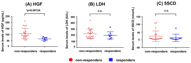
Melanoma is one of the most lethal and malignant cancers and its incidence is increasing worldwide, and Japan is not an exception. Although there are numerous therapeutic options for melanoma, the prognosis is still poor once it has metastasized. The main concern after removal of a primary melanoma is whether it has metastasized, and early detection of metastatic melanoma would be effective in improving the prognosis of patients. Thus, it is very important to identify reliable methods to detect metastases as early as possible. Although many prognostic biomarkers (mainly for metastases) of melanoma have been reported, there are very few effective for an early diagnosis. Serum and urinary biomarkers for melanoma diagnosis have especially received great interest because of the relative ease of sample collection and handling. Several serum and urinary biomarkers appear to have significant potential both as prognostic indicators and as targets for future therapeutic methods, but still there are no efficient serum and urinary biomarkers for early detection, accurate diagnosis and prognosis, efficient monitoring of the disease and reliable prediction of survival and recurrence. Levels of 5-S-cysteinyldopa (5SCD) in the serum or urine as biomarkers of melanoma have been found to be significantly elevated earlier and to reflect melanoma progression better than physical examinations, laboratory tests and imaging techniques, such as scintigraphy and echography. With recent developments in the treatment of melanoma, studies reporting combinations of 5SCD levels and new applications for the treatment of melanoma are gradually increasing. This review summarizes the usefulness of 5SCD, the most widely used and well-known melanoma marker in the serum and urine, compares 5SCD and other useful markers, and finally its application to other fields.
Keywords: 5-S-cysteinyldopa (5SCD); Melanoma inhibitory activity (MIA); S100 calcium-binding protein B (S100B); anti-programmed cell death protein (PD)-1 antibody; biomarker; early detection; hepatocyte growth factor (HGF); lactate dehydrogenase (LDH); melanoma.
Conflict of interest statement
The authors declare no conflict of interest.
Figures
References
Publication types
MeSH terms
Substances
Grants and funding
- Fujita Health University/Scholarship donation
- 02670487/Grants-in-Aid for Scientific Research from the Ministry of Education, Science, and Culture
- 11-7/the Ministry of Health and Welfare, no. 3-20, Japan, and for Cancer Research from the Ministry of Health, Labour and Welfare of Japan
- 18K08301/Japan Society for the Promotion of Science (JSPS)
LinkOut - more resources
Full Text Sources
Medical
Miscellaneous
