Review
. 2020 Jan 10;21(2):451.
doi: 10.3390/ijms21020451.

Neurogenesis and Specification of Retinal Ganglion Cells

Affiliations
Review

Neurogenesis and Specification of Retinal Ganglion Cells

Kim Tuyen Nguyen-Ba-Charvet et al. Int J Mol Sci. .

Abstract

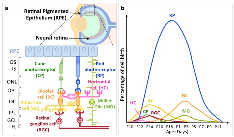
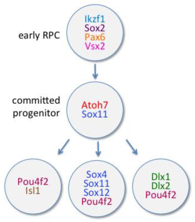
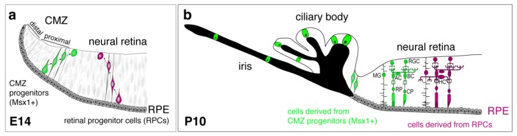
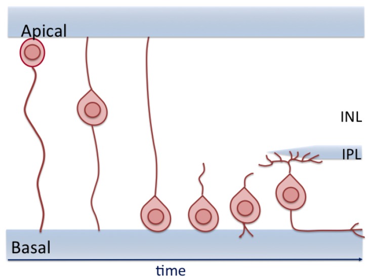
Across all species, retinal ganglion cells (RGCs) are the first retinal neurons generated during development, followed by the other retinal cell types. How are retinal progenitor cells (RPCs) able to produce these cell types in a specific and timely order? Here, we will review the different models of retinal neurogenesis proposed over the last decades as well as the extrinsic and intrinsic factors controlling it. We will then focus on the molecular mechanisms, especially the cascade of transcription factors that regulate, more specifically, RGC fate. We will also comment on the recent discovery that the ciliary marginal zone is a new stem cell niche in mice contributing to retinal neurogenesis, especially to the generation of ipsilateral RGCs. Furthermore, RGCs are composed of many different subtypes that are anatomically, physiologically, functionally, and molecularly defined. We will summarize the different classifications of RGC subtypes and will recapitulate the specification of some of them and describe how a genetic disease such as albinism affects neurogenesis, resulting in profound visual deficits.

Keywords: RGC subtype; albinism; competence; development; fate control; retinal progenitor cell; retinogenesis; stochastic.


Conflict of interest statement

The authors declare no conflict of interest.

Figures

Figure 1
Figure 1
Layer distribution and genesis of the different retinal cell types. (a) Schematic representation of the eye with an enlargement on the neural retina and the retinal pigmented epithelium (RPE) showing all the retinal cell types and their organization into layers. Inner segments (IS) and outer segments (OS) of photoreceptors. The outer nuclear layer (ONL) contains soma of cone photoreceptors (CP) and rod photoreceptors (RP). OPL: outer plexiform layer. The inner nuclear layer (INL) contains soma of Müller glia (MG) and different interneurons: amacrine cells (AC), horizontal cells (HC) and bipolar cells (BC). IPL: inner plexiform layer. The ganglion cell layer (GCL) contains retinal ganglion cells (RGC). The fiber layer (FL) contains RGC axons. Adapted from Cepko et al., 2014 [19]. (b) Time course of differentiation of the retinal cells generated during mouse development illustrating the early differentiation of postmitotic RGCs. E: embryonic days, P: postnatal days. Adapted from Young et al., 1985 [12].
Figure 2
Figure 2
Transcription factors participating in the specification of RGCs from early retinal progenitor cells (RPC) to committed progenitors that will later produce postmitotic RGCs.
Figure 3
Figure 3
Schematic representation of lineage tracing from progenitors of the ciliary marginal zone (CMZ) or neural retina in mouse. (a) At E14, retinal progenitor cells (RPCs) (magenta) are the main contributor to retinal neurogenesis, but Msx1+ progenitors (green) in the proximal CMZ migrate towards the neural retina and contribute to retinal neurogenesis at the periphery of the retina. (b) At P10, cells derived from Msx1+ progenitors (green) produce most retinal cell types, although with a different ratio than cells derived from RPCs (magenta). Also, Msx1+ progenitors (green) contribute to non-pigmented epithelial cells of the ciliary body and the cells in the iris. RPE: retinal pigmented epithelium, RGC: retinal ganglion cells, BC: bipolar cells, AC: amacrine cells, MG: Müller glia, RP: rod photoreceptors, CP: cone photoreceptors, HC: horizontal cells. Adapted from Marcucci et al., 2016 [151] and Bélanger et al., 2017 [152].
Figure 4
Figure 4
The newborn retinal ganglion cell (RGC) translocates from the apical part of the retina toward the future ganglion cell layer at the basal side. During the entire process, the cell is attached to both sides of the retina. After the migration, the RGC adjusts its position while losing its apical process and send its axon to the optic nerve. INL: inner nuclear layer, IPL: inner plexiform layer. Adapted from Amini et al. [159].
Figure 5
Figure 5
Examples of specific transcription factors that determine the identity of RGC subtypes or subclasses.

Similar articles

Cited by

References

    1. Sanes J.R., Masland R.H. The Types of Retinal Ganglion Cells: Current Status and Implications for Neuronal Classification. Annu. Rev. Neurosci. 2015;38:221–246. doi: 10.1146/annurev-neuro-071714-034120. - DOI - PubMed
    1. Rheaume B.A., Jereen A., Bolisetty M., Sajid M.S., Yang Y., Renna K., Sun L., Robson P., Trakhtenberg E.F. Single cell transcriptome profiling of retinal ganglion cells identifies cellular subtypes. Nat. Commun. 2018;9:2759. doi: 10.1038/s41467-018-05134-3. - DOI - PMC - PubMed
    1. Turner D.L., Cepko C.L. A common progenitor for neurons and glia persists in rat retina late in development. Nature. 1987;328:131–136. doi: 10.1038/328131a0. - DOI - PubMed
    1. Holt C.E., Bertsch T.W., Ellis H.M., Harris W.A. Cellular determination in the Xenopus retina is independent of lineage and birth date. Neuron. 1988;1:15–26. doi: 10.1016/0896-6273(88)90205-X. - DOI - PubMed
    1. Wetts R., Fraser S.E. Multipotent precursors can give rise to all major cell types of the frog retina. Science. 1988;239:1142–1145. doi: 10.1126/science.2449732. - DOI - PubMed

LinkOut - more resources