. 2020 Dec;26(14):1854-1865.
doi: 10.1177/1352458519893093. Epub 2020 Jan 15.

MRI differences between MOG antibody disease and AQP4 NMOSD

Affiliations

MRI differences between MOG antibody disease and AQP4 NMOSD

Sara Salama et al. Mult Scler. 2020 Dec.

Abstract

Background: MOG antibody and AQP4 antibody seropositive diseases are immunologically distinct subtypes of neuromyelitis optica spectrum disorders (NMOSD) with similar clinical presentations. MRI findings can be instrumental in distinguishing MOG antibody disease from AQP4 antibody NMOSD.

Objectives: The aim of this study is to characterize the neuroradiological differences between MOG antibody disease and AQP4 antibody NMOSD with the aim to distinguish between the two entities.

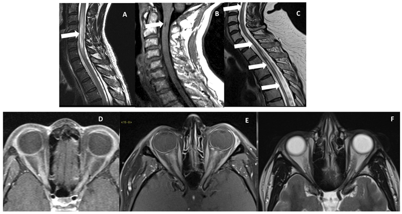
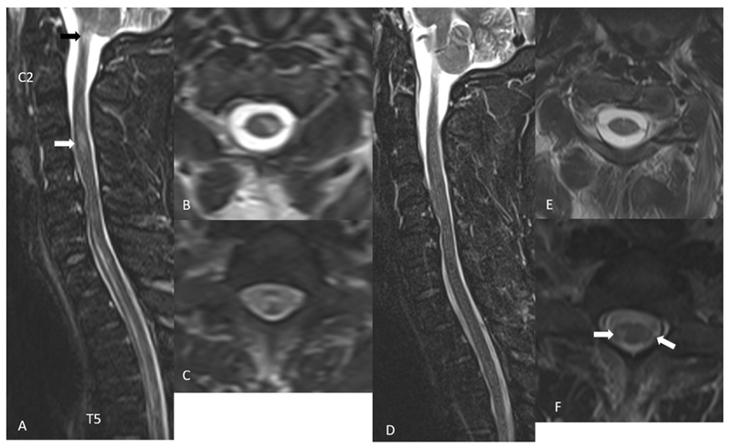
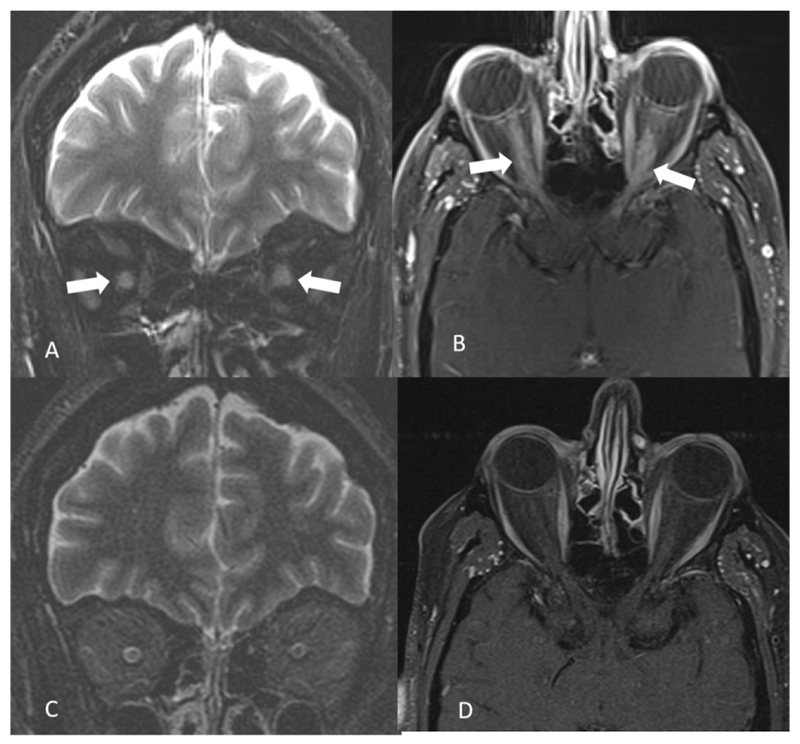
Methods: This is a retrospective study of 26 MOG and 25 AQP4 seropositive patients in which MRI features of the brain, spinal cord, and orbit were compared.

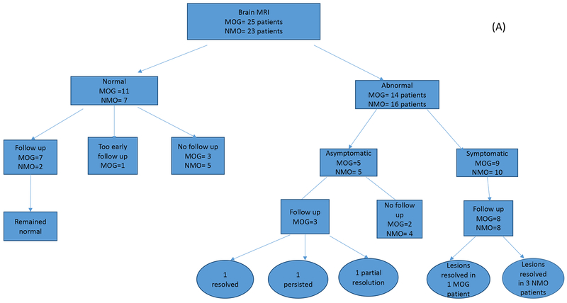
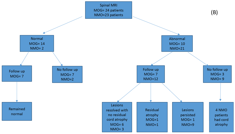
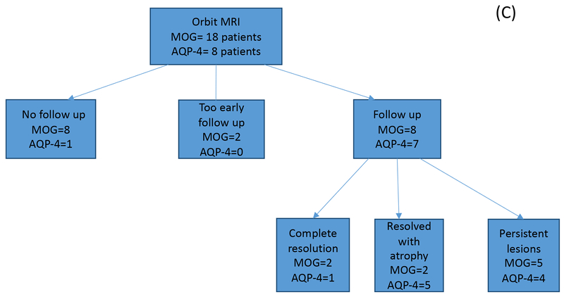
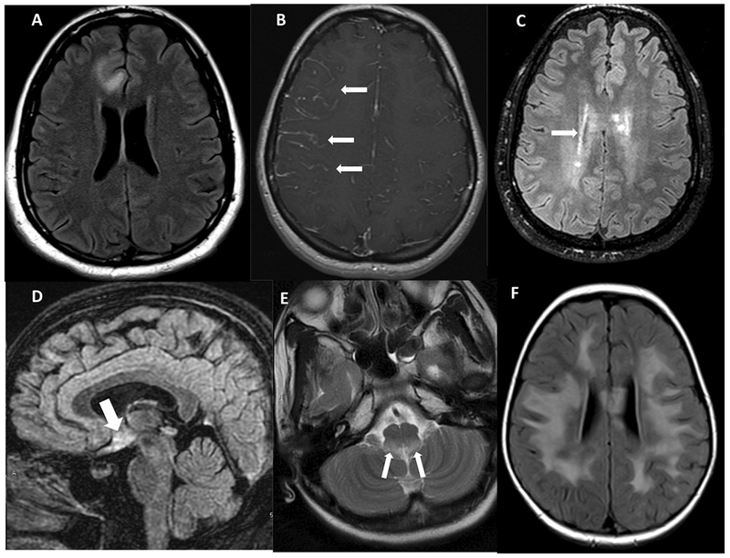
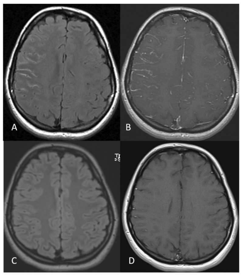
Results: The majority of the abnormal findings in the MOG cohort were located on orbital MRIs, while spinal cord magnetic resonance (MR) abnormalities were more common in the AQP4 cohort. Brain abnormalities showed some overlap, but cortical gray/juxtacortical white matter involvement was distinct to MOG patients, while area postrema involvement was a rare feature.

Conclusion: Cortical gray/juxtacortical white matter lesions on brain MRI might help distinguish MOG antibody disease from AQP4-positive NMOSD. These findings could be of value in distinguishing the two entities as early as the first presentation.

Keywords: AQP4 antibody; MOG antibody; NMOSD; magnetic resonance imaging.


Conflict of interest statement

None of the authors has any conflict of interests to disclose.

Figures

Figure 1.
Figure 1.
A. Number of brain MRIs analyzed for the MOG antibody (n=25) and matched AQP4 seropositive disease groups (n=23). Studies were first divided into abnormal versus normal studies, then by follow up status. Final outcomes are indicated in both cohorts. B. Number of spinal cord MRIs analyzed for the MOG antibody (n=24) and matched AQP4 seropositive disease groups (n=23). Studies were first divided into abnormal versus normal studies, then by follow up status. Final outcomes are indicated in both cohorts. C. Number of orbital MRIs analyzed for the MOG antibody (n=18) and matched AQP4 seropositive disease groups (n=8). Studies were first divided into abnormal versus normal studies, then by follow up status. Final outcomes are indicated in both cohorts.
Figure 1.
Figure 1.
A. Number of brain MRIs analyzed for the MOG antibody (n=25) and matched AQP4 seropositive disease groups (n=23). Studies were first divided into abnormal versus normal studies, then by follow up status. Final outcomes are indicated in both cohorts. B. Number of spinal cord MRIs analyzed for the MOG antibody (n=24) and matched AQP4 seropositive disease groups (n=23). Studies were first divided into abnormal versus normal studies, then by follow up status. Final outcomes are indicated in both cohorts. C. Number of orbital MRIs analyzed for the MOG antibody (n=18) and matched AQP4 seropositive disease groups (n=8). Studies were first divided into abnormal versus normal studies, then by follow up status. Final outcomes are indicated in both cohorts.
Figure 2.
Figure 2.
A. Axial FLAIR image showing cortical grey matter and juxta-cortical white matter involvement in MOG B. Axial post-contrast images showing leptomeningeal enhancement in a patient with MOG. C. Axial FLAIR image showing peri-ependymal involvement of supratentorial brain. D. Sagittal FLAIR showing signal changes from involvement of hypothalamus suggestive of diencephalon syndromic pattern of AQP4 NMOSD. E. Axial T2W-weighted image showing signal changes in the region of area postrema in a patient with NMOSD. F. Axial FLAIR image showing the encephalopathic pattern of involvement in a patient with MOG disease.
Figure 3:
Figure 3:
A 23 year- old MOGAD woman without past medical history presents with 5 days of right frontal headache and painful right eye movements. Orbital MRI shows right optic neuritis (not shown) and right sided non-suppression of CSF signal on the FLAIR sequence (A) and leptomeningeal enhancement in the right cerebral sulci (B). Follow up brain MRI after one month shows normal brain MRI findings (C and D).
Figure 4.
Figure 4.
A. Sagittal T2-weighted image showing a bright spot ventrally in the cervical cord in an NMOSD patient. B. Sagittal T1-weighted image shows a dark T1 spot in a patient with AQP4 NMOSD. C. Sagittal T2-weighted image showing cord atrophy and residual T2 hyperintensity in a patient with NMOSD. D. Axial post-contrast image showing longitudinally extensive optic neuritis in a patient with MOG. E. Axial post-contrast image showing longitudinally extensive bilateral optic neuritis in a patient with NMOSD, and on follow up scan as seen in F axial T2-weighted image showing bilateral optic atrophy.
Figure 5:
Figure 5:
A 32-year-old MOGAD male presents with leg weakness and numbness, bladder and bowel retention. STIR sagittal image (A) shows longitudinally extensive T2 hyperintense signal changes in the anterior cord extending from C7 to T5 vertebra and a smaller areas of T2 hyperintensity at C4 level (white arrow in A) and at C1 level (black arrow in A). Axial T2 weighted images show a left central and lateral cord hyperintensity (B) and almost entire cord signal abnormality at T1 level (C). Follow up imaging after 18 days show almost complete resolution of signal abnormalities on STIR sagittal image (D and E). There are a few small areas of residual T2 hyperintense signal changes in the bilateral lateral cord at T1 level (arrows in F).
Figure 6:
Figure 6:
A 53-year-old MOGAD male presents with bilateral orbital pain and gradual loss of vision within 2 weeks. Coronal fat saturated T2 weighted image shows bilateral optic nerve hyperintensity and swelling (arrows in A). On fat saturated contrast enhanced T1 weighted image longitudinally extensive contrast enhancement is noted in bilateral optic nerves (arrows in B). Three months follow up orbital MRI shows complete resolution of the findings (C and D).

References

    1. Oh J, Levy M. Neuromyelitis optica: an antibody-mediated disorder of the central nervous system. Neurol Res Int. 2012;2012:460825. - PMC - PubMed
    1. Wingerchuk DM, Banwell B, Bennett JL, Cabre P, Carroll W, Chitnis T, et al. International consensus diagnostic criteria for neuromyelitis optica spectrum disorders. Neurology. 2015;85(2):177–89. - PMC - PubMed
    1. Jiao Y, Fryer JP, Lennon VA, Jenkins SM, Quek AM, Smith CY, et al. Updated estimate of AQP4-IgG serostatus and disability outcome in neuromyelitis optica. Neurology. 2013;81(14):1197–204. - PMC - PubMed
    1. Jarius S, Ruprecht K, Kleiter I, Borisow N, Asgari N, Pitarokoili K, et al. MOG-IgG in NMO and related disorders: a multicenter study of 50 patients. Part 1: Frequency, syndrome specificity, influence of disease activity, long-term course, association with AQP4-IgG, and origin. J Neuroinflammation. 2016;13(1):279. - PMC - PubMed
    1. Probstel AK, Rudolf G, Dornmair K, Collongues N, Chanson JB, Sanderson NS, et al. Anti-MOG antibodies are present in a subgroup of patients with a neuromyelitis optica phenotype. J Neuroinflammation. 2015;12:46. - PMC - PubMed

Publication types

LinkOut - more resources