. 2020 Feb 21;295(8):2160-2174.
doi: 10.1074/jbc.RA119.010315. Epub 2020 Jan 14.

Oxygen-dependent asparagine hydroxylation of the ubiquitin-associated (UBA) domain in Cezanne regulates ubiquitin binding

Affiliations

Oxygen-dependent asparagine hydroxylation of the ubiquitin-associated (UBA) domain in Cezanne regulates ubiquitin binding

Julia Mader et al. J Biol Chem. .

Abstract

Deubiquitinases (DUBs) are vital for the regulation of ubiquitin signals, and both catalytic activity of and target recruitment by DUBs need to be tightly controlled. Here, we identify asparagine hydroxylation as a novel posttranslational modification involved in the regulation of Cezanne (also known as OTU domain-containing protein 7B (OTUD7B)), a DUB that controls key cellular functions and signaling pathways. We demonstrate that Cezanne is a substrate for factor inhibiting HIF1 (FIH1)- and oxygen-dependent asparagine hydroxylation. We found that FIH1 modifies Asn35 within the uncharacterized N-terminal ubiquitin-associated (UBA)-like domain of Cezanne (UBACez), which lacks conserved UBA domain properties. We show that UBACez binds Lys11-, Lys48-, Lys63-, and Met1-linked ubiquitin chains in vitro, establishing UBACez as a functional ubiquitin-binding domain. Our findings also reveal that the interaction of UBACez with ubiquitin is mediated via a noncanonical surface and that hydroxylation of Asn35 inhibits ubiquitin binding. Recently, it has been suggested that Cezanne recruitment to specific target proteins depends on UBACez Our results indicate that UBACez can indeed fulfill this role as regulatory domain by binding various ubiquitin chain types. They also uncover that this interaction with ubiquitin, and thus with modified substrates, can be modulated by oxygen-dependent asparagine hydroxylation, suggesting that Cezanne is regulated by oxygen levels.

Keywords: Cezanne; FIH1; OTU domain-containing protein 7B (OTUD7B); UBA domain; deubiquitinase (DUB); deubiquitylation (deubiquitination); hydroxylation; nuclear magnetic resonance (NMR); posttranslational modification (PTM); protein-protein interaction; ubiquitin.


Conflict of interest statement

The authors declare that they have no conflicts of interest with the contents of this article

Figures
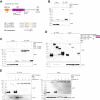
Figure 1.
Figure 1.
Cezanne contains a putative consensus site for FIH1-dependent hydroxylation. A, overview of the domain architecture of Cezanne and sequence alignment showing an FIH1 consensus sequence in Cezanne (where Φ indicates aliphatic amino acids and * represents the modified asparagine residue). B, co-IP of GFP-Cezanne and FIH1 from HEK293 cells. C, co-IP of GFP-Cezanne and catalytic inactive FIH1 (H199A) and dimerization-deficient FIH1 (L340R) mutants. D, co-IP of GFP-Cezanne truncated versions and FIH1 to narrow down the region in Cezanne that interacts with FIH1. E, co-IP of GFP-Cezanne and FIH1, assessing the effect of hypoxia on the interaction of the two proteins.
Figure 2.
Figure 2.
Cezanne is hydroxylated within its UBA-like domain. A, FIH1 was efficiently depleted by CRISPR/Cas9-mediated knockout in HEK293 cells. B, cartoon depicting design of SILAC-based MS experiment for the detection of asparagine hydroxylation in Cezanne. C, annotated MS2 spectrum showing hydroxylated Asn35 of Cezanne in HEK293 cells. D, FIH1- and oxygen-dependent enrichment of Asn35 hydroxylation. Data are represented as mean of SILAC ratios plus S.E. (error bars). *, p < 0.1; **, p < 0.01 by one-sample t test. The experiment was performed with three biological replicates.
Figure 3.
Figure 3.
Cezanne's UBA domain binds polyubiquitin of different linkage types. A, sequence alignment of various UBA domains revealed that UBACez lacks both the MGF motif and the dileucine motif. B, Western blotting showing in vitro GST pulldown assay. GST-UBACez co-precipitated Lys11-, Lys48-, and Lys63-linked tetraubiquitin. C, in vitro pulldown of WT and mutant UBACez and Halo-tagged tetraubiquitin (Met1-linked). Mutation of Asn35 in UBACez greatly reduced ubiquitin binding. D, semi-in cellulo pulldown of recombinant WT or mutant UBACez and ubiquitinated proteins from HEK293 cells treated with the proteasome inhibitor MG132.
Figure 4.
Figure 4.
Hydroxylation of Asn35 modulates UBA-ubiquitin interaction. A, cartoon depicting experimental strategy for assessing the effect of asparagine hydroxylation on ubiquitin binding. B, verification of FIH1-dependent enrichment of Asn35 hydroxylation of recombinant UBACez by MS analysis. Data are represented as mean of log2 -fold change intensity plus S.E. (error bars). **, p < 0.01; ***, p < 0.001 by one-sample t test. The experiment was performed with three biological replicates. C, in vitro GST pulldown assay comparing interaction of linear tetraubiquitin with unmodified and hydroxylated UBACez, respectively.
Figure 5.
Figure 5.
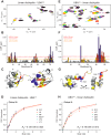
Binding between linear diubiquitin and the UBA domain of Cezanne. Shown are the results for NMR titrations of 15N-labeled linear diubiquitin with nonlabeled UBACez (A–D) and NMR titrations of 15N-labeled UBACez with nonlabeled linear diubiquitin (E–H). A, representative area of 15N,1H BEST-TROSY HSQC spectra for linear diubiquitin, to which UBACez was added in a specified molar ratio (1:8, 1:4, 1:2, 1, 2, 4, and 8), is shown. The rainbow color code indicates increased molar ratios upon titration from free linear diubiquitin (red) to full saturation upon 8-fold excess of UBACez (dark gray). B, CSP mapping on the ubiquitin sequence. Yellow and red lines indicate the 1× and 2× S.D. (δ) calculated from CSP values of all residues, respectively. Residues of linear diubiquitin, which showed double peaks (either in the free or UBACez-bound form) and significant line broadening, are colored in blue. C, CSP mapping on the linear diubiquitin structure (PDB code 2L2D). The diubiquitin is presented as a ribbon diagram; P and D symbols indicate proximal and distal ubiquitin moieties. Residues with intermediately (δ ≤ CSP ≤ 2×δ) and strongly (CSP ≥ 2×δ) perturbed backbone HN resonances are marked in yellow and red, respectively. Residues with anomalous exchange behavior (described above) are marked in blue. Side chains of residues from the ubiquitin hydrophobic patch (Leu8-Ile44-Val70) are shown as spherical atom models. D, KD calculated for the selected linear diubiquitin residues upon titration with UBACez. Normalized CSP values of residues I3p (where p represents proximal ubiquitin), Leu8, Ile44, Lys63p, Val70, and Leu71, showing significant CSP in the fast exchange mode, were used in the global fit. E, representative area of 15N,1H BEST-TROSY HSQC spectra for UBACez, to which linear diubiquitin was added in the specified molar ratio (1:8, 1:4, 1:2, 1, 2, 4, and 8), is shown. The rainbow color code indicates increased molar ratios upon titration from the free UBACez (red) to full saturation upon 8-fold excess of linear diubiquitin (dark gray). F, CSP mapping on the UBACez sequence. Yellow and red lines, 1× and 2× S.D. (δ) calculated from CSP values of all residues, respectively. Residues of UBACez, which showed intermediate exchange and significant line broadening, are colored in blue. *, CSP for the side chain of Gln43, which shows intermediate and big CSP upon binding of linear diubiquitin. G, CSP mapping on the modeled UBACez structure. The UBACez is presented as a ribbon diagram. Residues with intermediately (δ ≤ CSP ≤ 2×δ) and strongly (CSP ≥ 2×δ) perturbed backbone HN resonances are marked in yellow and red, respectively. Residues with anomalous exchange behavior (described above) are marked in blue. H, KD calculated for the selected UBACez residues upon titration with linear diubiquitin. Normalized CSP values of residues Ser10, Leu27, Glu42, Gln43, and Gly50, showing significant CSP in the fast exchange mode, were used in the global fit.

References

    1. Clague M. J., Urbé S., and Komander D. (2019) Breaking the chains: deubiquitylating enzyme specificity begets function. Nat. Rev. Mol. Cell Biol. 20, 338–352 10.1038/s41580-019-0099-1 - DOI - PubMed
    1. Sahtoe D. D., and Sixma T. K. (2015) Layers of DUB regulation. Trends Biochem. Sci. 40, 456–467 10.1016/j.tibs.2015.05.002 - DOI - PubMed
    1. Bremm A., Freund S. M. V., and Komander D. (2010) Lys11-linked ubiquitin chains adopt compact conformations and are preferentially hydrolyzed by the deubiquitinase Cezanne. Nat. Struct. Mol. Biol. 17, 939–947 10.1038/nsmb.1873 - DOI - PMC - PubMed
    1. Mevissen T. E. T., Kulathu Y., Mulder M. P. C., Geurink P. P., Maslen S. L., Gersch M., Elliott P. R., Burke J. E., van Tol B. D. M., Akutsu M., Oualid F. E., Kawasaki M., Freund S. M. V., Ovaa H., and Komander D. (2016) Molecular basis of Lys11-polyubiquitin specificity in the deubiquitinase Cezanne. Nature 538, 402–405 10.1038/nature19836 - DOI - PMC - PubMed
    1. Pareja F., Ferraro D. A., Rubin C., Cohen-Dvashi H., Zhang F., Aulmann S., Ben-Chetrit N., Pines G., Navon R., Crosetto N., Köstler W., Carvalho S., Lavi S., Schmitt F., Dikic I., et al. (2012) Deubiquitination of EGFR by Cezanne-1 contributes to cancer progression. Oncogene 31, 4599–4608 10.1038/onc.2011.587 - DOI - PMC - PubMed

Publication types