. 2020 Mar 31;94(13):e1386-e1394.
doi: 10.1212/WNL.0000000000008939. Epub 2020 Jan 14.

MRI vastus lateralis fat fraction predicts loss of ambulation in Duchenne muscular dystrophy

Affiliations

MRI vastus lateralis fat fraction predicts loss of ambulation in Duchenne muscular dystrophy

Karin J Naarding et al. Neurology. .

Abstract

Objective: We studied the potential of quantitative MRI (qMRI) as a surrogate endpoint in Duchenne muscular dystrophy by assessing the additive predictive value of vastus lateralis (VL) fat fraction (FF) to age on loss of ambulation (LoA).

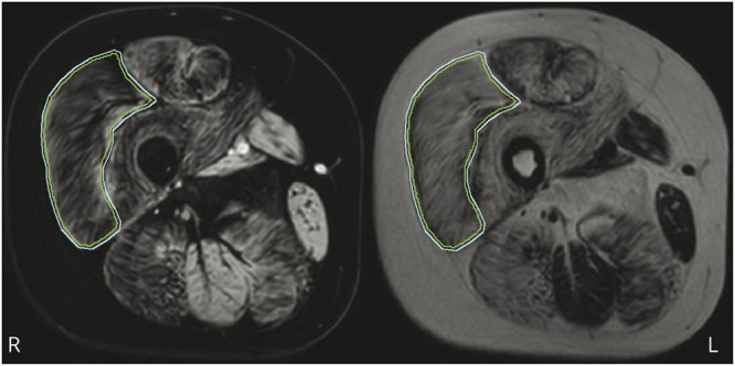
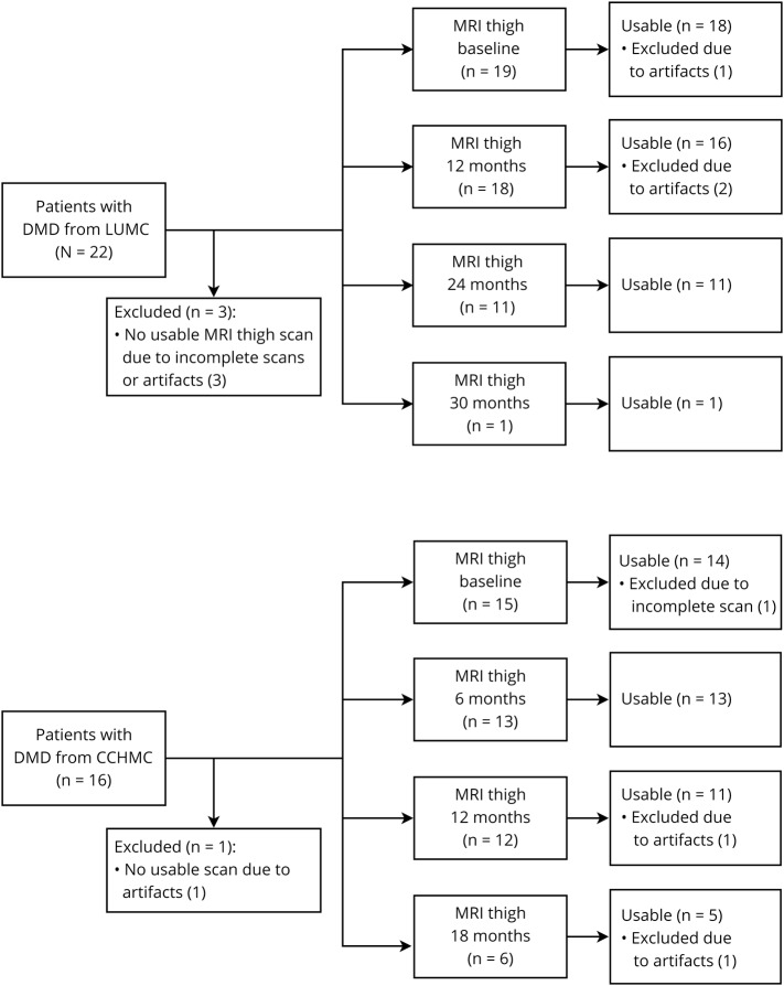
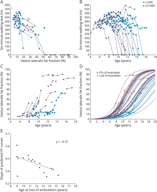
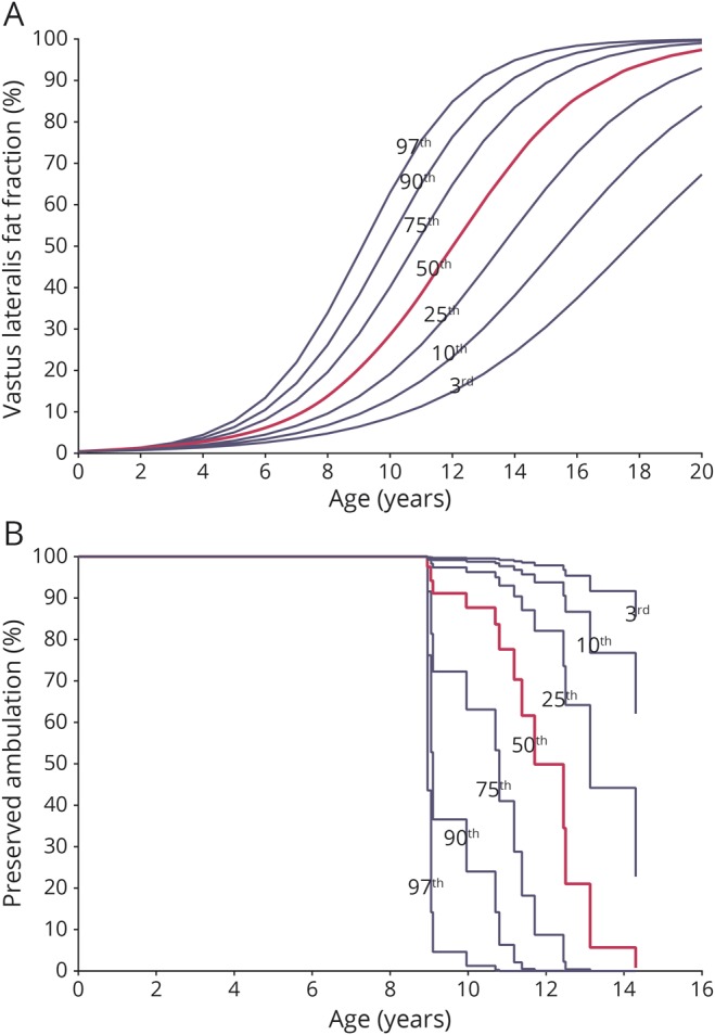
Methods: VL FFs were determined on longitudinal Dixon MRI scans from 2 natural history studies in Leiden University Medical Center (LUMC) and Cincinnati Children's Hospital Medical Center (CCHMC). CCHMC included ambulant patients, while LUMC included a mixed ambulant and nonambulant population. We fitted longitudinal VL FF values to a sigmoidal curve using a mixed model with random slope to predict individual trajectories. The additive value of VL FF over age to predict LoA was calculated from a Cox model, yielding a hazard ratio.

Results: Eighty-nine MRIs of 19 LUMC and 15 CCHMC patients were included. At similar age, 6-minute walking test distances were smaller and VL FFs were correspondingly higher in LUMC compared to CCHMC patients. Hazard ratio of a percent-point increase in VL FF for the time to LoA was 1.15 for LUMC (95% confidence interval [CI] 1.05-1.26; p = 0.003) and 0.96 for CCHMC (95% CI 0.84-1.10; p = 0.569).

Conclusions: The hazard ratio of 1.15 corresponds to a 4.11-fold increase of the instantaneous risk of LoA in patients with a 10% higher VL FF at any age. Although results should be confirmed in a larger cohort with prospective determination of the clinical endpoint, this added predictive value of VL FF to age on LoA supports the use of qMRI FF as an endpoint or stratification tool in clinical trials.


Figures

Figure 1
Figure 1. VL ROI
Example of a region of interest (ROI) drawn on the vastus lateralis (VL) (outer line) and the 2-mm inward erosion (inner line) on a water image (left) and corresponding fat image (right).
Figure 2
Figure 2. Flowchart of included thigh MRI datasets
Inclusion of patients with Duchenne muscular dystrophy (DMD) and thigh MRI scan data at Leiden University Medical Center (LUMC) and Cincinnati Children's Hospital Medical Center (CCHMC). Forty-six useable MRIs from 1 to 4 time points were available for 19 LUMC patients, and 43 useable MRIs from again 1 to 4 time points were available for 15 CCHMC patients.
Figure 3
Figure 3. Longitudinal 6MWT and VL FF data
Longitudinal data of patients with Duchenne muscular dystrophy (DMD) from Leiden University Medical Center (LUMC) (dark circles) and Cincinnati Children's Hospital Medical Center (CCHMC) (lighter squares). (A) 6-Minute walking test (6MWT) results plotted vs vastus lateralis (VL) fat fraction (FF). LUMC and CCHMC patients with similar 6MWT results have similar VL FFs. FFs from nonambulant patients are plotted as 0 m on the 6MWT. (B) 6MWT data plotted vs age. CCHMC patients on average walk longer distances at later ages than LUMC patients. Age at loss of ambulation (LoA) is plotted as 0 m. (C) Original VL FF results plotted vs age. On average VL FF results were higher and increased faster over time in LUMC patients compared to CCHMC patients. Data visually correspond to a sigmoid curve. (D) Original and predicted VL FF results plotted vs age. Patients with higher VL FFs at younger ages or faster FF increases had steeper predicted FF slopes. On average, LUMC patients showed steeper slopes than patients from CCHMC. Logistic curves from 2 LUMC patients at comparable ages are highlighted (pink lines) to illustrate the relationship with their LoA, depicted as an X, at 11.4 and 13.1 years of age. (E) Individual slope of the predicted FF curves plotted vs age at LoA. With the use of a Spearman correlation analysis on the LUMC cohort, there was a negative correlation between these variables (ρ = −0.72, p = 0.001).
Figure 4
Figure 4. VL FF growth chart and survival chart of preserved ambulation for the LUMC cohort
Growth charts based on data of patients with Duchenne muscular dystrophy (DMD) from Leiden University Medical Center (LUMC) plotted vs age. (A) We generated a vastus lateralis (VL) fat fraction (FF) growth chart with a 3rd, 10th, 25th, 50th, 75th, 90th, and 97th percentile curve from the predicted LUMC VL FF data. (B) Using the resulting hazard ratio from the LUMC cohort, we transformed the predicted LUMC VL FF growth curves to survival curves for preserved ambulation. A patient on the third percentile in the VL FF growth chart is also on the third percentile in the survival chart.

References

    1. Cros D, Harnden P, Pellissier JF, Serratrice G. Muscle hypertrophy in Duchenne muscular dystrophy: a pathological and morphometric study. J Neurol 1989;236:43–47. - PubMed
    1. Verhaart IEC, Aartsma-Rus A. Therapeutic developments for Duchenne muscular dystrophy. Nat Rev Neurol 2019;15:373–386. - PubMed
    1. Straub V, Balabanov P, Bushby K, et al. . Stakeholder cooperation to overcome challenges in orphan medicine development: the example of Duchenne muscular dystrophy. Lancet Neurol 2016;15:882–890. - PubMed
    1. Barnard AM, Willcocks RJ, Finanger EL, et al. . Skeletal muscle magnetic resonance biomarkers correlate with function and sentinel events in Duchenne muscular dystrophy. PLoS One 2018;13:e0194283. - PMC - PubMed
    1. Bonati U, Hafner P, Schädelin S, et al. . Quantitative muscle MRI: a powerful surrogate outcome measure in Duchenne muscular dystrophy. Neuromuscul Disord 2015;25:679–685. - PubMed

Publication types

MeSH terms