. 2020 Jan 14;11(1):264.
doi: 10.1038/s41467-019-13839-2.

Dorsal root ganglion macrophages contribute to both the initiation and persistence of neuropathic pain

Affiliations

Dorsal root ganglion macrophages contribute to both the initiation and persistence of neuropathic pain

Xiaobing Yu et al. Nat Commun. .

Abstract

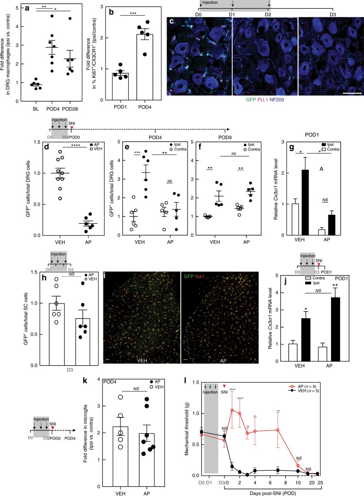
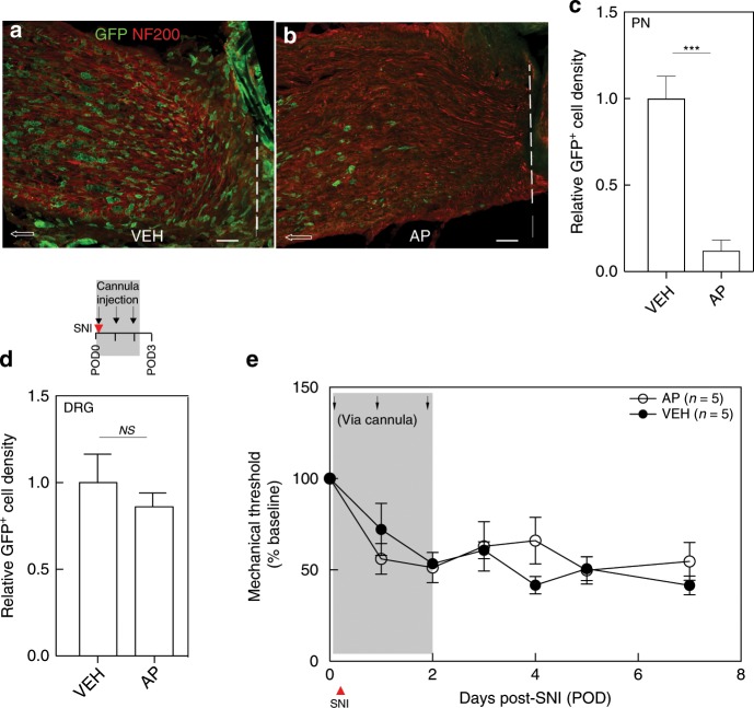
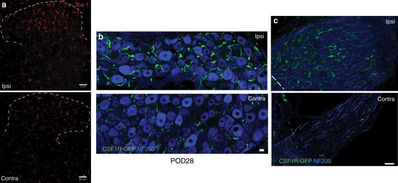
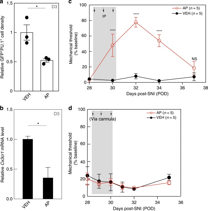
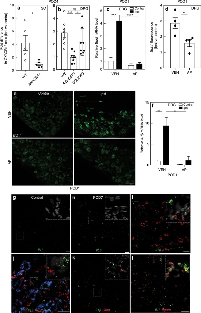
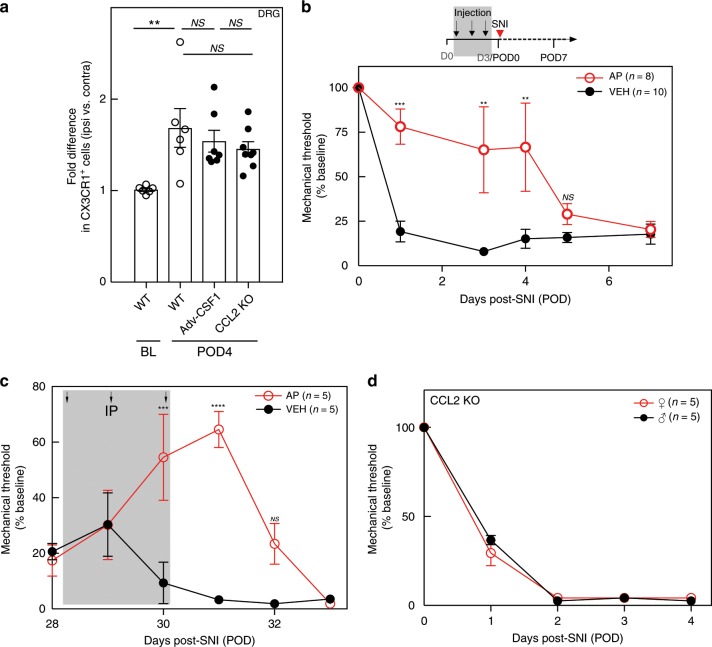
Paralleling the activation of dorsal horn microglia after peripheral nerve injury is a significant expansion and proliferation of macrophages around injured sensory neurons in dorsal root ganglia (DRG). Here we demonstrate a critical contribution of DRG macrophages, but not those at the nerve injury site, to both the initiation and maintenance of the mechanical hypersensitivity that characterizes the neuropathic pain phenotype. In contrast to the reported sexual dimorphism in the microglial contribution to neuropathic pain, depletion of DRG macrophages reduces nerve injury-induced mechanical hypersensitivity and expansion of DRG macrophages in both male and female mice. However, fewer macrophages are induced in the female mice and deletion of colony-stimulating factor 1 from sensory neurons, which prevents nerve injury-induced microglial activation and proliferation, only reduces macrophage expansion in male mice. Finally, we demonstrate molecular cross-talk between axotomized sensory neurons and macrophages, revealing potential peripheral DRG targets for neuropathic pain management.


Conflict of interest statement

The authors declare no competing interests.

Figures

Fig. 1
Fig. 1. Systemic AP treatment depletes macrophages in the DRG of MAFIA mice and delays mechanical allodynia.
a FACS analysis of CX3CR1+ macrophage expansion in the L4/L5 DRG of wild-type mice after SNI (n = 6–7 per group). BL, baseline. POD, post-operative day. b FACS analysis of Ki67 expression in CX3CR1+ macrophages in the L4/L5 DRG of wild-type mice after nerve injury (n = 5 per group). c Representative images illustrating AP-induced depletion of GFP+ (green)/PU.1+(red) macrophages in the L4/L5 DRG of MAFIA mice injected with systemic AP (1.0 mg kg−1) for 3 days. NF200 (blue) marks myelinated neurons; scale bar: 50 μm. d FACS analysis of GFP+ DRG macrophages 1 day after the 3rd AP injection (nVEH = 9; nAP = 6). e, f SNI 1 day after AP or VEH treatment followed by FACS analysis of DRG macrophages on POD4 (e) and POD9 (f) (n = 5–6 per group). g qPCR analysis of Cx3cr1 gene expression in the L4/5 DRG of mice pretreated with AP or VEH on POD1 (n = 3 per group). h FACS analysis of GFP+ spinal cord microglia after a 3-day AP or VEH (n = 6 per group). i GFP+ (green) and Iba1+ (red) immunoreactive spinal cord microglia after a 3-day AP or VEH. Scale bar: 50 µm. j qPCR analysis of Cx3cr1 gene expression on POD1 in the lumbar spinal cord in mice pretreated with AP or VEH (n = 3 per group). k FACS analysis of GFP+ microglia in the lumbar spinal cord of mice pretreated with AP or VEH (nVEH = 5; nAP = 7) on POD4. l Effect on mechanical thresholds of systemic AP or VEH followed by SNI (n = 5 per group). Gray shading indicates injection days. Data presented as mean ± SEM. One-way ANOVA with Tukey’s correction in a, Student’s t-test in b, d, h, and k, and two-way ANOVA with Sidak’s correction in eg, j, and l. *P < 0.05, **P < 0.01, ***P < 0.001, ****P < 0.0001, NS nonsignificant compared with control. Source data are available as a Source Data file.
Fig. 2
Fig. 2. Macrophages in the DRG, but not at the nerve injury site, are required for mechanical allodynia initiation.
ae Effect of SNI followed by cannula injection with VEH or AP on immunostaining of GFP+ macrophages at the peripheral nerve (PN) ligature site (ac) and DRG (d) (n = 3 per group) and on mechanical thresholds (e). Dashed lines in a and b denote ligature site; arrows point proximally. Scale bar: 50 µm. Gray shading indicates injection days. Data presented as mean ± SEM. Student’s t-test in c and d, and two-way repeated-measures ANOVA with Sidak’s correction in e. ***P < 0.001, NS nonsignificant compared with control. Source data are available as a Source Data file.
Fig. 3
Fig. 3. Microglia activation and peripheral macrophage infiltration persist at 28 days post SNI.
ac Immunostaining of Iba1+ microglia in the dorsal horn (a), CSF1R-GFP+ macrophages (green) in the DRG (b), and at the nerve injury site (c), 28 days after SNI (POD28). NF200 (blue) labels neuronal cell bodies (b) and peripheral nerve myelinated axons (c). Dashed line in c denotes nerve ligature site. Scale bar: 50 µm in a; 15 µm in b, c.
Fig. 4
Fig. 4. Macrophages in the DRG, but not at the peripheral nerve injury site, are required for mechanical allodynia maintenance.
Four weeks after SNI (POD28), MAFIA mice received either systemic (ac) or cannula (d) injections of AP or VEH. a, b GFP+/PU.1+ macrophage density (a) and qPCR of Cx3cr1 gene expression (b) in the ipsilateral DRG 1 day after the 3rd AP injection (D3, n = 3 per group). c, d Mechanical thresholds after 3-day systemic (c) or cannula (d) injections (n = 5 per group). Gray shading indicates injection days. Data presented as mean ± SEM. Student’s t-test in a and b, two-way repeated-measures ANOVA with Sidak’s correction in c and d. *P < 0.05, ****P < 0.0001, NS nonsignificant compared with control. Source data are available as a Source Data file.
Fig. 5
Fig. 5. Nerve injury triggers reciprocal molecular interactions between DRG macrophages and sensory neurons.
a FACS analysis of CX3CR1+ microglia in the lumbar spinal cord of WT and Adv-Csf1 mice 4 days after SNI (POD4, n = 5 per group). b FACS analysis of CX3CR1+ macrophages in the L4/L5 DRG of WT (n = 7), Adv-Csf1 (n = 8), and CCL2 knockout (KO) mice (n = 8) on POD4. ce qPCR (c, n = 3 per group), or in situ hybridization (ISH) (d, e, n = 4 per group) analysis of Bdnf expression in the DRG of mice pretreated for 3 days with systemic AP or VEH on POD1. Scale bar: 15 µm in e. f qPCR for Il1β on POD1 in the DRG of mice pretreated with AP or VEH. gl DRG expression of Il1β mRNA (green) in control (g) or 7 days after nerve injury (POD7, hl). il Coexpression of Il1β with neuronal markers Atf3 (i), Prph (red, j), Nefh (blue, j); a satellite cell marker, Gfap (red, k); and a macrophage marker, Itgam (red, l). Scale bars: 50 µm. Insets illustrate high magnification of labeled cells. Data presented as mean ± SEM. Student’s t-test in a and d, one-way ANOVA with Tukey’s correction in b, and two-way ANOVA with Sidak’s correction in c and f. *P < 0.05, **P < 0.01, ***P < 0.001, ****P < 0.0001, NS nonsignificant compared with control. Source data are available as a Source Data file.
Fig. 6
Fig. 6. The DRG macrophage contribution to neuropathic pain initiation and maintenance is not sexually dimorphic.
a FACS analysis of ipsilateral CX3CR1+ macrophage expansion in the L4/L5 DRG of WT (n = 6), Adv-Csf1 (n = 7), and CCL2 KO (n = 8) female mice on POD4. b Effect on mechanical thresholds of systemic AP (n = 8) or VEH (n = 10) followed by SNI in female MAFIA mice. c Effect on mechanical thresholds of systemic AP or VEH (n = 5 per group) 4 weeks after SNI in female MAFIA mice. d Effect of CCL2 deletion on mechanical thresholds in male and female mice (n = 5 per group). Gray shading indicates injection days. Data presented as mean ± SEM. One-way ANOVA with Tukey’s correction in a and two-way repeated-measures ANOVA with Sidak’s correction in bd. *P < 0.05, **P < 0.01, ***P < 0.001, ****P < 0.0001, NS nonsignificant compared with control. Source data are available as a Source Data file.

References

    1. Ji RR, Chamessian A, Zhang YQ. Pain regulation by non-neuronal cells and inflammation. Science. 2016;354:572–577. doi: 10.1126/science.aaf8924. - DOI - PMC - PubMed
    1. Inoue K, Tsuda M. Microglia in neuropathic pain: cellular and molecular mechanisms and therapeutic potential. Nat. Rev. Neurosci. 2018;19:138–152. doi: 10.1038/nrn.2018.2. - DOI - PubMed
    1. Hu P, McLachlan EM. Distinct functional types of macrophage in dorsal root ganglia and spinal nerves proximal to sciatic and spinal nerve transections in the rat. Exp. Neurol. 2003;184:590–605. doi: 10.1016/S0014-4886(03)00307-8. - DOI - PubMed
    1. Simeoli R, et al. Exosomal cargo including microRNA regulates sensory neuron to macrophage communication after nerve trauma. Nat. Commun. 2017;8:1778. doi: 10.1038/s41467-017-01841-5. - DOI - PMC - PubMed
    1. Cobos EJ, et al. Mechanistic differences in neuropathic pain modalities revealed by correlating behavior with global expression profiling. Cell Rep. 2018;22:1301–1312. doi: 10.1016/j.celrep.2018.01.006. - DOI - PMC - PubMed

Publication types

MeSH terms