

MicroRNA-142-3p inhibits high-glucose-induced endothelial-to-mesenchymal transition through targeting TGF-β1/Smad pathway in primary human aortic endothelial cells
- PMID: 31938215
- PMCID: PMC6958121
MicroRNA-142-3p inhibits high-glucose-induced endothelial-to-mesenchymal transition through targeting TGF-β1/Smad pathway in primary human aortic endothelial cells
Abstract
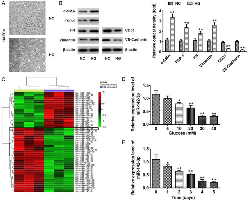
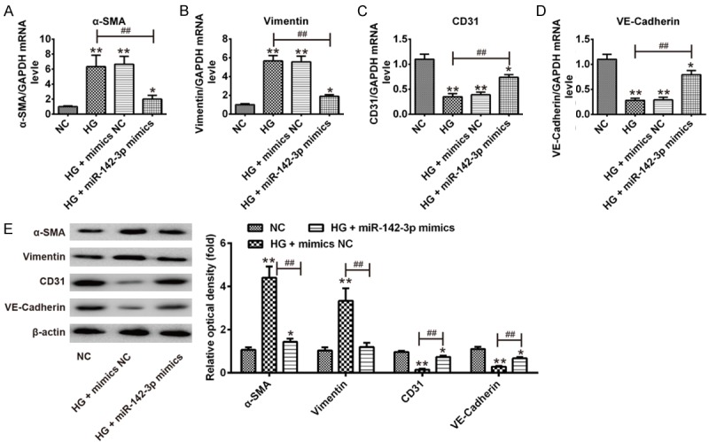
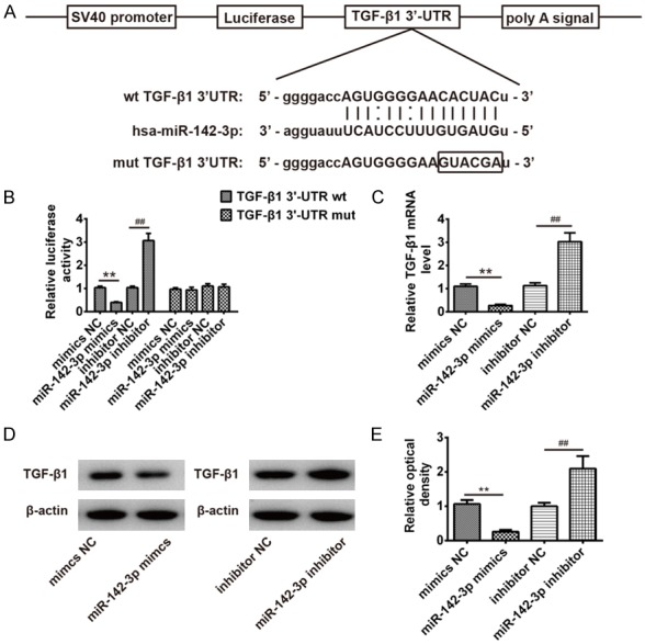
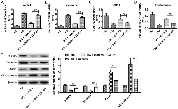
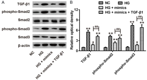
Myocardial fibrosis is an important pathological feature of diabetic cardiomyopathy (DCM) and endothelial-to-mesenchymal transition (EndMT) is an essential process for myocardial fibrosis. Recent studies have demonstrated an association between miRs and DCM. Therefore, the aim of this study is to investigate the role and the mechanism of miRNAs in the process of EndMT. We simulated the conditions occurring in EndMT by application of high glucose in primary human aortic endothelial cells (HAECs). Firstly, we compared the expression profiles of miRNAs in HAECs with or without HG treatment using microarray. Then, after addition of miR-142-3p mimics, the expression levels of EndMT markers were assessed by qRT-PCR and Western Blot. Moreover, bioinformatics analysis and luciferase assay were used to confirm the direct regulation of miR-142-3p to TGF-β1. Furthermore, the role of TGF-β1 in the inhibitory effect of miR-142-3p on EndMT was evaluated. In addition, the expressions of TGF-β1/Smad signaling signatures were measured by Western Blot. MiR-142-3p screened by miRNA microarray was significantly down-regulated in HAECs under HG stimulation in a dose and time dependent manner. Subsequently, we found that overexpression of miR-142-3p could inhibit HG-induced EndMT, as evidenced by decreased α-SMA and vimentin expression, and increased CD31 and VE-cadherin expression. Of note, transforming growth factor beta 1 (TGF-β1), one of the molecular mediators implicated in the progression of EndMT, was confirmed to be downstream target gene of miR-142-3p in HAECs. Moreover, TGF-β1 overexpression remarkably abolished the inhibitory effects of miR-142-3p overexpression on HG induced EndMT. Finally, miR-142-3p also mediated its anti-EndMT action by inactivation of TGF-β1/Smad pathway, as demonstrated by downregulation of TGF-β1, phospho-Smad2 and phospho-Smad2. Our findings demonstrated that miR-142-3p could attenuate HG-induced EndMT in HAECs, the mechanism of which may be at least partly through blocking TGF-β1/Smad signaling pathway. This might provide a potential therapeutic target for DCM in future.
Keywords: Diabetic cardiomyopathy; EndMT; TGF-β1/Smad signaling pathway; miR-142-3p.
IJCEP Copyright © 2018.
Conflict of interest statement
None.
Figures
Similar articles
-
MicroRNA-195-5p Downregulation Inhibits Endothelial Mesenchymal Transition and Myocardial Fibrosis in Diabetic Cardiomyopathy by Targeting Smad7 and Inhibiting Transforming Growth Factor Beta 1-Smads-Snail Pathway.Front Physiol. 2021 Sep 30;12:709123. doi: 10.3389/fphys.2021.709123. eCollection 2021. Front Physiol. 2021. PMID: 34658906 Free PMC article.
-
MiR-200a modulates TGF-β1-induced endothelial-to-mesenchymal shift via suppression of GRB2 in HAECs.Biomed Pharmacother. 2017 Nov;95:215-222. doi: 10.1016/j.biopha.2017.07.104. Epub 2017 Sep 12. Biomed Pharmacother. 2017. PMID: 28846982
-
MALAT1 Modulates TGF-β1-Induced Endothelial-to-Mesenchymal Transition through Downregulation of miR-145.Cell Physiol Biochem. 2017;42(1):357-372. doi: 10.1159/000477479. Epub 2017 May 25. Cell Physiol Biochem. 2017. PMID: 28535533
-
Modulation of EndMT by Hydrogen Sulfide in the Prevention of Cardiovascular Fibrosis.Antioxidants (Basel). 2021 Jun 3;10(6):910. doi: 10.3390/antiox10060910. Antioxidants (Basel). 2021. PMID: 34205197 Free PMC article. Review.
-
TGF-β-Induced Endothelial to Mesenchymal Transition in Disease and Tissue Engineering.Front Cell Dev Biol. 2020 Apr 21;8:260. doi: 10.3389/fcell.2020.00260. eCollection 2020. Front Cell Dev Biol. 2020. PMID: 32373613 Free PMC article. Review.
Cited by
-
MicroRNA-195-5p Downregulation Inhibits Endothelial Mesenchymal Transition and Myocardial Fibrosis in Diabetic Cardiomyopathy by Targeting Smad7 and Inhibiting Transforming Growth Factor Beta 1-Smads-Snail Pathway.Front Physiol. 2021 Sep 30;12:709123. doi: 10.3389/fphys.2021.709123. eCollection 2021. Front Physiol. 2021. PMID: 34658906 Free PMC article.
-
Roles of non-coding RNA in diabetic cardiomyopathy.Cardiovasc Diabetol. 2024 Jun 29;23(1):227. doi: 10.1186/s12933-024-02252-9. Cardiovasc Diabetol. 2024. PMID: 38951895 Free PMC article. Review.
-
MicroRNAs as Biomarkers for Coronary Artery Disease Related to Type 2 Diabetes Mellitus-From Pathogenesis to Potential Clinical Application.Int J Mol Sci. 2022 Dec 29;24(1):616. doi: 10.3390/ijms24010616. Int J Mol Sci. 2022. PMID: 36614057 Free PMC article. Review.
-
EndMT Regulation by Small RNAs in Diabetes-Associated Fibrotic Conditions: Potential Link With Oxidative Stress.Front Cell Dev Biol. 2021 May 19;9:683594. doi: 10.3389/fcell.2021.683594. eCollection 2021. Front Cell Dev Biol. 2021. PMID: 34095153 Free PMC article. Review.
-
Interplay Between TGF-β Signaling and MicroRNA in Diabetic Cardiomyopathy.Cardiovasc Drugs Ther. 2025 Jun;39(3):633-641. doi: 10.1007/s10557-023-07532-2. Epub 2023 Dec 20. Cardiovasc Drugs Ther. 2025. PMID: 38117422 Review.
References
-
- Aneja A, Tang WH, Bansilal S, Garcia MJ, Farkouh ME. Diabetic cardiomyopathy: insights into pathogenesis, diagnostic challenges, and therapeutic options. Am J Med. 2008;121:748–757. - PubMed
-
- Asbun J, Villarreal FJ. The pathogenesis of myocardial fibrosis in the setting of diabetic cardiomyopathy. J Am Coll Cardiol. 2006;47:693–700. - PubMed
LinkOut - more resources
Full Text Sources