. 2019 Dec 13;11(1):56-64.
doi: 10.1021/acsmedchemlett.9b00471. eCollection 2020 Jan 9.

Class I/IIb-Selective HDAC Inhibitor Exhibits Oral Bioavailability and Therapeutic Efficacy in Acute Myeloid Leukemia

Affiliations

Class I/IIb-Selective HDAC Inhibitor Exhibits Oral Bioavailability and Therapeutic Efficacy in Acute Myeloid Leukemia

Andrew E Shouksmith et al. ACS Med Chem Lett. .

Abstract

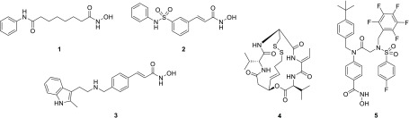
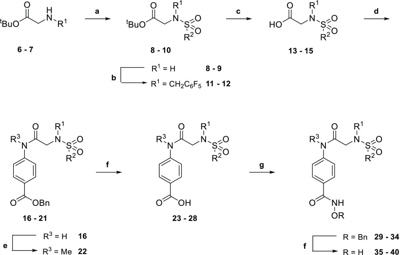
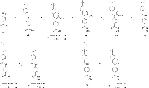
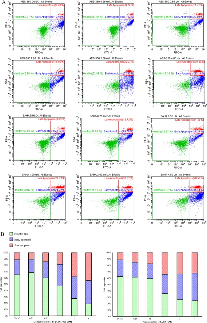
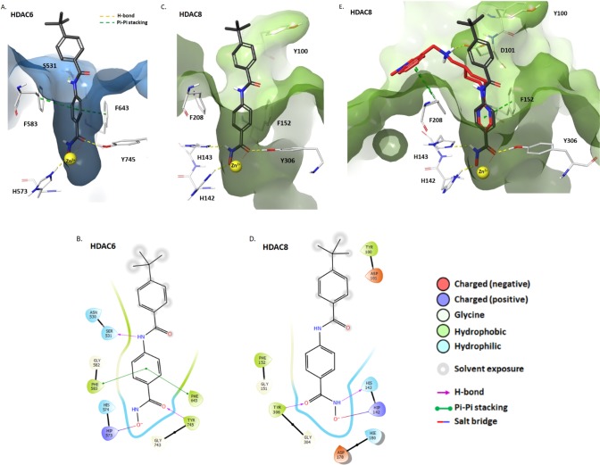
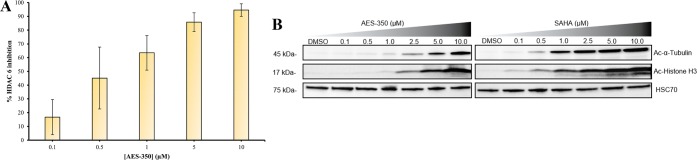
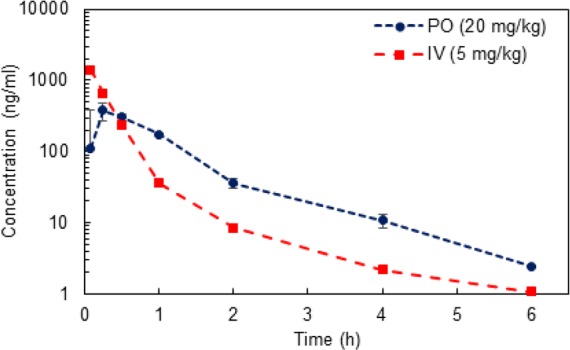
The HDAC inhibitor 4-tert-butyl-N-(4-(hydroxycarbamoyl)phenyl)benzamide (AES-350, 51) was identified as a promising preclinical candidate for the treatment of acute myeloid leukemia (AML), an aggressive malignancy with a meagre 24% 5-year survival rate. Through screening of low-molecular-weight analogues derived from the previously discovered novel HDAC inhibitor, AES-135, compound 51 demonstrated greater HDAC isoform selectivity, higher cytotoxicity in MV4-11 cells, an improved therapeutic window, and more efficient absorption through cellular and lipid membranes. Compound 51 also demonstrated improved oral bioavailability compared to SAHA in mouse models. A broad spectrum of experiments, including FACS, ELISA, and Western blotting, were performed to support our hypothesis that 51 dose-dependently triggers apoptosis in AML cells through HDAC inhibition.


Conflict of interest statement

The authors declare no competing financial interest.

Figures

Figure 1
Figure 1
Vorinostat (1), belinostat (2), panobinostat (3), romidepsin (4), and AES-135 (5).
Scheme 1
Scheme 1. Synthesis of Compounds 3540
Key: (a) R2SO2Cl, iPr2NEt, CH2Cl2, 16 h, rt, N2; (b) pentafluorobenzyl bromide, Cs2CO3, MeCN, 16 h, rt; (c) CF3CO2H/CHCl3 (1:3), 24 h, rt; (d) appropriate aniline, PPh3Cl2, CHCl3, 90 min, 100 °C, MW, N2; (e) MeI, Cs2CO3, MeCN, 20 h, rt; (f) H2, 10% Pd/C, THF/MeOH (2:1), 16 h, rt; (g) (i) (COCl)2, THF, DMF, 2 h, 0 °C, N2; (ii) O-benzylhydroxylamine, iPr2NEt, THF, 16 h, rt, N2. R1, R2, and R3 are variable depending on the molecule and shown below in Table 1.
Scheme 2
Scheme 2. Synthesis of Compounds 46, 47, 51, and 54
Key: (a) (i) 4-tert-butylbenzaldehyde, THF/TFE (4:1), 16 h, rt, (ii) NaBH4, MeOH, 6 h, rt; (b) (Boc)2O, DMAP, MeCN, 2 h, 80 °C, MW; (c) H2, 10% Pd/C, THF/MeOH (2:1), 16 h, rt; (d) (i) (COCl)2, THF, DMF, 2 h, 0 °C, N2; (ii) O-benzylhydroxylamine, iPr2NEt, THF, 16 h, rt, N2; (e) CF3CO2H/CHCl3 (1:3), 18 h, rt; (f) 4-tert-butylbenzoic acid, PPh3Cl2, CHCl3, 90 min, 100 °C, MW, N2; (g) AcCl, CH2Cl2, 24 h, 0 °C to rt.
Figure 2
Figure 2
(A) Dot plots and (B) stacked bar graphs representing the distribution of MV4-11 cells classed as healthy, early apoptosis, and late apoptosis 18 h postdosing with varying concentrations of SAHA and 51 (AES-350) using FACS.
Figure 3
Figure 3
(A) Docking of 51 with zebra fish HDAC6 CD2 (light blue) (PDB: 6CSR). (B) Ligand interaction diagram depicting the key interactions of 51 in HDAC6 CD2. (C) Docking of 51 in the human HDAC8 catalytic domain (green) (PDB: 6HSK). (D) Ligand interaction diagram depicting the key interactions of 51 in the HDAC8 catalytic domain. (E) Overlap of 51 (gray) with quisinostat (red) in the human HDAC8 catalytic domain (PDB: 6HSK). In A, C, and E, 51 is illustrated by gray (C), white (H), blue (N), red (O), and yellow (Zn2+). Other interactions are shown as follows: π-stacking (green dashed lines), H-bonds and salt bridges (yellow dashed lines), and Zn2+ chelation (yellow/red dots).
Figure 4
Figure 4
(A) HDAC6 inhibition profile with varying concentrations of 51 in HeLa cell lysates (IC50 = 0.58 ± 0.13 μM, n = 2). (B) Western blots probing for Ac-α-tubulin, Ac-Histone H3, and HSC70 from MV4–11 after 6 h with varying concentrations of 51 (AES-350) and SAHA.
Figure 5
Figure 5
Mean plasma concentrations of 51 in CD-1 mice following PO (20 mg/kg, n = 3) and IV administration (5 mg/kg, n = 3).

References

    1. Khan O.; La Thangue N. B. HDAC Inhibitors in Cancer Biology: Emerging Mechanisms and Clinical Applications. Immunol. Cell Biol. 2012, 90, 85–94. 10.1038/icb.2011.100. - DOI - PubMed
    1. Ceccacci E.; Minucci S. Inhibition of Histone Deacetylases in Cancer Therapy: Lessons from Leukaemia. Br. J. Cancer 2016, 114 (6), 605–611. 10.1038/bjc.2016.36. - DOI - PMC - PubMed
    1. Kumagai T.; Wakimoto N.; Yin D.; Gery S.; Kawamata N.; Takai N.; Komatsu N.; Chumakov A.; Imai Y.; Koeffler H. P. Histone Deacetylase Inhibitor, Suberoylanilide Hydroxamic Acid (Vorinostat, SAHA) Profoundly Inhibits the Growth of Human Pancreatic Cancer Cells. Int. J. Cancer 2007, 121 (3), 656–665. 10.1002/ijc.22558. - DOI - PubMed
    1. Qian X.; Ara G.; Mills E.; LaRochelle W. J.; Lichenstein H. S.; Jeffers M. Activity of the Histone Deacetylase Inhibitor Belinostat (PXD101) in Preclinical Models of Prostate Cancer. Int. J. Cancer 2008, 122 (6), 1400–1410. 10.1002/ijc.23243. - DOI - PubMed
    1. Ellis L.; Pan Y.; Smyth G. K.; George D. J.; McCormack C.; Williams-Truax R.; Mita M.; Beck J.; Burris H.; Ryan G.; et al. Histone Deacetylase Inhibitor Panobinostat Induces Clinical Responses with Associated Alterations in Gene Expression Profiles in Cutaneous T-Cell Lymphoma. Clin. Cancer Res. 2008, 14 (14), 4500–4510. 10.1158/1078-0432.CCR-07-4262. - DOI - PubMed

LinkOut - more resources