Case Reports
. 2019 Nov 15;5(4):20190030.
doi: 10.1259/bjrcr.20190030. eCollection 2019 Dec.

Transthoracic ultrasound sign in severe asthmatic patients: a lack of "gliding sign" mimic pneumothorax

Affiliations
Case Reports

Transthoracic ultrasound sign in severe asthmatic patients: a lack of "gliding sign" mimic pneumothorax

Anna Del Colle et al. BJR Case Rep. .

Abstract

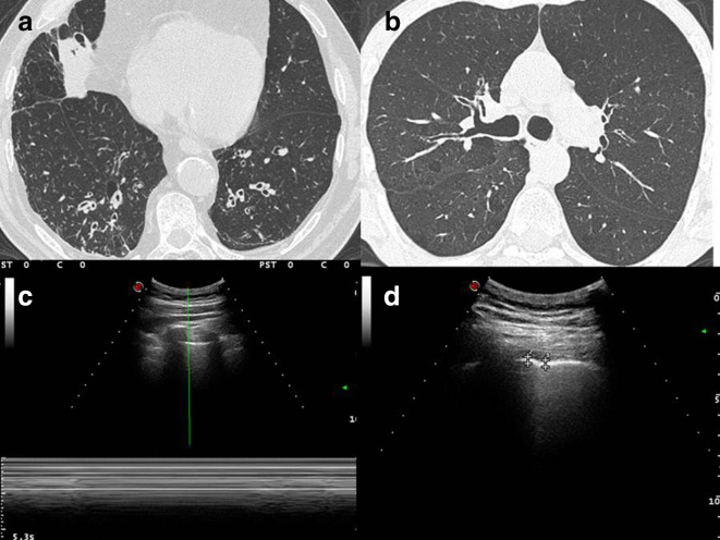
Transthoracic ultrasound (TUS) is a validate complementary technique widely used in everyday medical practice. TUS is the gold-standard for studying pleural effusion and for echo-guided thoracentesis, moreover, it is employed in detection of pleural and pulmonary lesions adherent to pleural surface and their ccho-guided percutaneous needle biopsy (PTNB).1 We used TUS technique to study severe asthma patients. We found that several patterns are constant in these patients. One of these patterns, i.e. lack of gliding sign, mimic pneumothorax (PNX). In this study, we attempted an echographic approach to asthma, trying to lay the first stone for the individuation of common ultrasound patterns in this disease.


Figures

Figure 1.
Figure 1.
(A, B) HRCT imaging shows overdistension and the airway wall thickening with bronchiectasis of lung bases; in addition, it shows also centrilobular micronodular lesions which state distal airway inflammation. (C) Ultrasound imaging, taken with convex probe (5 MHz), indicates the absence of gliding sign, which was confirmed by Barcode sign in M-mode. (D) Ultrasound imaging, with convex probe (5 MHz) shows an irregular thickening of the hyperechoic pleural line (3.1–3.6 mm). HRCT, high resolution CT.
Figure 2.
Figure 2.
(A) HRCT imaging shows bronchial wall thickening, endobronchial nodules in segmental and subsegmentary bronchi of the lower left lobe. (B.) The more caudal scan shows the presence of focal areas of ground glass hyperdensity in the peribronchiolar area in the basal pyramid of the lower right lobe, in relation to flogistic alterations of distal airways. (C) Ultrasound imaging with convex probe (5 MHz) shows an irregular, limited but multiple focal thickening of the hyperechoic pleural line (3.2–4 mm) (D). the lack of gliding sign is confirmed by Barcode sign in M-mode with linear probe (10 MHz) (Figure D2). HRCT, high resolution CT.
Figure 3.
Figure 3.
(A, B) HRCT scans show minimal thickening of the bronchial walls and a slightly inhomogeneous appearance of the lung parenchyma (a mild picture of “mosaic oligohemia”). (C) Lack of gliding sign is confirmed by Barcode sign in M-mode with convex probe (5 MHz). (D) Ultrasound imaging with convex probe (5 MHz) shows an irregular thickening of the hyperechoic pleural line (3.1–4.4 mm). HRCT, high resolution CT.

References

    1. Sperandeo M, Del Colle A, Frongillo E, Rea G, Dimitri L, Cipriani C, et al. ;in press Percutaneous transthoracic needle biopsy with ultrasound guide in subpleural lesions in the evaluation of pulmonary consolidation. can we maximize safety. Respiratory Research 2019. - PMC - PubMed
    1. Chung KF, Wenzel SE, Brozek JL, Bush A, Castro M, Sterk PJ, et al. . International ERS/ATS guidelines on definition, evaluation and treatment of severe asthma. Eur Respir J 2014; 43: 343–7310.1183/09031936.00202013 - DOI - PubMed
    1. Global Initiative for Asthma Global Strategy for Asthma Management and Prevention. 2018. Available from: www.ginasthma.org.
    1. Sperandeo M, Rotondo A, Guglielmi G, Catalano D, Feragalli B, Trovato GM. Transthoracic ultrasound in the assessment of pleural and pulmonary diseases: use and limitations. Radiol Med 2014; 119: 729–4010.1007/s11547-014-0385-0 - DOI - PubMed
    1. Tinti MG, Frongillo E, Sperandeo M. Lung fissures detection with transthoracic ultrasound: is it simply a matter of artifacts? Chest 2018; 154: 453–510.1016/j.chest.2018.03.049 - DOI - PubMed

Publication types