. 2020 Jan 14;9(1):4.
doi: 10.1186/s40169-020-0257-2.

Detection of bladder cancer using urinary cell-free DNA and cellular DNA

Affiliations

Detection of bladder cancer using urinary cell-free DNA and cellular DNA

Zhenyu Ou et al. Clin Transl Med. .

Abstract

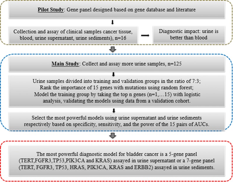
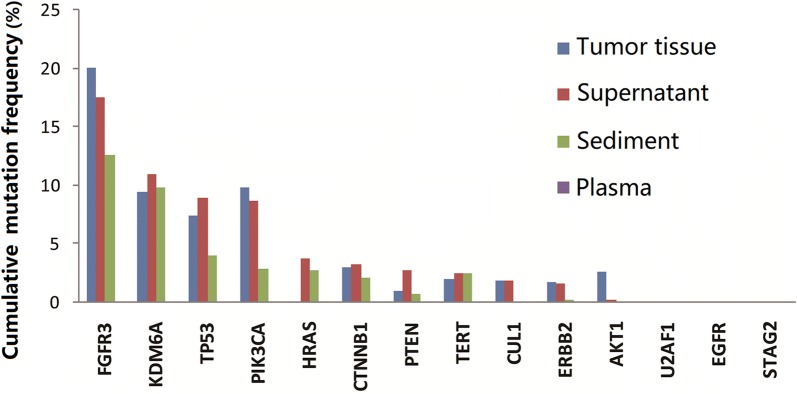
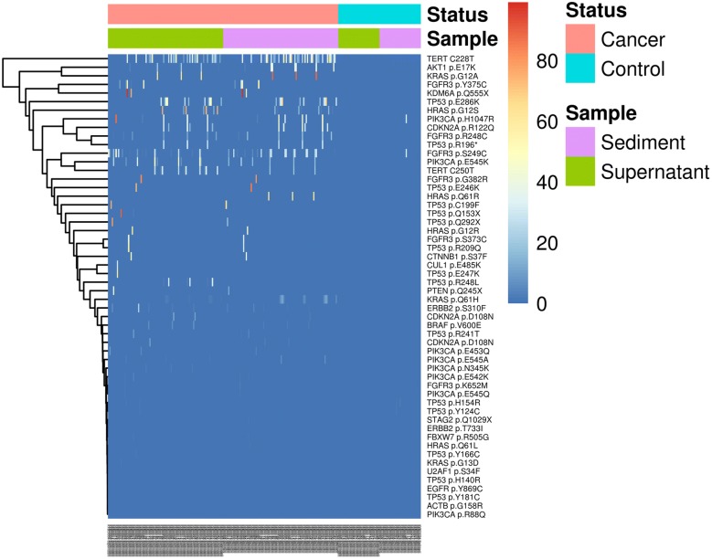
Background: The present study sought to identify a panel of DNA markers for noninvasive diagnosis using cell-free DNA (cfDNA) from urine supernatant or cellular DNA from urine sediments of hematuria patients. A panel of 48 bladder cancer-specific genes was selected. A next-generation sequencing-based assay with a cfDNA barcode-enabled single-molecule test was employed. Mutation profiles of blood, urine, and tumor sample from 16 bladder cancer patients were compared. Next, urinary cellular DNA and cfDNA were prospectively collected from 125 patients (92 bladder cancer cases and 33 controls) and analyzed using the 48-gene panel. The individual gene markers and combinations of markers were validated according to the pathology results. The mean areas under the receiver operating characteristic (ROC) curves (AUCs) obtained with the various modeling approaches were calculated and compared.

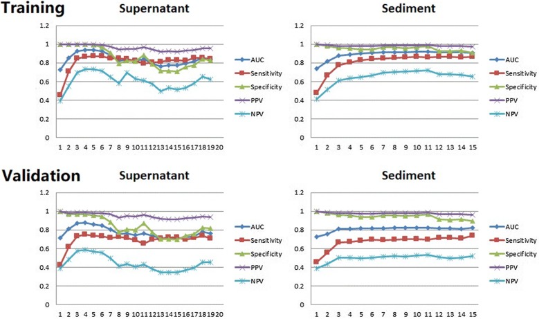
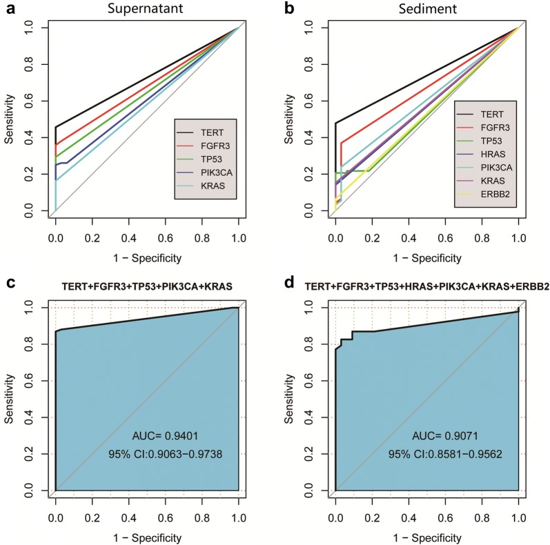
Results: This pilot study of 16 bladder cancer patients demonstrated that gene mutations in urine supernatant and sediments had better concordance with cancer tissue as compared with plasma. Logistic analyses suggested two powerful combinations of genes for genetic diagnostic modeling: five genes for urine supernatant (TERT, FGFR3, TP53, PIK3CA, and KRAS) and seven genes for urine sediments (TERT, FGFR3, TP53, HRAS, PIK3CA, KRAS, and ERBB2). The accuracy of the five-gene panel and the seven-gene panel in the validation cohort yielded AUCs of 0.94 [95% confidence interval (CI) 0.91-0.97] and 0.91 (95% CI 0.86-0.96), respectively. With the addition of age and gender, the diagnostic power of the urine supernatant five-gene model and the urine sediment seven-gene model improved as the revised AUCs were 0.9656 (95% CI 0.9368-0.9944) and 0.9587 (95% CI 0.9291-0.9883).

Conclusions: cfDNA from urine bears great diagnostic potential. A five-gene panel for urine supernatant and a seven-gene panel for urine sediments are promising options for identifying bladder cancer in hematuria patients.

Keywords: Bladder cancer; Cell-free DNA; Hematuria; Mutation; Next-generation sequencing.


Conflict of interest statement

The authors declare that they have no competing interests.

Figures

Fig. 1
Fig. 1
Overview of the study plan. This study consisted of three phases: a pilot study, the main study, and the finalization of the diagnostic modeling, which are shown circumscribed in grey, blue, and red, respectively
Fig. 2
Fig. 2
Average mutation frequencies in 14 bladder cancer biomarker genes in 16 cases in the pilot study. The cumulative frequencies of mutations identified in urine supernatant and urine sediments were comparable with those in tumor tissue and significantly higher than those in plasma
Fig. 3
Fig. 3
Heatmap illustrating the distribution of mutation rates identified in the DNA of urine supernatant and urine sediments. In cancer tissue, high mutation rates were identified in KDM6A p.Q555X, KRAS pG2A, TP53 p.Q153X, TERT promoter, FGFR3 p.Y375C, and TP53 p.E246K. The Y-axis lists the mutation locations and the X-axis shows the identification number of each sample. The mutation rates from low to high are represented by their colors from blue (low) to red (high) accordingly
Fig. 4
Fig. 4
Boxplot illustrating the frequency of mutations in the urine supernatant and sediments from 92 cases. The boxplot shows the number of positive mutant genes among 92 tumor patients, whose urine supernatant and sediment samples were screened using the 48-gene panel. The points in the graph represent discrete points that were statistically distant from the median. The Y-axis represents the number of mutated genes in the sample. A t-test analysis revealed that there was no statistically significant difference in the number of mutated genes between the urine supernatant and sediments obtained from the same subjects (p = 0.201)
Fig. 5
Fig. 5
Averaged diagnostic parameters of the logistic models for the genetic diagnosis of bladder cancer. For urine supernatant, all parameters of the models are satisfactory when gene combinations having three, four, and five genes (TERT, FGFR3, TP53, PIK3CA, and KRAS) were used. In contrast, the models using urine sediments require the combination of seven (TERT, FGFR3, TP53, HRAS, PIK3CA, KRAS, and ERBB2) or more genes to attain satisfactory predictive potential. The X-axis represents the number of genes factored into the model. The Y-axis plots diagnostic potential, with 1.0 representing perfect disease discrimination
Fig. 6
Fig. 6
Diagnostic models based on mutations of a single gene or gene combination found in all 125 urine supernatant or urine sediment samples for discriminating cancer from controls. a Individual AUCs based on the five genes TERT, FGFR3, TP53, PIK3CA, and KRAS assayed in urine supernatant; b individual AUCs based on the seven genes TERT, FGFR3, TP53, HRAS, PIK3CA, KRAS, and ERBB2 assayed in urine sediments; c, d AUCs of the five-gene and seven-gene combinations using urine supernatant and sediments, respectively

References

    1. Jemal A, et al. Cancer statistics, 2006. CA Cancer J Clin. 2006;56:106–130. doi: 10.3322/canjclin.56.2.106. - DOI - PubMed
    1. Ferlay J, et al. Cancer incidence and mortality worldwide: sources, methods and major patterns in GLOBOCAN 2012. Int J Cancer. 2015;136:E359–386. doi: 10.1002/ijc.29210. - DOI - PubMed
    1. Price SJ, Shephard EA, Stapley SA, Barraclough K, Hamilton WT. Non-visible versus visible haematuria and bladder cancer risk: a study of electronic records in primary care. Br J Gen Pract. 2014;64:584–589. doi: 10.3399/bjgp14X681409. - DOI - PMC - PubMed
    1. Khadra MH, Pickard RS, Charlton M, Powell PH, Neal DE. A prospective analysis of 1,930 patients with hematuria to evaluate current diagnostic practice. J Urol. 2000;163:524–527. doi: 10.1016/S0022-5347(05)67916-5. - DOI - PubMed
    1. Ward DG, Bryan RT. Liquid biopsies for bladder cancer. Transl Androl Urol. 2017;6:331–335. doi: 10.21037/tau.2017.03.08. - DOI - PMC - PubMed