. 2020 Jan 21;49(2):433-464.
doi: 10.1039/c9cs00285e. Epub 2020 Jan 15.

Catechol-functionalized hydrogels: biomimetic design, adhesion mechanism, and biomedical applications

Affiliations

Catechol-functionalized hydrogels: biomimetic design, adhesion mechanism, and biomedical applications

Wei Zhang et al. Chem Soc Rev. .

Abstract

Hydrogels are a unique class of polymeric materials that possess an interconnected porous network across various length scales from nano- to macroscopic dimensions and exhibit remarkable structure-derived properties, including high surface area, an accommodating matrix, inherent flexibility, controllable mechanical strength, and excellent biocompatibility. Strong and robust adhesion between hydrogels and substrates is highly desirable for their integration into and subsequent performance in biomedical devices and systems. However, the adhesive behavior of hydrogels is severely weakened by the large amount of water that interacts with the adhesive groups reducing the interfacial interactions. The challenges of developing tough hydrogel-solid interfaces and robust bonding in wet conditions are analogous to the adhesion problems solved by marine organisms. Inspired by mussel adhesion, a variety of catechol-functionalized adhesive hydrogels have been developed, opening a door for the design of multi-functional platforms. This review is structured to give a comprehensive overview of adhesive hydrogels starting with the fundamental challenges of underwater adhesion, followed by synthetic approaches and fabrication techniques, as well as characterization methods, and finally their practical applications in tissue repair and regeneration, antifouling and antimicrobial applications, drug delivery, and cell encapsulation and delivery. Insights on these topics will provide rational guidelines for using nature's blueprints to develop hydrogel materials with advanced functionalities and uncompromised adhesive properties.


Conflict of interest statement

Conflicts of interest

There are no conflicts to declare.

Figures

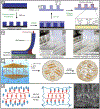
Figure 1.
Figure 1.
Design and development of adhesive hydrogels based on biomimetic principles, and their potential applications in a wide range of fields.
Figure 2.
Figure 2.
Hydrogel adhesion in air (left panel) and in water (right panel), and typical interfacial interactions at the contact points.
Figure 3.
Figure 3.
The multiple roles of catechol groups in wet adhesion: (a) instantaneous adhesion, (b) coacervation formation, and (c) wet adhesive curing.
Figure 4.
Figure 4.
Effect of pH on molecular adhesion to the mica surface. (a) Asymmetric and symmetric tests of mussel foot proteins carried out using surface force apparatus (SFA). Asymmetric configuration is used to measure the interfacial adhesion, while the symmetric configuration tests the cohesion between protein monolayers (blue). D, distance; R, radius. Reproduced with permission from ref. 91. Copyright 2017 The Company of Biologists Ltd. (b) Asymmetric adhesion of mfp-3 at different pH values (3, 5.5, and 7.5). Reproduced with permission from ref. 100. Copyright 2011 Springer Nature Limited.
Figure 5.
Figure 5.
(a) pH-dependent transition from interfacial hydrogen bonding to coordinate bonds on the surfaces of metal oxide. (b) catechol-Fe3+ stoichiometry varied from acidic pH ~2.0 to basic pH ~8.0. Reproduced with permission from ref. 91. Copyright 2017 The Company of Biologists Ltd.
Figure 6.
Figure 6.
(a) Cation-π interaction driven like-charge complex coacervate formation. (b) The obtained coacervate under light microscopy, and (c) its bulk phase separation. Reproduced with permission from ref. 96. Copyright 2016 National Academy of Sciences.
Figure 7.
Figure 7.
Oxidative cross-linking pathways for catechol-containing molecules. R group represents the backbone of the polymer. Reproduced with permission from ref. 98. Copyright 2019 Wiley-VCH.
Figure 8.
Figure 8.
(A) Schematic representation of the dip-coating of an object in dopamine solution at pH 8.5. (B) Thickness evolution of the formed polydopamine films. (C) XPS characterization of 25 polydopamine-coated surfaces. The bar graph represents the intensity of the characteristic substrate signal before (hatched) and after (solid) coating with polydopamine. Reproduced with permission from ref. 42. Copyright 2007 Macmillan Publishers Limited.
Figure 9.
Figure 9.
Chemical structures of (a) N-carboxyanhydride co-polypeptides, (b) styrene-based copolymers, (c) DOPA-modified PEGs with four different ligands.
Figure 10.
Figure 10.
Synthetic approaches for the preparation of catechol-functionalized adhesive hydrogels through (a) catechol cross-linking, (b) assembly or cross-linking of catechol-containing copolymers, and (c) catechol-metal coordination chemistry.
Figure 11.
Figure 11.
The synthesis and chemical structure of (a) catechol-conjugated hyaluronic acid and (b) cysteamine-conjugated Pluronic F-127. Reproduced with permission from ref. 147. Copyright 2010 The Royal Society of Chemistry. (c) In situ preparation of cross-linkable CHI-C/Plu-SH hydrogels. Reproduced with permission from ref. 148. Copyright 2011 American Chemical Society.
Figure 12.
Figure 12.
(a) Synthetic pathways for DOPA-modified Pluroinc block copolymers. Reproduced with permission from ref. 149. Copyright 2002 American Chemical Society. (b) Chemical structure of DOPA-modified methacrylic triblock copolymer and (c) its self-assembly into hydrogel when exposed to saturated water. Reproduced with permission from ref. 150. Copyright 2008 American Chemical Society.
Figure 13.
Figure 13.
(a) Photopolymerization of DOPA-modified triblock copolymer consisted of a PEG mid-block and PLA end-blocks. Concentrating the polymerizable methacrylate groups in the hydrophobic micelle core promoted rapid photo-initiated polymerization. Reproduced with permission from ref. 151. Copyright 2006 American Chemical Society. (b) Photo-initiated polymerization of cross-linked poly(DMA-co-MEA). Reproduced with permission from ref. 152. Copyright 2011 American Chemical Society. (c) Chemical structures of highly branched PEG-catechol copolymers. Reproduced with permission from ref. 153. Copyright 2015 Wiley-VCH.
Figure 14.
Figure 14.
(a) Catechol forms pH dependent mono-, bis-, and tris-DOPA-Fe3+ complexes with increasing pH. Complexation associated with elevated stoichiometry resulted in the formation of hydrogel. Reproduced with permission from ref. 154. Copyright 2011 National Academy of Sciences. (b) Photographs of the precursor solutions (left) and cross-linked hydrogels (right) formed by mixing three trivalent metal ion (V3+, Fe3+, Al3+) with PEG-catechol. Reproduced with permission from ref. 155. Copyright 2014 The Royal Society of Chemistry. (c) Photo-initiated degradation of nitrodopamine-PEG hydrogel cross-linked with metal ion. Reproduced with permission from ref. 156. Copyright 2012 Wiley-VCH. (d) pH-responsive hydrogel based on the interaction of PEG-catechol with 1,3-benzenediboronic acid though the formation of catechol-bonronate complexation. Reproduced with permission from ref. 108. Copyright 2011 The Royal Society of Chemistry.
Figure 15.
Figure 15.
Illustration of a mussel-inspired hydrogel adhesive: surface functionalization of the substrates immobilizes dopamine, which forms coordinate bonds with the ferric ions and alginate solution is injected in between to provide bulk cohesion. Adapted with permission from ref. 157. Copyright 2019 Elsevier.
Figure 16.
Figure 16.
(a) Fabrication of p(DMA-coMEA)-coated nanopillar using electron-beam lithography. Adapted with permission from ref. 43. Copyright 2007 Springer Nature Limited. (b) Strong hydrogel-elastomer interaction induces a cohesive failure near the interface during the peeling test. Adapted with permission from ref. 163. Copyright 2016 Springer Nature Limited. (c) The formation of PDA-clay-PAM hydrogel via in situ polymerization. Adapted with permission from ref. 23. Copyright 2017 American Chemical Society. (d) The design mechanism for a double-network adhesive hydrogel and the interconnected microfibrils in the 3D nanostructure. Adapted with permission from ref. 164. Copyright 2018 American Chemical Society.
Figure 17.
Figure 17.
Schematic representation of catechol-functionalized hydrogels for tissue adhesives. (a) Adhesive designed for dermal adhesive repair consisted of interpenetrated network with self-healing property and the ability to enhance cellular infiltration. Reproduced with permission from ref. 164. Copyright 2018 American Chemical Society. (b) For fetal membrane repair, catechol cross-linking was used to design a hydrophilic hydrogel that prevented cellular infiltration. Reproduced with permission from ref. 168. Copyright 2013 Elsevier. (c) For myocardial tissue adhesive, catechol-metal ion complexation and electro conductive PPy nanoparticles were used to develop a nanocomposite self-healable electroconductive hydrogel. Reproduced with permission from ref. 169. Copyright 2018 Wiley-VCH. (d) For hepatic tissue repair, catechol-PEG-EPL was used to design an adhesive that rapidly cured on blood contact. Reproduced with permission from ref. 170. Copyright 2017 Wiley-VCH.
Figure 18.
Figure 18.
(a) Proposed mechanism of catechol oxidation and H2O2 generation. (b) Schematic illustration of recyclable catechol-containing microgels. The dried microgels were hydrated in neutral to alkaline solution to generate H2O2. Reproduced with permission from ref. 175. Copyright 2019 Elsevier.
Figure 19.
Figure 19.
Schematic representation of catechol-functionalized hydrogels for drug delivery applications: (a) A catechol-containing drug carrier demonstrated increased mucoadhesive properties. Reproduced with permission from ref. 179. Copyright 2019 Elsevier. (b) Reversible catechol boronate complexation was used to design a hydrogel with pH-responsive drug release behavior. Reproduced with permission from ref. 183. Copyright 2018 American Chemical Society. (c) Catechol-metal ion complexation was used to develop a metal-phenolic network (MPN) capsule. Reproduced with permission from ref. 187. Copyright 2016 American Chemical Society.

References

    1. Yuk H, Lu B and Zhao X, Chem. Soc. Rev, 2019, 48, 1642–1667. - PubMed
    1. Zhang YS and Khademhosseini A, Science, 2017, 356, 3627. - PMC - PubMed
    1. Caccavo D, Cascone S, Lamberti G and Barba AA, Chem. Soc. Rev, 2018, 47, 2357–2373. - PubMed
    1. Thoniyot P, Tan MJ, Karim AA, Young DJ and Loh XJ, Adv. Sci, 2015, 2, 1400010. - PMC - PubMed
    1. Yang JA, Yeom J, Hwang BW, Hoffman AS and Hahn SK, Prog. Polym. Sci, 2014, 39, 1973–1986.