. 2020 Feb 5;31(2):394-404.
doi: 10.1021/jasms.9b00024. Epub 2020 Jan 13.

Signature Fragment Ions of Biotinylated Peptides

Affiliations

Signature Fragment Ions of Biotinylated Peptides

Santosh Renuse et al. J Am Soc Mass Spectrom. .

Abstract

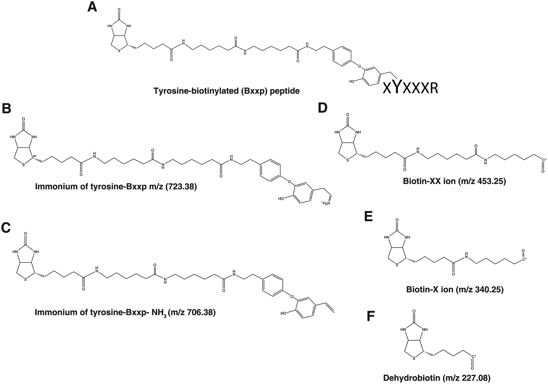
The use of biotin or biotin-containing reagents is an essential component of many protein purification and labeling technologies. Owing to its small size and high affinity to the avidin family of proteins, biotin is a versatile molecular handle that permits both enrichment and purity that is not easily achieved by other reagents. Traditionally, the use of biotinylation to enrich for proteins has not required the detection of the site of biotinylation. However, newer technologies for discovery of protein-protein interactions, such as APEX and BioID, as well as some of the click chemistry-based labeling approaches have underscored the importance of determining the exact residue that is modified by biotin. Anti-biotin antibody-based enrichment of biotinylated peptides (e.g., BioSITe) coupled to LC-MS/MS permit large-scale detection and localization of sites of biotinylation. As with any chemical modification of peptides, understanding the fragmentation patterns that result from biotin modification is essential to improving its detection by LC-MS/MS. Tandem mass spectra of biotinylated peptides has not yet been studied systematically. Here, we describe the various signature fragment ions generated with collision-induced dissociation of biotinylated peptides. We focused on biotin adducts attached to peptides generated by BioID and APEX experiments, including biotin, isotopically heavy biotin, and biotin-XX-phenol, a nonpermeable variant of biotin-phenol. We also highlight how the detection of biotinylated peptides in high-throughput studies poses certain computational challenges for accurate quantitation which need to be addressed. Our findings about signature fragment ions of biotinylated peptides should be helpful in the confirmation of biotinylation sites.

Keywords: marker ions; mass spectrometry; post-translational modifications; protein−protein interactions.


Conflict of interest statement

The authors declare no competing financial interest.

Figures

Figure 1.
Figure 1.
Structure of biotin (A), heavy-biotin (B), and biotin-XX-phenol (C) and signature fragment ions of lysine biotinylated peptides biotinylated lysine immonium ion, ImKbio at m/z 327.185, its derivative, ImKbio- NH3 at m/z 310.158 and dehydrobiotin at m/z 227.085 (D).
Figure 2.
Figure 2.
Representative MS/MS spectra of lysine biotinylated peptides, AGDSLMVMIAMkMEHTIK from protein methylcrotonoyl-CoA carboxylase subunit α, mitochondrial (Mccc1) (A), AVGTQALSGAGLLkMFNK from protein sorting nexin-1 (Snx1) (B), KFFNkEFLSKPTV from protein cytosolic phospholipase A2 (Pla2g4a) (C) and GLVkVNDkEVSDR from protein breakpoint cluster region protein (Bcr) (D). showing the signature fragment ions - biotinylated lysine immonium ion, ImKbio at m/z 327.185, its derivative, ImKbio- NH3 at m/z 310.158 and dehydrobiotin at m/z 227.085.
Figure 3.
Figure 3.
Relative intensity of signature fragment ions, ImKbio (m/z, 327.158), ImKbiotin-NH3 at m/z 310.158, and dehydrobiotin at m/z 227.085 of identified lysine biotinylated peptides (A), distribution of Andromeda score for identified biotinylated peptides with/without inclusion of diagnostic ions for MaxQuant database searches (B), quantitative BioSITe experiment with relative intensity of signature fragment ions for light and heavy biotinylated peptides (C), and signature fragment ions from tyrosine biotinylated peptides with BxxP (D); ImYbxxp (m/z, 723.388), ImYbxxp-NH3 at m/z 706.361, biotin-XX at m/z, 453.251, biotin-X at m/z 340.168, and dehydrobiotin at m/z 227.085
Figure 4.
Figure 4.
MS1 spectrum showing light and heavy biotin labeled peptide indicating delta mass of +4 Da (A). MS/MS spectrum of light biotinylated lysine-containing peptide (B) and heavy biotin labeled peptide (C) showing light and heavy (+4 Da) version of biotinylated lysine immonium ion, ImKbio at m/z 327.185 and 331.214 and its derivatives, ImKbio-NH3 at m/z 310.158 and 314.178.
Figure 5.
Figure 5.
Structure of signature fragment ions identified in tyrosine biotinylated peptides with biotin-XX-phenol, ImYbio (m/z, 723.388), ImYbio- NH3 at m/z 706.361 and dehydrobiotin at m/z 227.085.
Figure 6.
Figure 6.
Representative MS/MS spectra of tyrosine biotinylated peptides, ESQAyYDGRR from major prion protein precursor (Prnp) (A), IIELVPDGAPyITCITK from Sodium/potassium-transporting ATPase subunit beta-3 (Atp1b3) (B), GFQIyDGPIHLTK from transmembrane protein 2 (Tmem2) (C) and YHySSATIPR from transmembrane protein 178B (Tmem178b) (D)-biotinylated tyrosine immonium ion, ImYbio at m/z 723.388, its derivative, ImYbio-NH3 at m/z 706.361 and dehydrobiotin at m/z 227.085.

References

    1. Chaiet L; Wolf FJ The Properties of Streptavidin, a Biotin-Binding Protein Produced by Streptomycetes. Arch. Biochem. Biophys 1964, 106, 1–5. - PubMed
    1. Cronan JE Jr. Biotination of proteins in vivo. A post-translational modification to label, purify, and study proteins. J. Biol. Chem 1990, 265 (18), 10327–33. - PubMed
    1. Roux KJ; et al. A promiscuous biotin ligase fusion protein identifies proximal and interacting proteins in mammalian cells. J. Cell Biol 2012, 196 (6), 801–10. - PMC - PubMed
    1. Kim DI; et al. An improved smaller biotin ligase for BioID proximity labeling. Mol. Biol. Cell 2016, 27 (8), 1188–96. - PMC - PubMed
    1. Lobingier BT; et al. An Approach to Spatiotemporally Resolve Protein Interaction Networks in Living Cells. Cell 2017, 169 (2), 350–360.e12. - PMC - PubMed