

Suppression of Superoxide-Hydrogen Peroxide Production at Site IQ of Mitochondrial Complex I Attenuates Myocardial Stunning and Improves Postcardiac Arrest Outcomes
- PMID: 31939812
- PMCID: PMC6964871
- DOI: 10.1097/CCM.0000000000004095
Suppression of Superoxide-Hydrogen Peroxide Production at Site IQ of Mitochondrial Complex I Attenuates Myocardial Stunning and Improves Postcardiac Arrest Outcomes
Abstract
Objectives: Cardiogenic shock following cardiopulmonary resuscitation for sudden cardiac arrest is common, occurring even in the absence of acute coronary artery occlusion, and contributes to high rates of postcardiopulmonary resuscitation mortality. The pathophysiology of this shock is unclear, and effective therapies for improving clinical outcomes are lacking.
Design: Laboratory investigation.
Setting: University laboratory.
Subjects: C57BL/6 adult female mice.
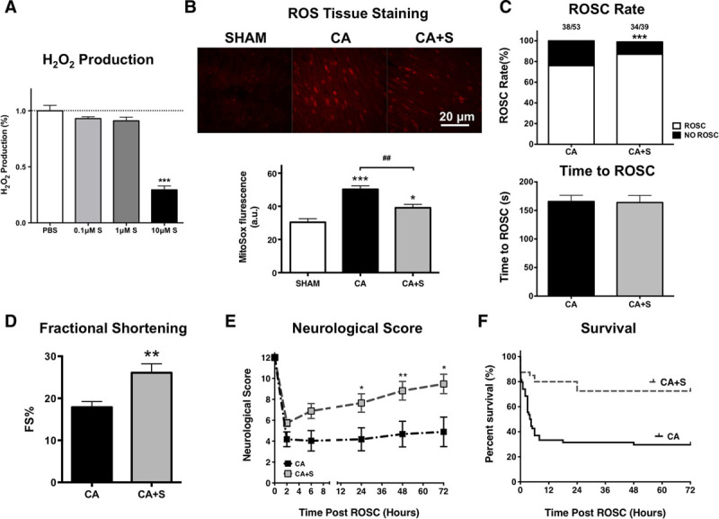
Interventions: Anesthetized and ventilated adult female C57BL/6 wild-type mice underwent a 4, 8, 12, or 16-minute potassium chloride-induced cardiac arrest followed by 90 seconds of cardiopulmonary resuscitation. Mice were then blindly randomized to a single IV injection of vehicle (phosphate-buffered saline) or suppressor of site IQ electron leak, an inhibitor of superoxide production by complex I of the mitochondrial electron transport chain. Suppressor of site IQ electron leak and vehicle were administered during cardiopulmonary resuscitation.
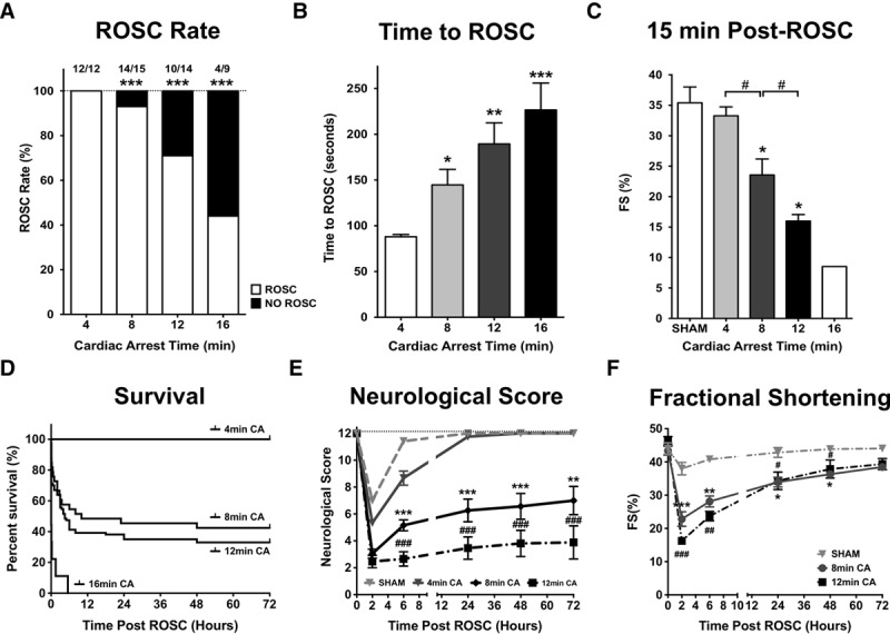
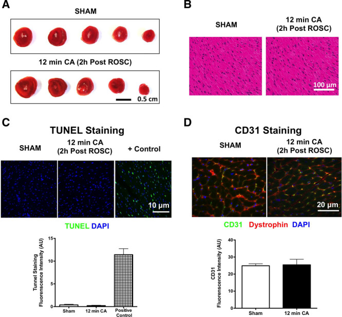
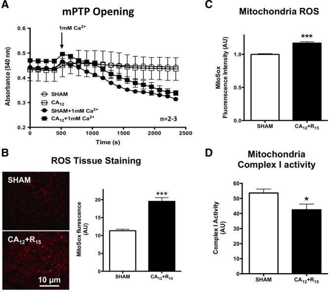
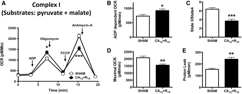
Measurements and main results: Using a murine model of asystolic cardiac arrest, we discovered that duration of cardiac arrest prior to cardiopulmonary resuscitation determined postresuscitation success rates, degree of neurologic injury, and severity of myocardial dysfunction. Post-cardiopulmonary resuscitation cardiac dysfunction was not associated with myocardial necrosis, apoptosis, inflammation, or mitochondrial permeability transition pore opening. Furthermore, left ventricular function recovered within 72 hours of cardiopulmonary resuscitation, indicative of myocardial stunning. Postcardiopulmonary resuscitation, the myocardium exhibited increased reactive oxygen species and evidence of mitochondrial injury, specifically reperfusion-induced reactive oxygen species generation at electron transport chain complex I. Suppressor of site IQ electron leak, which inhibits complex I-dependent reactive oxygen species generation by suppression of site IQ electron leak, decreased myocardial reactive oxygen species generation and improved postcardiopulmonary resuscitation myocardial function, neurologic outcomes, and survival.
Conclusions: The severity of cardiogenic shock following asystolic cardiac arrest is dependent on the length of cardiac arrest prior to cardiopulmonary resuscitation and is mediated by myocardial stunning resulting from mitochondrial electron transport chain complex I dysfunction. A novel pharmacologic agent targeting this mechanism, suppressor of site IQ electron leak, represents a potential, practical therapy for improving sudden cardiac arrest resuscitation outcomes.
Figures
Similar articles
-
Inhibition of the mitochondrial fission protein dynamin-related protein 1 improves survival in a murine cardiac arrest model.Crit Care Med. 2015 Feb;43(2):e38-47. doi: 10.1097/CCM.0000000000000817. Crit Care Med. 2015. PMID: 25599491 Free PMC article.
-
Nitrite therapy after cardiac arrest reduces reactive oxygen species generation, improves cardiac and neurological function, and enhances survival via reversible inhibition of mitochondrial complex I.Circulation. 2009 Sep 8;120(10):897-905. doi: 10.1161/CIRCULATIONAHA.109.853267. Epub 2009 Aug 24. Circulation. 2009. PMID: 19704094 Free PMC article.
-
Early mitochondrial dysfunction in electron transfer activity and reactive oxygen species generation after cardiac arrest.Crit Care Med. 2008 Nov;36(11 Suppl):S447-53. doi: 10.1097/ccm.0b013e31818a8a51. Crit Care Med. 2008. PMID: 20449909 Free PMC article.
-
delta-Opioid-induced pharmacologic myocardial hibernation during cardiopulmonary resuscitation.Crit Care Med. 2006 Dec;34(12 Suppl):S486-9. doi: 10.1097/01.CCM.0000246015.05214.5A. Crit Care Med. 2006. PMID: 17114982 Review.
-
Pathophysiology and pathogenesis of post-resuscitation myocardial stunning.Heart Fail Rev. 2012 Jan;17(1):117-28. doi: 10.1007/s10741-011-9255-1. Heart Fail Rev. 2012. PMID: 21584712 Review.
Cited by
-
Methodological Issue of Mitochondrial Isolation in Acute-Injury Rat Model: Asphyxia Cardiac Arrest and Resuscitation.Front Med (Lausanne). 2021 Apr 12;8:666735. doi: 10.3389/fmed.2021.666735. eCollection 2021. Front Med (Lausanne). 2021. PMID: 33912580 Free PMC article.
-
Hyperoxygenation With Cardiopulmonary Resuscitation and Targeted Temperature Management Improves Post-Cardiac Arrest Outcomes in Rats.J Am Heart Assoc. 2020 Oct 20;9(19):e016730. doi: 10.1161/JAHA.120.016730. Epub 2020 Sep 23. J Am Heart Assoc. 2020. PMID: 32964774 Free PMC article.
-
Prostaglandin E1 attenuates post‑cardiac arrest myocardial dysfunction through inhibition of mitochondria‑mediated cardiomyocyte apoptosis.Mol Med Rep. 2021 Feb;23(2):110. doi: 10.3892/mmr.2020.11749. Epub 2020 Dec 10. Mol Med Rep. 2021. PMID: 33300050 Free PMC article.
-
Attenuation of mitochondrial dysfunction in a ventricular fibrillation swine model of cardiac arrest treated with carbon monoxide.Resuscitation. 2025 Aug;213:110647. doi: 10.1016/j.resuscitation.2025.110647. Epub 2025 May 16. Resuscitation. 2025. PMID: 40383501 Free PMC article.
-
Protective Effect of Mitochondria-Targeted Antioxidants against Inflammatory Response to Lipopolysaccharide Challenge: A Review.Pharmaceutics. 2021 Jan 22;13(2):144. doi: 10.3390/pharmaceutics13020144. Pharmaceutics. 2021. PMID: 33499252 Free PMC article. Review.
References
-
- Benjamin EJ, Virani SS, Callaway CW, et al. ; American Heart Association Council on Epidemiology and Prevention Statistics Committee and Stroke Statistics Subcommittee: Heart disease and stroke statistics-2018 update: A report from the American Heart Association. Circulation 2018; 137:e67–e492 - PubMed
-
- Larsen MP, Eisenberg MS, Cummins RO, et al. Predicting survival from out-of-hospital cardiac arrest: A graphic model. Ann Emerg Med 1993; 22:1652–1658 - PubMed
-
- Kleinman ME, Brennan EE, Goldberger ZD, et al. Part 5: Adult basic life support and cardiopulmonary resuscitation quality: 2015 American Heart Association guidelines update for cardiopulmonary resuscitation and emergency cardiovascular care. Circulation 2015; 132:S414–S435 - PubMed
-
- Babini G, Grassi L, Russo I, et al. Duration of untreated cardiac arrest and clinical relevance of animal experiments: The relationship between the “No-Flow” duration and the severity of post-cardiac arrest syndrome in a porcine model. Shock 2018; 49:205–212 - PubMed
-
- Roberts BW, Kilgannon JH, Chansky ME, et al. Multiple organ dysfunction after return of spontaneous circulation in postcardiac arrest syndrome. Crit Care Med 2013; 41:1492–1501 - PubMed
Publication types
MeSH terms
Substances
Grants and funding
LinkOut - more resources
Full Text Sources
Medical
