

POLR3A variants with striatal involvement and extrapyramidal movement disorder
- PMID: 31940116
- PMCID: PMC7064625
- DOI: 10.1007/s10048-019-00602-4
POLR3A variants with striatal involvement and extrapyramidal movement disorder
Abstract
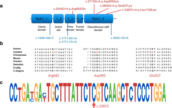
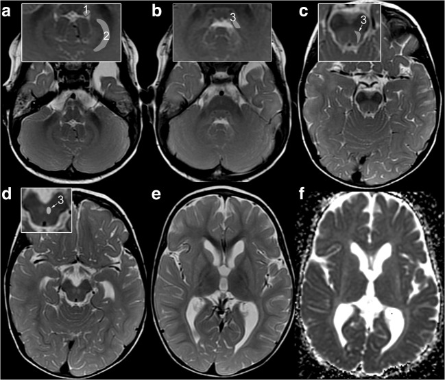
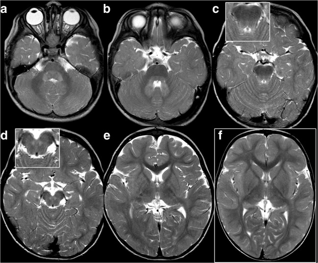
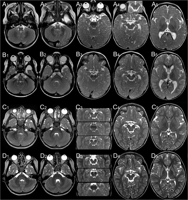
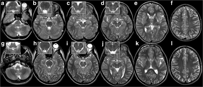
Biallelic variants in POLR3A cause 4H leukodystrophy, characterized by hypomyelination in combination with cerebellar and pyramidal signs and variable non-neurological manifestations. Basal ganglia are spared in 4H leukodystrophy, and dystonia is not prominent. Three patients with variants in POLR3A, an atypical presentation with dystonia, and MR involvement of putamen and caudate nucleus (striatum) and red nucleus have previously been reported. Genetic, clinical findings and 18 MRI scans from nine patients with homozygous or compound heterozygous POLR3A variants and predominant striatal changes were retrospectively reviewed in order to characterize the striatal variant of POLR3A-associated disease. Prominent extrapyramidal involvement was the predominant clinical sign in all patients. The three youngest children were severely affected with muscle hypotonia, impaired head control, and choreic movements. Presentation of the six older patients was milder. Two brothers diagnosed with juvenile parkinsonism were homozygous for the c.1771-6C > G variant in POLR3A; the other seven either carried c.1771-6C > G (n = 1) or c.1771-7C > G (n = 7) together with another variant (missense, synonymous, or intronic). Striatal T2-hyperintensity and atrophy together with involvement of the superior cerebellar peduncles were characteristic. Additional MRI findings were involvement of dentate nuclei, hila, or peridentate white matter (3, 6, and 4/9), inferior cerebellar peduncles (6/9), red nuclei (2/9), and abnormal myelination of pyramidal and visual tracts (6/9) but no frank hypomyelination. Clinical and MRI findings in patients with a striatal variant of POLR3A-related disease are distinct from 4H leukodystrophy and associated with one of two intronic variants, c.1771-6C > G or c.1771-7C > G, in combination with another POLR3A variant.
Keywords: Basal ganglia; Brainstem; Hypomyelination; Inferior cerebellar peduncle; MRI; POLR3A; Striatum; Superior cerebellar peduncle.
Conflict of interest statement
All authors declare no conflicts of interest in the publication of this manuscript.
Figures
Similar articles
-
POLR3A variants in striatal involvement without diffuse hypomyelination.Brain Dev. 2020 Apr;42(4):363-368. doi: 10.1016/j.braindev.2019.12.012. Epub 2020 Jan 10. Brain Dev. 2020. PMID: 31932101
-
Exome sequencing reveals a novel WDR45 frameshift mutation and inherited POLR3A heterozygous variants in a female with a complex phenotype and mixed brain MRI findings.Eur J Med Genet. 2015 Aug;58(8):381-6. doi: 10.1016/j.ejmg.2015.05.009. Epub 2015 Jun 19. Eur J Med Genet. 2015. PMID: 26096995
-
A novel homozygous mutation in POLR3A gene causing 4H syndrome: a case report.BMC Pediatr. 2018 Apr 4;18(1):126. doi: 10.1186/s12887-018-1108-9. BMC Pediatr. 2018. PMID: 29618326 Free PMC article.
-
Persistent basal ganglia involvement in aminoacylase-1 deficiency: expanding imaging findings and review of literature.Ir J Med Sci. 2024 Feb;193(1):449-456. doi: 10.1007/s11845-023-03452-0. Epub 2023 Jul 31. Ir J Med Sci. 2024. PMID: 37523070 Review.
-
Chapter 33: the history of movement disorders.Handb Clin Neurol. 2010;95:501-46. doi: 10.1016/S0072-9752(08)02133-7. Handb Clin Neurol. 2010. PMID: 19892136 Review.
Cited by
-
Biallelic POLR3A variants cause Wiedemann-Rautenstrauch syndrome with atypical brain involvement.Clin Exp Pediatr. 2023 Mar;66(3):142-144. doi: 10.3345/cep.2022.01144. Epub 2022 Dec 30. Clin Exp Pediatr. 2023. PMID: 36596744 Free PMC article. No abstract available.
-
POLR3-related leukodystrophy caused by biallelic POLR3A and 1C pathogenic variants: a single-center experience.Front Neurol. 2024 Mar 14;15:1355484. doi: 10.3389/fneur.2024.1355484. eCollection 2024. Front Neurol. 2024. PMID: 38550343 Free PMC article.
-
Comprehensive genotype-phenotype analysis in POLR3-related disorders.HGG Adv. 2025 Jul 18;6(4):100481. doi: 10.1016/j.xhgg.2025.100481. Online ahead of print. HGG Adv. 2025. PMID: 40684265 Free PMC article.
-
POLR3-related leukodystrophy: How do mutations affecting RNA polymerase III subunits cause hypomyelination?Fac Rev. 2021 Feb 5;10:12. doi: 10.12703/r/10-12. eCollection 2021. Fac Rev. 2021. PMID: 33659930 Free PMC article. Review.
-
The Genetic Basis of the First Patient with Wiedemann-Rautenstrauch Syndrome in the Russian Federation.Genes (Basel). 2024 Jan 29;15(2):180. doi: 10.3390/genes15020180. Genes (Basel). 2024. PMID: 38397171 Free PMC article.
References
-
- Saitsu H, Osaka H, Sasaki M, Takanashi J, Hamada K, Yamashita A, Shibayama H, Shiina M, Kondo Y, Nishiyama K, Tsurusaki Y, Miyake N, Doi H, Ogata K, Inoue K, Matsumoto N. Mutations in POLR3A and POLR3B encoding RNA polymerase III subunits cause an autosomal-recessive hypomyelinating leukoencephalopathy. Am J Hum Genet. 2011;89(5):644–651. doi: 10.1016/j.ajhg.2011.10.003. - DOI - PMC - PubMed
-
- Bernard G, Chouery E, Putorti ML, Tetreault M, Takanohashi A, Carosso G, Clement I, Boespflug-Tanguy O, Rodriguez D, Delague V, Abou Ghoch J, Jalkh N, Dorboz I, Fribourg S, Teichmann M, Megarbane A, Schiffmann R, Vanderver A, Brais B. Mutations of POLR3A encoding a catalytic subunit of RNA polymerase Pol III cause a recessive hypomyelinating leukodystrophy. Am J Hum Genet. 2011;89(3):415–423. doi: 10.1016/j.ajhg.2011.07.014. - DOI - PMC - PubMed
-
- Tetreault M, Choquet K, Orcesi S, Tonduti D, Balottin U, Teichmann M, Fribourg S, Schiffmann R, Brais B, Vanderver A, Bernard G. Recessive mutations in POLR3B, encoding the second largest subunit of Pol III, cause a rare hypomyelinating leukodystrophy. Am J Hum Genet. 2011;89(5):652–655. doi: 10.1016/j.ajhg.2011.10.006. - DOI - PMC - PubMed
-
- Thiffault I, Wolf NI, Forget D, Guerrero K, Tran LT, Choquet K, Lavallee-Adam M, Poitras C, Brais B, Yoon G, Sztriha L, Webster RI, Timmann D, van de Warrenburg BP, Seeger J, Zimmermann A, Mate A, Goizet C, Fung E, van der Knaap MS, Fribourg S, Vanderver A, Simons C, Taft RJ, Yates JR, 3rd, Coulombe B, Bernard G. Recessive mutations in POLR1C cause a leukodystrophy by impairing biogenesis of RNA polymerase III. Nat Commun. 2015;6:7623. doi: 10.1038/ncomms8623. - DOI - PMC - PubMed
-
- Wolf NI, Vanderver A, van Spaendonk RM, Schiffmann R, Brais B, Bugiani M, Sistermans E, Catsman-Berrevoets C, Kros JM, Pinto PS, Pohl D, Tirupathi S, Stromme P, de Grauw T, Fribourg S, Demos M, Pizzino A, Naidu S, Guerrero K, van der Knaap MS, Bernard G. Clinical spectrum of 4H leukodystrophy caused by POLR3A and POLR3B mutations. Neurology. 2014;83(21):1898–1905. doi: 10.1212/WNL.0000000000001002. - DOI - PMC - PubMed
