Case Reports
. 2020 Jan 16;382(3):256-265.
doi: 10.1056/NEJMoa1905633.

JAK Inhibitor Therapy in a Child with Inherited USP18 Deficiency

Affiliations
Case Reports

JAK Inhibitor Therapy in a Child with Inherited USP18 Deficiency

Fahad Alsohime et al. N Engl J Med. .

Abstract

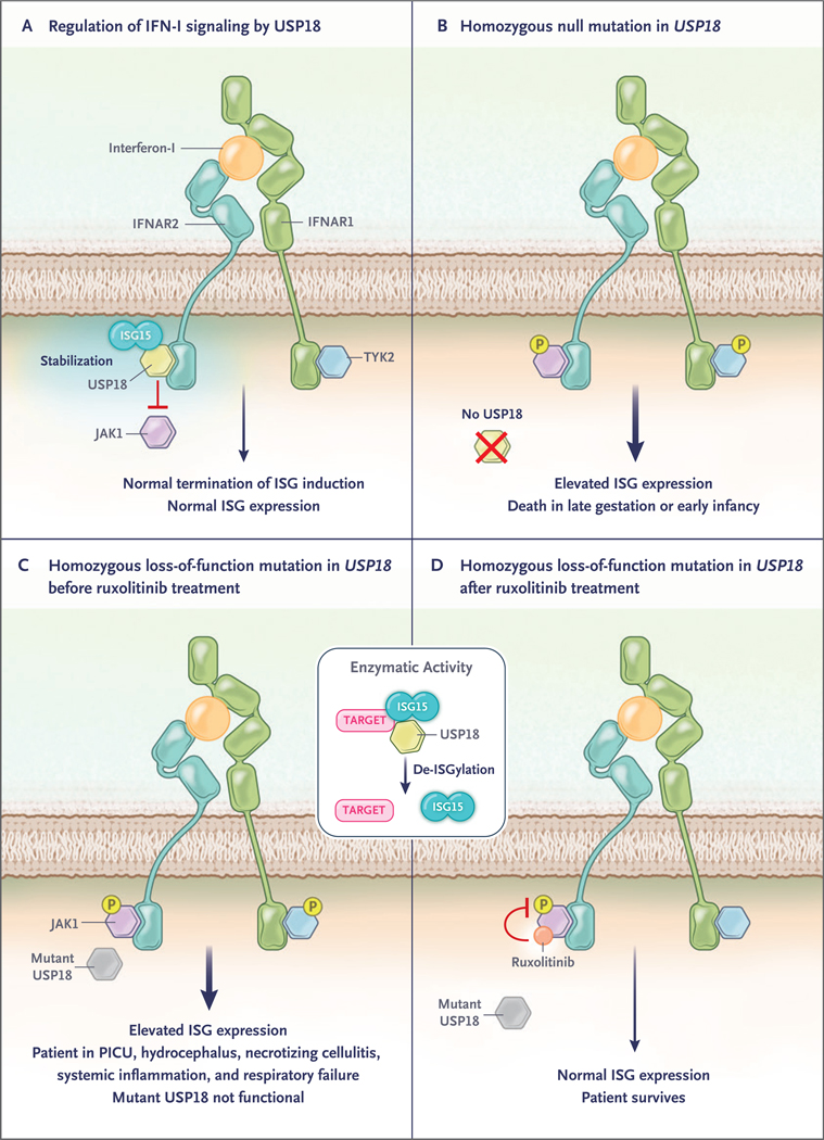
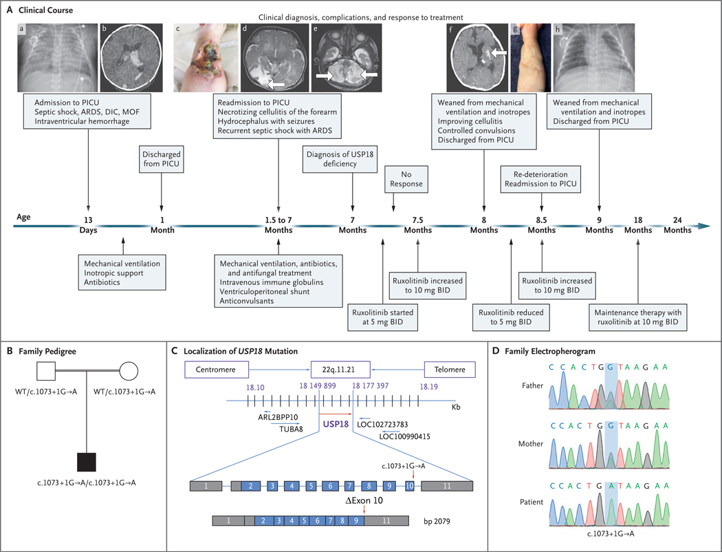
Deficiency of ubiquitin-specific peptidase 18 (USP18) is a severe type I interferonopathy. USP18 down-regulates type I interferon signaling by blocking the access of Janus-associated kinase 1 (JAK1) to the type I interferon receptor. The absence of USP18 results in unmitigated interferon-mediated inflammation and is lethal during the perinatal period. We describe a neonate who presented with hydrocephalus, necrotizing cellulitis, systemic inflammation, and respiratory failure. Exome sequencing identified a homozygous mutation at an essential splice site on USP18. The encoded protein was expressed but devoid of negative regulatory ability. Treatment with ruxolitinib was followed by a prompt and sustained recovery. (Funded by King Saud University and others.).


Figures

Figure 1 (facing page).
Figure 1 (facing page).. Molecular Functions of USP18.
Type I interferon (IFN–I) binds IFN–I receptor, consisting of two subunits, IFN–I receptor 1 (IFNAR1) and IFN–I receptor 2 (IFNAR2). It signals through activation of two receptor–associated protein tyrosine kinases, Janus kinase 1 (JAK1) and tyrosine kinase 2 (TYK2), which phosphorylate (P) signal transducer and activator of transcription 1 (STAT1) and 2 (STAT2) to induce the transcription of hundreds of interferon–stimulated genes (ISGs). Panel A shows the activity of USP18 (an ISG) as a negative–feedback regulator of IFN–I signaling through the inhibition of JAK1 recruitment to IFNAR2. ISG15 stabilizes USP18 and protects it against proteasomal degradation, resulting in normal ISG expression. Panel B shows that homozygous mutations resulting in no synthesis of USP18 cause unrestrained IFN–I signaling and are lethal. Panels C and D show the outcome in the patient in the case report before and after the initiation of ruxolitinib, a JAK1 inhibitor. The box in the middle of the figure shows that in addition to inhibiting JAK1, USP18 has catalytic activity: it cleaves the ubiquitin–like ISG15 protein from conjugated proteins in a process called de–ISGylation. PICU denotes pediatric intensive care unit.
Figure 2 (facing page).
Figure 2 (facing page).. Clinical Course of the Patient and Identification of a USP18 Mutation.
In Panel A, at the age of 13 days, the patient was admitted to the pediatric intensive care unit (PICU), where he underwent chest radiography, which showed patches of space opacification with an air bronchogram of both lungs characteristic of severe acute respiratory distress syndrome (ARDS) (subpanel a). At that time, multiple organ failure (MOF) and disseminated intravascular coagulopathy (DIC) had also developed, along with septic shock. Computed tomography (CT) of the brain at the age of 23 days showed mildly dilated lateral ventricles with intraventricular hyperdense acute hemorrhage more marked on the left, corresponding to grade II intraventricular hemorrhage (subpanel b). At the age of 45 days, the patient was readmitted with necrotizing cellulitis at the site of a peripheral venous catheter in the right forearm (subpanel c). Magnetic resonance imaging (MRI) of the brain at the age of 6 weeks showed a well–defined high–intensity signal in the right occipital region (subpanel d, arrow). MRI of the brain at the age of 11 weeks showed a well–defined high–intensity signal in the bilateral cerebellum owing to subacute infarction (subpanel e, arrows). One month after the diagnosis of USP18 deficiency and the initiation of ruxolitinib (at a dose of 5 mg twice daily [BID]) at the age of 7 months, CT of the brain showed resolution of hydrocephalus, hemorrhage, and ischemia with only small areas of residual calcifications in the left putamen (subpanel f, arrow) and the formation of scar tissue on the forearm after healing of the necrotizing cellulitis (subpanel g). Chest radiography after 2 months of ruxolitinib therapy showed sufficient improvement for weaning from mechanical ventilation (subpanel h). Panel B shows familial segregation of the c.1073+1G→A USP18 allele. The unaffected parents, who were first cousins (as indicated by the double horizontal line), were heterozygous for the mutation, and the patient was homozygous. Panel C shows the localization of the USP18 mutation in the genomic DNA and messenger RNA, with coding regions in blue, untranslated regions in gray, and the mutation indicated by red arrows, with the predicted transcript skipping exon 10. Panel D shows the electro-pherographic results of the homozygous c.1073+1G→A USP18 mutation in the patient and the heterozygous mutation in the unaffected parents.
Figure 3.
Figure 3.. Characterization of the USP18 Allele.
Panel A shows the relative levels of messenger RNA (mRNA), as normalized with the 18S housekeeper gene, in wild-type (WT) USP18 or the c.1073+1G→A (ΔEx10) variant and in a control with lentiviral particles expressing luciferase (Luc), as assayed on quantitative reverse–transcriptase–polymerase chain reaction (RT–PCR) in three independent experiments, each with technical triplicates; the results of a representative experiment are shown. Panel B shows a representative experiment involving cell lysates that were analyzed by Western blotting for the USP18 protein. Panel C shows HEK293T cells that were transfected with wild–type USP18 or the ΔEx10 variant, together with various amounts of pTRIP–ISG15 (graph at the left). Cell lysates were analyzed by Western blotting with the antibodies indicated. The graph at the right shows the densitometric analysis of USP18 protein expression levels normalized over an endogenous control. T bars represent the standard error of three independent experiments. NT denotes not transfected. Panel D shows HEK293T cells that were transduced with wild–type USP18, the ΔEx10 variant, or Luc. Twenty–four hours later, the cells were treated with the indicated concentrations of interferon–α2b for 20 minutes. Cell lysates were analyzed by Western blotting with the antibodies indicated. Samples were run on the same gel, and extra lanes were deleted to remove irrelevant data, as indicated by the vertical dashed line. Panel E shows HEK293T cells that were cotransfected with ISG15 and E1, E2, and E3 enzymes for ISG15 (Ube1L, UbcH8, and Herc5) in combination with the different variants of USP18. The graph at the right shows the densitometric analysis of ISGylated proteins normalized over an endogenous control. Cell lysates were analyzed by Western blotting with the antibodies indicated. The results of a representative experiment are shown. EV denotes empty vector.

References

    1. Rodero MP, Decalf J, Bondet V, et al. Detection of interferon alpha protein reveals differential levels and cellular sources in disease. J Exp Med 2017; 214: 1547–55. - PMC - PubMed
    1. Rodero MP, Crow YJ. Type I interferon-mediated monogenic autoinflammation: the type I interferonopathies, a conceptual overview. J Exp Med 2016; 213: 2527–38. - PMC - PubMed
    1. Crow YJ. Type I interferonopathies: mendelian type I interferon up-regulation. Curr Opin Immunol 2015; 32: 7–12. - PubMed
    1. Hermann M, Bogunovic D. ISG15: in sickness and in health. Trends Immunol 2017; 38: 79–93. - PubMed
    1. Malakhova OA, Kim KI, Luo JK, et al. UBP43 is a novel regulator of interferon signaling independent of its ISG15 isopeptidase activity. EMBO J 2006; 25: 2358–67. - PMC - PubMed

Publication types

MeSH terms