Review
. 2020 Jan 11;8(1):0.
doi: 10.3390/microorganisms8010105.

Rules and Exceptions: The Role of Chromosomal ParB in DNA Segregation and Other Cellular Processes

Affiliations
Review

Rules and Exceptions: The Role of Chromosomal ParB in DNA Segregation and Other Cellular Processes

Adam Kawalek et al. Microorganisms. .

Abstract

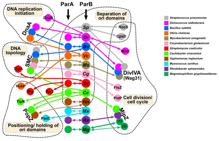
The segregation of newly replicated chromosomes in bacterial cells is a highly coordinated spatiotemporal process. In the majority of bacterial species, a tripartite ParAB-parS system, composed of an ATPase (ParA), a DNA-binding protein (ParB), and its target(s) parS sequence(s), facilitates the initial steps of chromosome partitioning. ParB nucleates around parS(s) located in the vicinity of newly replicated oriCs to form large nucleoprotein complexes, which are subsequently relocated by ParA to distal cellular compartments. In this review, we describe the role of ParB in various processes within bacterial cells, pointing out interspecies differences. We outline recent progress in understanding the ParB nucleoprotein complex formation and its role in DNA segregation, including ori positioning and anchoring, DNA condensation, and loading of the structural maintenance of chromosome (SMC) proteins. The auxiliary roles of ParBs in the control of chromosome replication initiation and cell division, as well as the regulation of gene expression, are discussed. Moreover, we catalog ParB interacting proteins. Overall, this work highlights how different bacterial species adapt the DNA partitioning ParAB-parS system to meet their specific requirements.

Keywords: ParB; cell division; chromosome segregation; gene expression regulation; partitioning proteins; segrosome.


Conflict of interest statement

The authors declare no conflict of interest. The funders had no role in the design of the study; in the collection, analyses, or interpretation of data; in the writing of the manuscript, or in the decision to publish the results.

Figures

Figure 1
Figure 1
Involvement of partition ParB protein in ori domain re-locations, structuring and positioning during bacterial cell cycle.simplified scheme for P. aeruginosa-like longitudinal chromosome rearrangements is presented, in which the replisome is located in the cell centre, and the ParB-bound ori domains are anchored close to the cell poles before division [30].
Figure 2
Figure 2
ParB complex assembly at parS. (a) Schematic representation of chromosomally encoded ParB protein (dimer) with the indicated functions of individual domains. *- only confirmed for B. subtilis Spo0J. (b) Models of the ParB–ParB interactions involved in formation of the ParB nucleoprotein complexes around parS. (I) Adjacent ParB dimers may interact with each other to form 1D filaments around parS. (II) Interactions between ParB dimers associated with distal DNA fragments may lead to DNA bridging and looping. (III) ParB self-interactions provide a scaffold (cage), attracting and trapping additional ParB molecules. (c) A model illustrating ParB loading at parS and sliding [122]. Free CTP-ParB exists as a dimer in an open conformation. Binding to parS induces conformational changes involving the N-terminal ParB domains and the formation of “closed” ring-shaped molecules. Steric hindrance between HTH motifs interacting with parS in such a closed conformation may prompt the release of ParB rings from parS via their sliding on adjacent DNA and the loading of new ParB dimers at parS. Finally, switching from a closed to open conformation by an unknown mechanism (possibly involving CTP hydrolysis) may lead to ParB’s dissociation from the DNA. The parS sites are indicated in red.
Figure 3
Figure 3
Interactions of ParBs with half-parS sites. (a) Outline of the analysis of ParB binding to half-parS using the available ParB ChIP-seq data. (b) The binding of ParB proteins from various bacterial species to half-parSs assessed by the enrichment of half-parS (GTTCCAC and GTTTCAC) containing genomic DNA fragments in the ChIP samples. Heatmaps represent the read coverage for the ParB ChIP samples, calculated for each nucleotide of a ±300 bp region around all the indicated motifs in the corresponding reference genomes. Plots represent median coverage. A central increase of the coverage indicates enrichment of the DNA containing motif during chromatin immunoprecipitation for the corresponding ParB protein; hence, ParB binds to these sequences. The ChIP-seq data for 17 ParBs from different species produced in E. coli [Gene Expression Omnibus GSE129285 [142]], V. cholerae ParB1 [GSM3161909, GSM3161911 [129]], and C. glutamicum ParB [SRX5581454, SRX5581458, SRX5581460 [72]] were included in the analysis. Raw data were downloaded from the sequence read archive (SRA) and quality-controlled using fastp [143]. Reads were mapped to the reference genomes of E. coli K-12 substr. MG1655 (U00096.3), V. cholerae O1 biovar El Tor str. N16961 (only chrI, NC_002505.1), and C. glutamicum ATCC 13032 (BX927147), respectively, using Bowtie [144] with the --sensitive-local option. Samtools was used to exclude duplicate reads and sort the .bam files [145]. Coverage (.bigwig) files were generated with bamCoverage [146], using the --normalizeUsing RPGC option, without binning and smoothing. Half-parS motifs (GTTCCAC and GTTTCAC) were identified in the corresponding genomes using fuzznuc (Emboss 6.6.0). Heatmap displaying coverage with reads in the ParB ChIP data around the identified motifs were generated using plotHeatmap from deepTools [146]. Each line in the heatmap represents the normalized read counts for each nucleotide of a ±300 bp region around one motif, sorted in the descending order of the mean coverage value and colored according to scale. The median coverage score for the two sets of motifs is presented on plots above the heatmap. For V. cholerae and C. glutamicum, the data from the biological replicates were averaged. (c) Hypothetical model engaging the half-parS sites in DNA structuring. I—The ParB complex loaded at parS interacts with an “open” dimeric ParB bound to a half-parS site. II—Interactions between two ParB dimers bound to separate half-parSs. III—Both monomers in a ParB dimer interact with separate half-parSs. All scenarios result in the formation of DNA bridges. In this model, we assume that the binding of ParB to half-parS involves the HTH in the central domain.
Figure 4
Figure 4
Network of ParBs interactions with ParAs and other partners coordinating chromosome segregation with various cellular functions in the analyzed bacterial species. The dashed arrows indicate interactions not yet confirmed experimentally.

References

    1. Dame R.T., Rashid F.-Z.M., Grainger D.C. Chromosome organization in bacteria: Mechanistic insights into genome structure and function. Nat. Rev. Genet. 2019 doi: 10.1038/s41576-019-0185-4. - DOI - PubMed
    1. Badrinarayanan A., Le T.B., Laub M.T. Bacterial chromosome organization and segregation. Annu. Rev. Cell Dev. Biol. 2015;31:171–199. doi: 10.1146/annurev-cellbio-100814-125211. - DOI - PMC - PubMed
    1. Dorman C.J. Genome architecture and global gene regulation in bacteria: Making progress towards a unified model? Nat. Rev. Microbiol. 2013;11:349–355. doi: 10.1038/nrmicro3007. - DOI - PubMed
    1. Dame R.T., Tark-Dame M. Bacterial chromatin: Converging views at different scales. Curr. Opin. Cell Biol. 2016;40:60–65. doi: 10.1016/j.ceb.2016.02.015. - DOI - PubMed
    1. Gruber S., Errington J. Recruitment of condensin to replication origin regions by ParB/Spo0J promotes chromosome segregation in B. subtilis. Cell. 2009;137:685–696. doi: 10.1016/j.cell.2009.02.035. - DOI - PubMed

LinkOut - more resources