Review
. 2020 Jan 12;10(1):128.
doi: 10.3390/biom10010128.

The Bcl-2 Family: Ancient Origins, Conserved Structures, and Divergent Mechanisms

Affiliations
Review

The Bcl-2 Family: Ancient Origins, Conserved Structures, and Divergent Mechanisms

Suresh Banjara et al. Biomolecules. .

Abstract

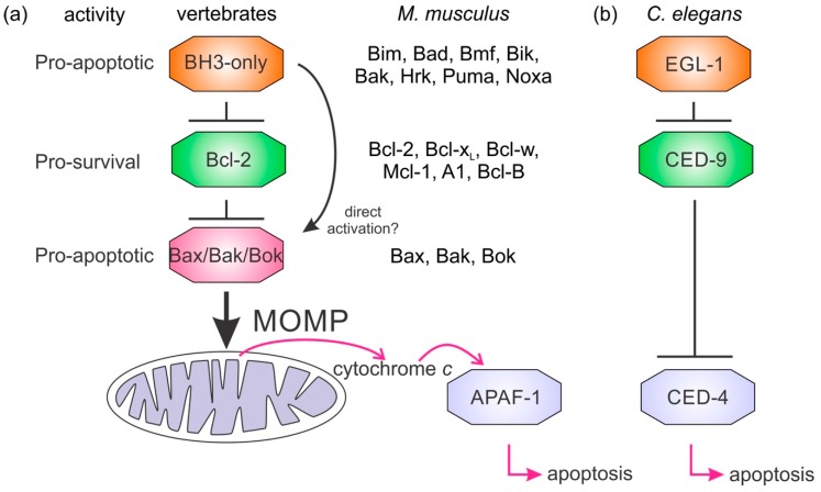
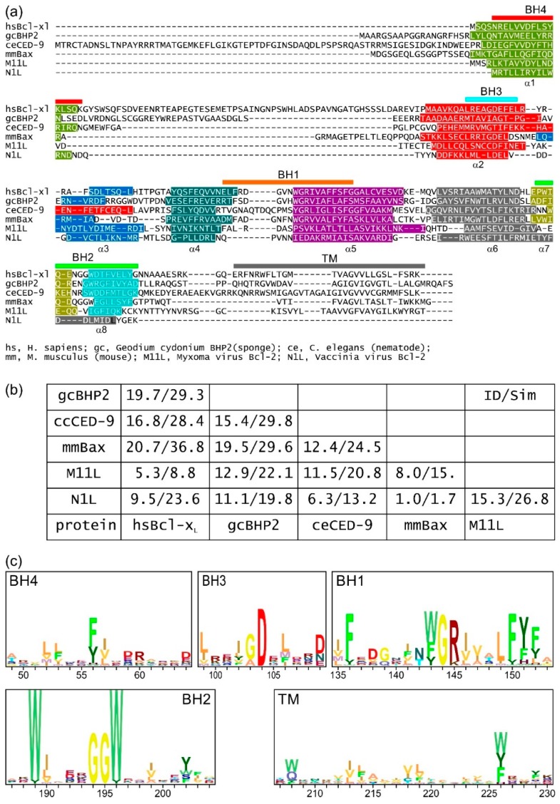
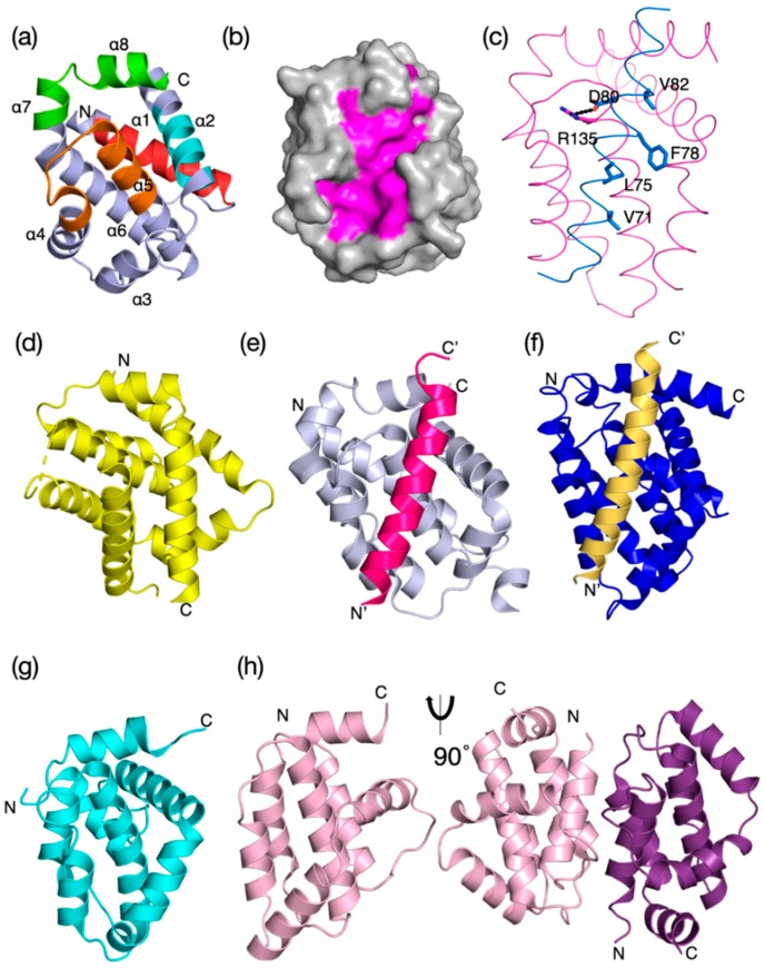
Intrinsic apoptosis, the response to intracellular cell death stimuli, is regulated by the interplay of the B-cell lymphoma 2 (Bcl-2) family and their membrane interactions. Bcl-2 proteins mediate a number of processes including development, homeostasis, autophagy, and innate and adaptive immune responses and their dysregulation underpins a host of diseases including cancer. The Bcl-2 family is characterized by the presence of conserved sequence motifs called Bcl-2 homology motifs, as well as a transmembrane region, which form the interaction sites and intracellular location mechanism, respectively. Bcl-2 proteins have been recognized in the earliest metazoans including Porifera (sponges), Placozoans, and Cnidarians (e.g., Hydra). A number of viruses have gained Bcl-2 homologs and subvert innate immunity and cellular apoptosis for their replication, but they frequently have very different sequences to their host Bcl-2 analogs. Though most mechanisms of apoptosis initiation converge on activation of caspases that destroy the cell from within, the numerous gene insertions, deletions, and duplications during evolution have led to a divergence in mechanisms of intrinsic apoptosis. Currently, the action of the Bcl-2 family is best understood in vertebrates and nematodes but new insights are emerging from evolutionarily earlier organisms. This review focuses on the mechanisms underpinning the activity of Bcl-2 proteins including their structures and interactions, and how they have changed over the course of evolution.

Keywords: Bcl-2; apoptosis; evolution; mechanism; structure analysis.


Conflict of interest statement

The authors declare no conflict of interest.

Figures

Figure 1
Figure 1
Mechanisms of Bcl-2 regulated apoptosis. Simplified apoptosis schemes showing the role of Bcl-2 proteins in apoptosis initiation differ between mammals (a) and nematodes (b). The Bcl-2 family members are indicated for M. musculus. Nematodes have a simplified activation of apoptosis where the BH3-only protein EGL–1 binds the sole prosurvival protein in the C. elegans genome CED-9. This event releases the caspase activating protein CED–4 to initiate the caspase cascade. In mammalian apoptosis, the BH3-only group of proteins antagonize the prosurvival Bcl-2 proteins releasing Bax, Bak, or Bok to oligomerize and form pores in mitochondria causing mitochondrial outer membrane permeabilization (MOMP). Cytochrome c release from the mitochondrion triggers the mammalian equivalent of CED-4, APAF-1, to oligomerize and initiate the activation of downstream caspases.
Figure 2
Figure 2
Sequence-structure analysis of Bcl-2 family members from sponges to man. (a) Structure-based sequence alignment of metazoan and viral Bcl-2 family members. Structurally equivalent residues are aligned. In (a), sequence–structure alignment shows prosurvival and proapoptotic Bcl-2 proteins share key sequence features. Sequences aligned: H. sapiens Bcl-xL; G. cydonium BHP2; C. elegans CED-9; Mus musculus Bax; Myxoma virus M11L; Vaccinia virus N1L. Sequence–structure alignment was performed using Dali [85] and the secondary structure is indicated by the colored bars. The extent of the Bcl-2 homology (BH) motifs and transmembrane region (TM) is indicated by bars above the sequence and the helices below the sequences. (b) Table of sequence identities and similarities for the sequences in (a) given as percentage sequence identity/sequence similarity in each entry. Notably, the viral Bcl-2 proteins have little recognizable shared sequence identity with mammalian Bcl-2 proteins. (c) Profiles of BH and TM regions from Bax sequences representing bilaterians (Lepisosteus oculatis, Strongylocentrotus purpuratus, Ciona intestinalis), cnidarians (H. vulgaris, Acropora digitifera), placozoa (T. adhaerens), and porifera (A queenslandica). The height of each stack represents the conservation and the residue frequencies are represented by their height as determined by the program Skylign [86]. These indicate that the BH4 motif is a relatively weak and poorly conserved motif when compared to the BH1–BH3 motifs. Uniprot sequence and PDB IDs, hsBcl-xL: Q07817, 1R2D; gcBHP2: Q967D2, 5TWA; ceCED-9: P41958, 1OHU; mmBax: Q07813, 5W62; Myxoma virus M11L: Q77PA8, 2JBX; Vaccinia virus N1L: P21054, 2I39.
Figure 3
Figure 3
Evolutionary structure conservation in the Bcl-2 family and their complexes. Ribbon representation of the 3D structures of prosurvival and proapoptotic Bcl-2 family members and their complexes are shown. The helical bundle Bcl-2 structure occurred early in evolution and changed little over evolutionary time scales. (a) H. sapiens Bcl-xL (PDB 1R2D) with BH1–4 motifs colored in orange, green, cyan, and red as shown in the sequence–structure alignment of Figure 2a, (b) H. sapiens Bcl–xL (PDB 1R2D) shown as grey surface with canonical ligand-binding groove shaded in magenta, (c) BHP2 from the sponge G. cydonium Bcl-2, BHP2 (PDB 5TWA) magenta, LB–Bak sky blue. The canonical ionic interaction between the conserved Arg from prosurvival Bcl-2 and conserved Asp from the BH3 motif of prodeath Bcl-2 as well as the four conserved hydrophobic residues from the Bak BH3 motif are shown as sticks. (d) M. musculus Bax (PDB 5W62) yellow, (e) H. sapiens Bcl-b:Bim complex (PDB 4B4S), (f) C. elegans CED-9: EGL-1 complex, CED-9 (navy) with EGL–1 (sand) in the binding groove (PDB 1TY4). (g) Myxoma virus Bcl-2 M11L (PDB 2JBX) cyan, (h) Vaccinia virus N1L (PDB 2I39) salmon. Monomeric N1 is shown in the same orientation as in (a), and the functionally relevant dimer is shown rotated by 90o around the vertical axis. In (a), the extent of the BH motifs is indicated as ribbon colored as in Figure 2a and the helices α1–α8 are also indicated. The structures were aligned on human Bcl-xL and the orientation for all structures is the same as that in (a). The N and C termini are indicated.
Figure 4
Figure 4
Bcl-2-like proteins in basal metazoan clades and potential apoptosis model. (a) Metazoan phylogenetic relationships and the presence or absence of Bcl-2 proteins. The presence or absence of Bcl-2 family members is indicated. (b) A simple model for intrinsic apoptosis in Porifera and Placozoa where BH3-only proteins have not been identified. Experiments have yet to delineate the roles of MOMP and adaptor protein initiation of caspases in their entirety.

References

    1. Koonin E.V., Aravind L. Origin and evolution of eukaryotic apoptosis: The bacterial connection. Cell Death Differ. 2002;9:394–404. doi: 10.1038/sj.cdd.4400991. - DOI - PubMed
    1. Huettenbrenner S., Maier S., Leisser C., Polgar D., Strasser S., Grusch M., Krupitza G. The evolution of cell death programs as prerequisites of multicellularity. Mutat. Res. 2003;543:235–249. doi: 10.1016/S1383-5742(02)00110-2. - DOI - PubMed
    1. Singh R., Letai A., Sarosiek K. Regulation of apoptosis in health and disease: The balancing act of BCL-2 family proteins. Nat. Rev. Mol. Cell Biol. 2019;20:175–193. doi: 10.1038/s41580-018-0089-8. - DOI - PMC - PubMed
    1. Strasser A., Vaux D.L. Viewing BCL2 and cell death control from an evolutionary perspective. Cell Death Differ. 2018;25:13–20. doi: 10.1038/cdd.2017.145. - DOI - PMC - PubMed
    1. Shamas-Din A., Kale J., Leber B., Andrews D.W. Mechanisms of action of bcl-2 family proteins. Cold Spring Harb. Perspect. Biol. 2013;5 doi: 10.1101/cshperspect.a008714. - DOI - PMC - PubMed

Publication types

Substances

LinkOut - more resources