. 2020 Jan 12;18(1):52.
doi: 10.3390/md18010052.

Pre-Treatment with Laminarin Protects Hippocampal CA1 Pyramidal Neurons and Attenuates Reactive Gliosis Following Transient Forebrain Ischemia in Gerbils

Affiliations

Pre-Treatment with Laminarin Protects Hippocampal CA1 Pyramidal Neurons and Attenuates Reactive Gliosis Following Transient Forebrain Ischemia in Gerbils

Tae-Kyeong Lee et al. Mar Drugs. .

Abstract

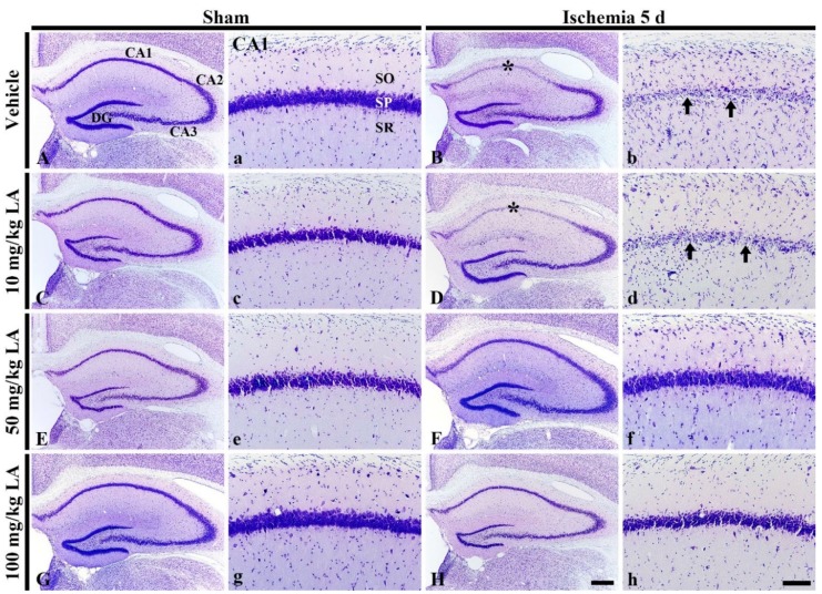
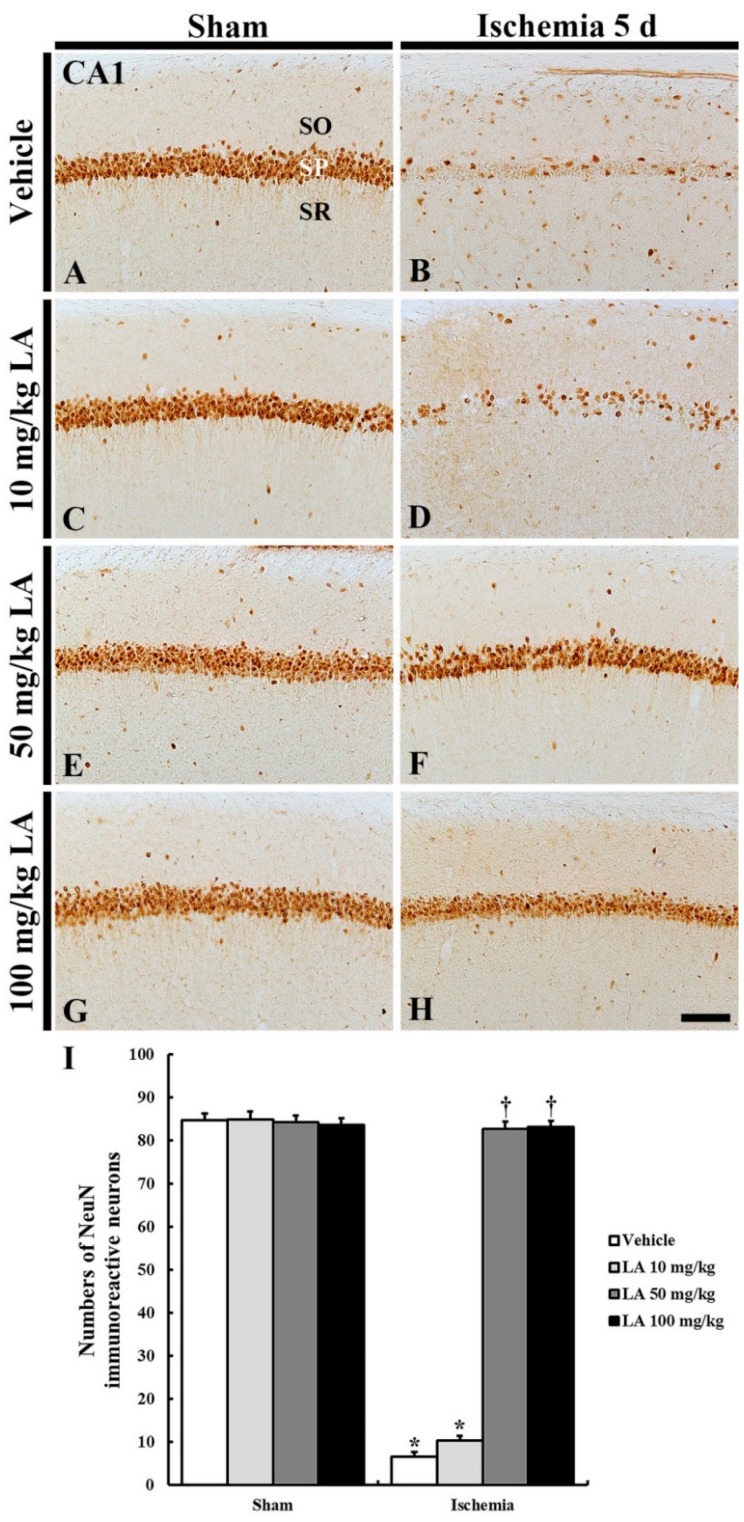
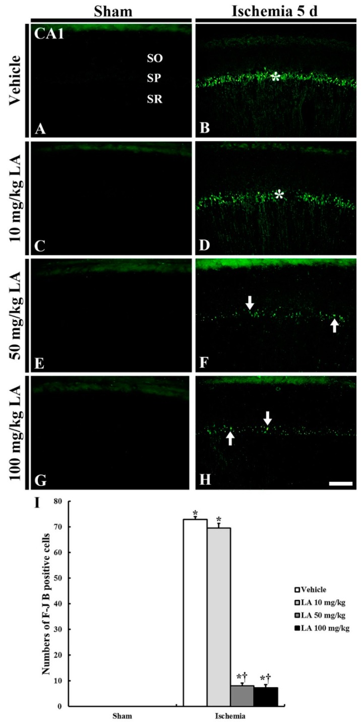
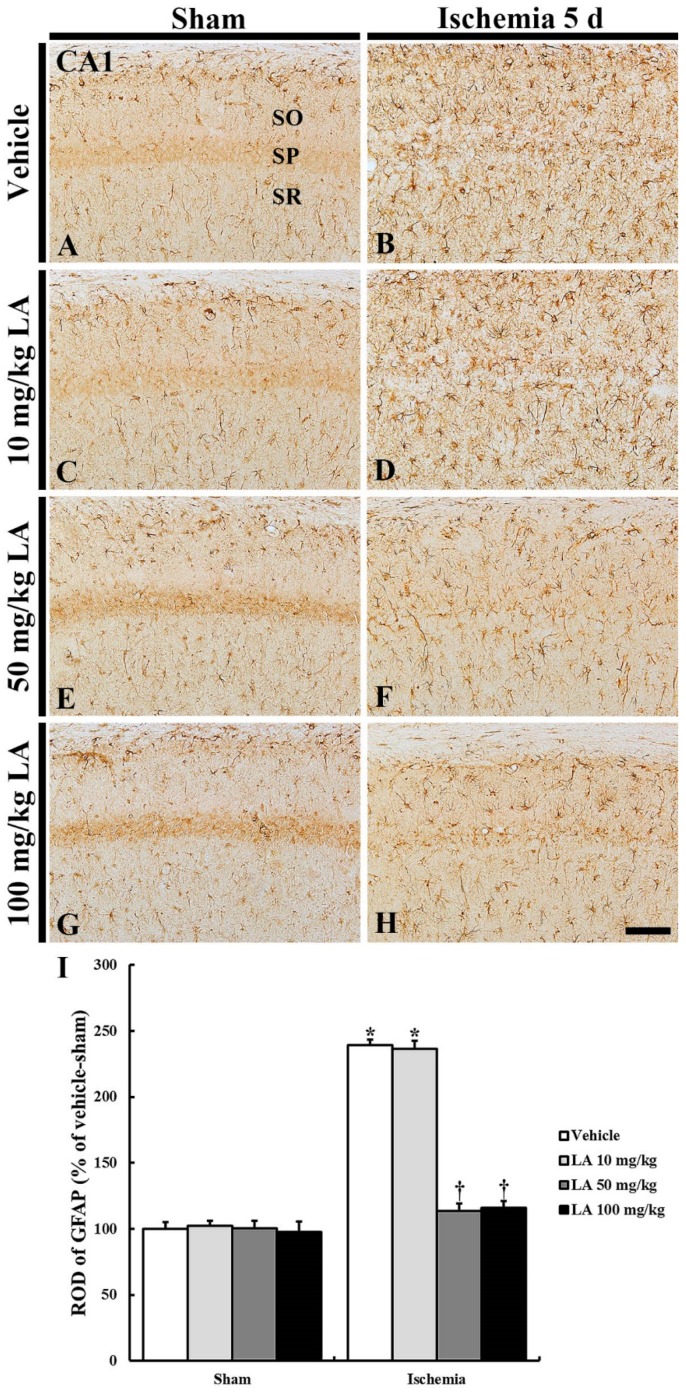
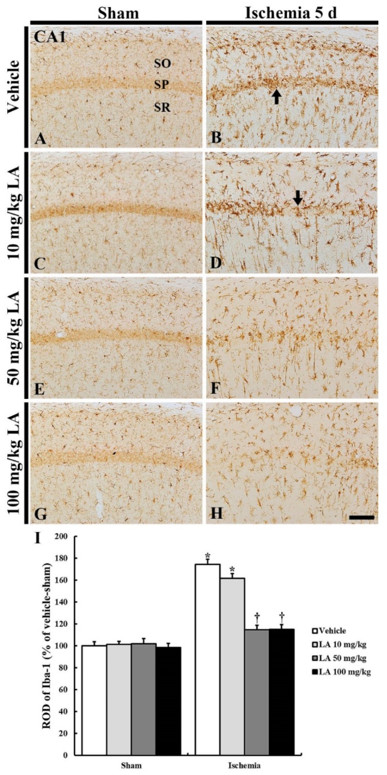
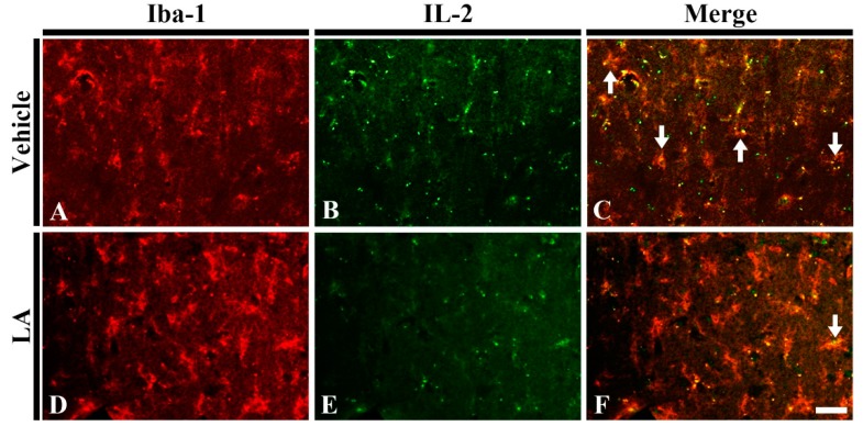
Transient brain ischemia triggers selective neuronal death/loss, especially in vulnerable regions of the brain including the hippocampus. Laminarin, a polysaccharide originating from brown seaweed, has various pharmaceutical properties including an antioxidant function. To the best of our knowledge, few studies have been conducted on the protective effects of laminarin against ischemic injury induced by ischemic insults. In this study, we histopathologically investigated the neuroprotective effects of laminarin in the Cornu Ammonis 1 (CA1) field of the hippocampus, which is very vulnerable to ischemia-reperfusion injury, following transient forebrain ischemia (TFI) for five minutes in gerbils. The neuroprotective effect was examined by cresyl violet staining, Fluoro-Jade B histofluorescence staining and immunohistochemistry for neuronal-specific nuclear protein. Additionally, to study gliosis (glial changes), we performed immunohistochemistry for glial fibrillary acidic protein to examine astrocytes, and ionized calcium-binding adaptor molecule 1 to examine microglia. Furthermore, we examined alterations in pro-inflammatory M1 microglia by using double immunofluorescence. Pretreatment with 10 mg/kg laminarin failed to protect neurons in the hippocampal CA1 field and did not attenuate reactive gliosis in the field following TFI. In contrast, pretreatment with 50 or 100 mg/kg laminarin protected neurons, attenuated reactive gliosis and reduced pro-inflammatory M1 microglia in the CA1 field following TFI. Based on these results, we firmly propose that 50 mg/kg laminarin can be strategically applied to develop a preventative against injuries following cerebral ischemic insults.

Keywords: gliosis; ischemia-reperfusion; polysaccharide; prevention; pyramidal neurons; rodents.


Conflict of interest statement

The authors of this article declare that there are no conflicts of interest.

Figures

Figure 1
Figure 1
Cresyl Violet (CV) staining in the hippocampus (AH) and its Cornu Ammonis 1 (CA1) field (ah) of the vehicle/sham (A,a), 10, 50 and 100 mg/kg laminarin (LA)/sham (C,c,E,e,G,g), vehicle/ischemia (B,b) and 10, 50 and 100 mg/kg LA/ischemia (D,d,F,f,H,h) groups at 5 days after sham or transient forebrain ischemia (TFI) operation. In the vehicle/ischemia group, CV dyeability is remarkably reduced in the stratum pyramidale (SP, arrows) of the CA1 field (asterisks). In the 10 mg/kg LA/ischemia group, the distribution pattern of CV stained cells is similar to that in the vehicle/ischemia group. However, in the 50 mg/kg and 100 mg/kg LA/ischemia groups, CV stainability is conserved. DG, dentate gyrus; SO, stratum oriens; SR stratum radiatum. Scale bars = 400 μm (AH) and 100 μm (ah).
Figure 2
Figure 2
NeuN immunohistochemistry in the CA1 field of the vehicle/sham (A), 10, 50 and 100 mg/kg LA/sham (C,E,G), vehicle/ischemia (B) and 10, 50 and 100 mg/kg LA/ischemia (D,F,H) groups at 5 days after sham or TFI operation. Numerous NeuN immunoreactive CA1 pyramidal neurons can be observed in the vehicle/sham group. In the vehicle/ischemia and 10 mg/kg LA/ischemia groups, significant decreases in NeuN immunoreactive CA1 pyramidal neurons were detected. In the 50 mg/kg and 100 mg/kg LA/ischemia groups, CA1 pyramidal neurons show strong NeuN immunoreactivity. Scale bar = 100 μm. (I) Mean number of NeuN immunoreactive pyramidal cells in the CA1 field at 5 days after TFI (n = 7 in each group, * p < 0.05 versus vehicle/sham group, † p < 0.05 versus vehicle/ischemia group). The bars indicate the means ± SEM.
Figure 3
Figure 3
F-J B histofluorescence staining in the CA1 field of the vehicle/sham (A), 10, 50 and 100 mg/kg LA/sham (C,E,G), vehicle-ischemia (B) and 10, 50 and 100 mg/kg LA/ischemia (D,F,H) groups at 5 days after sham or TFI operation. In all the sham groups, no F-J B positive cells are found in the CA1 field; numerous F-J B positive cells are shown in the SP (asterisks) in the vehicle/ and 10 mg/kg LA/ischemia groups. However, in the 50 mg/kg and 100 mg/kg LA/ischemia groups, F-J B positive cells (arrows) are significantly decreased. Scale bar = 100 μm. (I) Mean number of F-J B positive pyramidal cells in the CA1 field at 5 days after TFI (n = 7 in each group, * p < 0.05 versus vehicle/sham group, † p < 0.05 versus vehicle/ischemia group). The bars indicate the means ± SEM.
Figure 4
Figure 4
Glial fibrillary acidic protein (GFAP) immunohistochemistry in the CA1 field of the vehicle/sham (A), 10, 50 and 100 mg/kg LA/sham (C,E,G), vehicle/ischemia (B) and 10, 50 and 100 mg/kg LA/ischemia (D,F,H) groups at 5 days after sham or TFI operation. In all the sham groups, typical GFAP immunoreactive astrocytes are generally distributed in the stratum oriens (SO) and radiatum (SR). In the vehicle/ischemia group, GFAP immunoreactive astrocytes are hypertrophied. In the 10 mg/kg LA/ischemia group, GFAP immunoreactive astrocytes are similar to those in the vehicle/ischemia group. In the 50 mg/kg and 100 mg/kg LA/ischemia groups, hypertrophy of GFAP immunoreactive astrocytes is apparently attenuated. Scale bar = 100 μm. (I) ROD (percentage) of GFAP immunoreactive structures in the CA1 field at 5 days after TFI (n = 7 in each group, * p < 0.05 versus vehicle/sham group, † p < 0.05 versus vehicle/ischemia group). The bars indicate the means ± SEM.
Figure 5
Figure 5
Ionized calcium-binding adapter molecule 1 (Iba-1) immunohistochemistry in the CA1 field of the vehicle/sham (A), 10, 50 and 100 mg/kg LA/sham (C,E,G), vehicle/ischemia (B) and 10, 50 and 100 mg/kg LA/ischemia (D,F,H) groups at 5 days after sham or TFI operation. Iba-1 immunoreactive microglia are in a resting state in all the sham groups. In the vehicle/ischemia and 10 mg/kg LA/ischemia groups, Iba-1 immunoreactive microglia are hypertrophied, showing that many activated microglia gather in the SP (arrows). In the 50 mg/kg and 100 mg/kg LA/ischemia groups, activation of Iba-1 immunoreactive microglia is markedly attenuated, showing that they are evenly distributed in the CA1 field. Scale bar = 100 μm. (I) ROD (percentage) of Iba-1 immunoreactive structures in the CA1 field at 5 days after TFI (n = 7 in each group, * p < 0.05 versus vehicle/sham group, † p < 0.05 versus vehicle/ischemia group). The bars indicate the means ± SEM.
Figure 6
Figure 6
Double immunofluorescence staining for Iba-1 (red), interleukin 2 (IL-2) (green) and merged images in the hippocampal CA1 field of the vehicle/ischemia (AC) and 50 mg/kg LA/ischemia (DF) groups at 5 days after TFI. Many IL-2 immunoreactive microglia (arrows) are shown in the vehicle/ischemia group. However, in the 50 mg/kg LA/ischemia group, a few IL-2 immunoreactive microglia are detected. Scale bar = 40 μm (n = 7 in each group).

References

    1. Ahn J.H., Shin B.N., Park J.H., Lee T.K., Park Y.E., Lee J.C., Yang G.E., Shin M.C., Cho J.H., Lee K.C., et al. Pre- and Post-Treatment with Novel Antiepileptic Drug Oxcarbazepine Exerts Neuroprotective Effect in the Hippocampus in a Gerbil Model of Transient Global Cerebral Ischemia. Brain Sci. 2019;9:279. doi: 10.3390/brainsci9100279. - DOI - PMC - PubMed
    1. Kapoor M., Sharma S., Sandhir R., Nehru B. Temporal changes in physiological and molecular markers in various brain regions following transient global ischemia in rats. Mol. Biol. Rep. 2019 doi: 10.1007/s11033-019-05060-7. - DOI - PubMed
    1. Lee T.K., Kim H., Song M., Lee J.C., Park J.H., Ahn J.H., Yang G.E., Ohk T.G., Shin M.C., Cho J.H., et al. Time-course pattern of neuronal loss and gliosis in gerbil hippocampi following mild, severe, or lethal transient global cerebral ischemia. Neural Regen. Res. 2019;14:1394–1403. doi: 10.4103/1673-5374.253524. - DOI - PMC - PubMed
    1. Kirino T., Sano K. Selective vulnerability in the gerbil hippocampus following transient ischemia. Acta Neuropathol. 1984;62:201–208. doi: 10.1007/BF00691853. - DOI - PubMed
    1. Kirino T. Delayed neuronal death in the gerbil hippocampus following ischemia. Brain Res. 1982;239:57–69. doi: 10.1016/0006-8993(82)90833-2. - DOI - PubMed

MeSH terms

LinkOut - more resources