Review
. 2020 Jan 13;9(1):198.
doi: 10.3390/cells9010198.

Targeting Aberrant RAS/RAF/MEK/ERK Signaling for Cancer Therapy

Affiliations
Review

Targeting Aberrant RAS/RAF/MEK/ERK Signaling for Cancer Therapy

Ufuk Degirmenci et al. Cells. .

Abstract

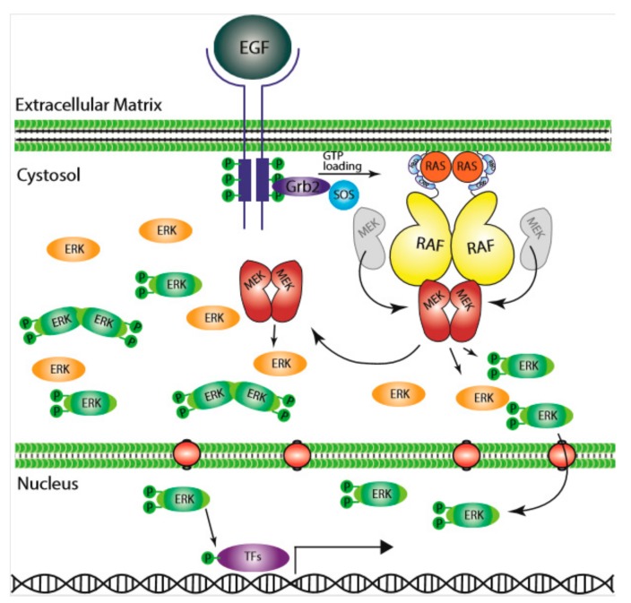
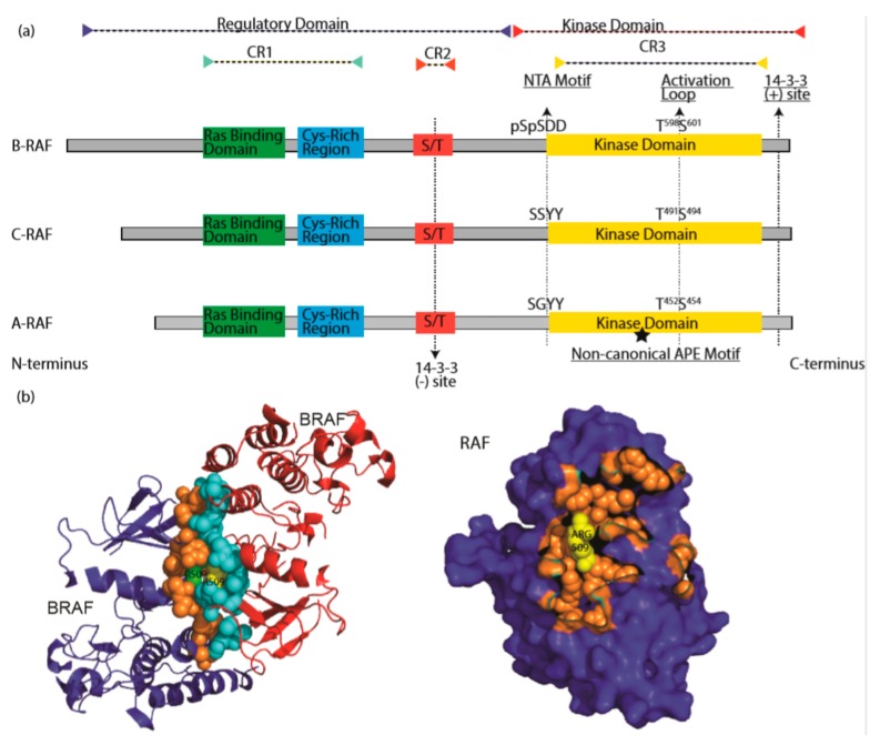
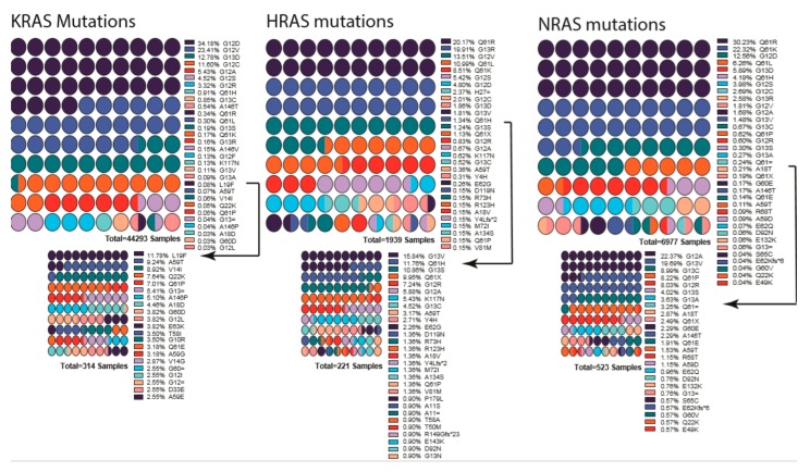
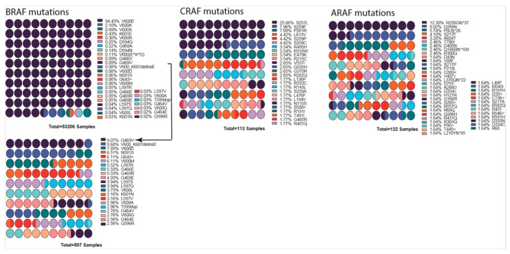
The RAS/RAF/MEK/ERK (MAPK) signaling cascade is essential for cell inter- and intra-cellular communication, which regulates fundamental cell functions such as growth, survival, and differentiation. The MAPK pathway also integrates signals from complex intracellular networks in performing cellular functions. Despite the initial discovery of the core elements of the MAPK pathways nearly four decades ago, additional findings continue to make a thorough understanding of the molecular mechanisms involved in the regulation of this pathway challenging. Considerable effort has been focused on the regulation of RAF, especially after the discovery of drug resistance and paradoxical activation upon inhibitor binding to the kinase. RAF activity is regulated by phosphorylation and conformation-dependent regulation, including auto-inhibition and dimerization. In this review, we summarize the recent major findings in the study of the RAS/RAF/MEK/ERK signaling cascade, particularly with respect to the impact on clinical cancer therapy.

Keywords: BRAF(V600E); RAF inhibitors; RAS GTPases, RAF family kinases; Ras/RAF/MEK/ERK signaling; neoplasm; paradoxical activation; protein–protein interactions; synthetic lethal.


Conflict of interest statement

The authors declare no conflicts of interest. The funders had no role in the design of the study; in the collection, analyses, or interpretation of data; in the writing of the manuscript, or in the decision to publish the results.

Figures

Figure 1
Figure 1
The Ras/RAF/MEK/ERK signaling pathway. Epidermal growth factor (EGF) initiates the signal on the cell surface through the EGF receptor (EGFR) (receptor tyrosine kinase), which activates guanine exchange factor to load RAS with GTP. RAS–GTP dimers/nanoclusters recruit RAFs or RAF/MEK heterodimers to plasma membranes, where RAF and MEK assemble transient tetramers that facilitate RAF activation through a back-to-back dimerization. MEKs docking on active RAF dimers further form face-to-face homodimers that are turned on by RAF. Activated MEKs phosphorylate ERKs, which generate response to the signal. CRR; Cys-rich region, RBD; Ras-binding domain.
Figure 2
Figure 2
The RAF family kinases. (a) Conserved domains on three RAF proteins are shown in panel (a). CR1 contains a Ras binding domain and a Cys-rich region while CR2 includes a S/T phosphorylation site. The 14-3-3 binding at this region inhibits RAF. CR3 contains a putative kinase domain adjacent to an acidic N-terminus (NTA) and a regulatory C-terminus. At the C terminus, there is a secondary 14-3-3 binding site which promotes dimerization. The non-canonical APE motif of ARAF is labeled with a star. ((b), left) Dimer interface of BRAF is shown, crystallography data was obtained from [86], PDB ID: 4MNE. Blue and red color indicates two separate BRAF molecules. Orange (Blue BRAF) and turquoise colored (Red BRAF) sphere-shaped amino acids indicate the dimer interface. While R509 from both RAF molecule located at the center of the dimer interface, green R509 belongs to red BRAF, while yellow R509 does to blue BRAF. ((b), right) Crystal structure of dimer interface from plane of interaction with only blue BRAF chain is visible. Orange spheres indicate the amino acids at the dimer interface, with exception of Arg509, which is labeled with yellow color. Structure was drawn by using pyMOL. CR1: conserved region 1; CR2: conserved region 2; CR3: conserved region 3.
Figure 3
Figure 3
List of mutations detected in each RAS gene isoform. Percentages are indicated next to the mutation, and colors indicate the mutation. Low-percentage mutations were shown as a smaller graph underneath due to their being almost invisible in first graph. Arrows indicate from which point onward graphs were cut into two. Color scheme repeats every ten mutations and should be interpreted in combination of percentages and order. Graphs were drawn by using Prism 8.
Figure 4
Figure 4
List of mutations detected in each RAF gene isoform. Percentages are indicated next to the mutation, and colors indicate the mutation. BRAF low percentage mutations are shown as a smaller graph underneath due to their being almost invisible in the first graph. Arrow indicates from which point onward graph was cut into two. Mutation data were obtained from COSMIC database. Color scheme repeats every ten mutations and should be interpreted in combination of percentages and order. Graphs were drawn by using Prism 8.
Figure 5
Figure 5
An optimal ERK signaling is required for tumor growth. (a) First-generation RAF inhibitors paradoxically agonist ERK signaling in cancer cells with active Ras. (b) Drug-resistant tumors rely on the presence of the drug for optimal growth, and a removal of the drug delays tumor progression. (c) ERK signaling levels in response to phenomenon in panel (b). RAF inhibitor keeps ERK signaling within optimal zone.

References

    1. Rapp U.R., Todaro C. Generation of new mouse sarcoma viruses in cell culture. Science. 1978;201:821–824. doi: 10.1126/science.210501. - DOI - PubMed
    1. Jansen H.W., Lurz R., Bister K., Bonner T.I., Mark G.E., Rapp U.R. Homologous cell-derived oncogenes in avian carcinoma virus MH2 and murine sarcoma virus 3611. Nature. 1984;307:281–284. doi: 10.1038/307281a0. - DOI - PubMed
    1. Jansen H.W., Rückert B., Lurz R., Bister K. Two unrelated cell-derived sequences in the genome of avian leukemia and carcinoma inducing retrovirus MH2. EMBO J. 1983;2:1969–1975. doi: 10.1002/j.1460-2075.1983.tb01686.x. - DOI - PMC - PubMed
    1. Bonner T., O’Brien S., Nash W., Rapp U., Morton C., Leder P. The human homologs of the raf (mil) oncogene are located on human chromosomes 3 and 4. Science. 1984;223:71–74. doi: 10.1126/science.6691137. - DOI - PubMed
    1. Kozak C., Gunnell M.A., Rapp U.R. A new oncogene, c-raf, is located on mouse chromosome 6. J. Virol. 1984;49:297–299. doi: 10.1128/JVI.49.1.297-299.1984. - DOI - PMC - PubMed

Publication types

MeSH terms