. 2020 Jan 31;7(1):ENEURO.0448-19.2020.
doi: 10.1523/ENEURO.0448-19.2020. Print 2020 Jan/Feb.

Hsc70 Ameliorates the Vesicle Recycling Defects Caused by Excess α-Synuclein at Synapses

Affiliations

Hsc70 Ameliorates the Vesicle Recycling Defects Caused by Excess α-Synuclein at Synapses

Susan M L Banks et al. eNeuro. .

Abstract

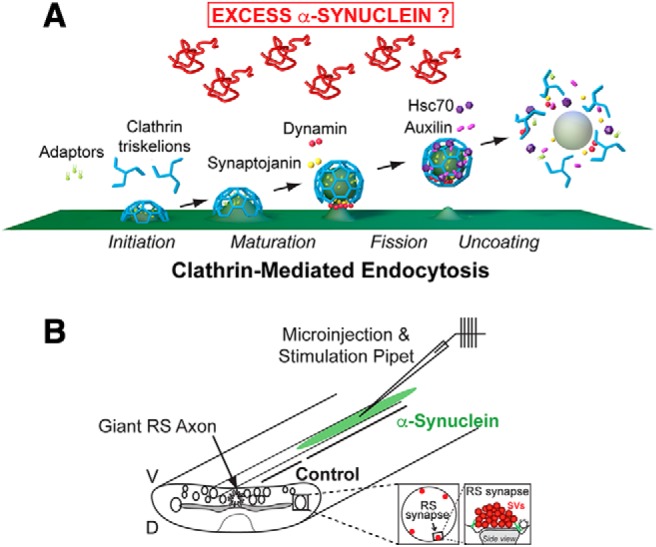
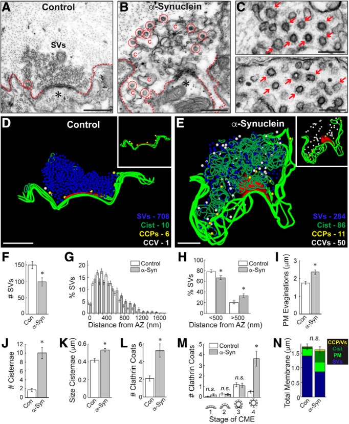
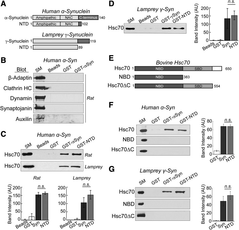
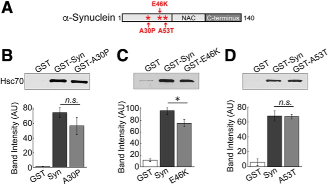
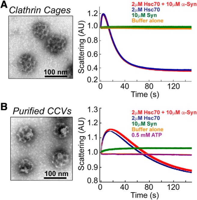
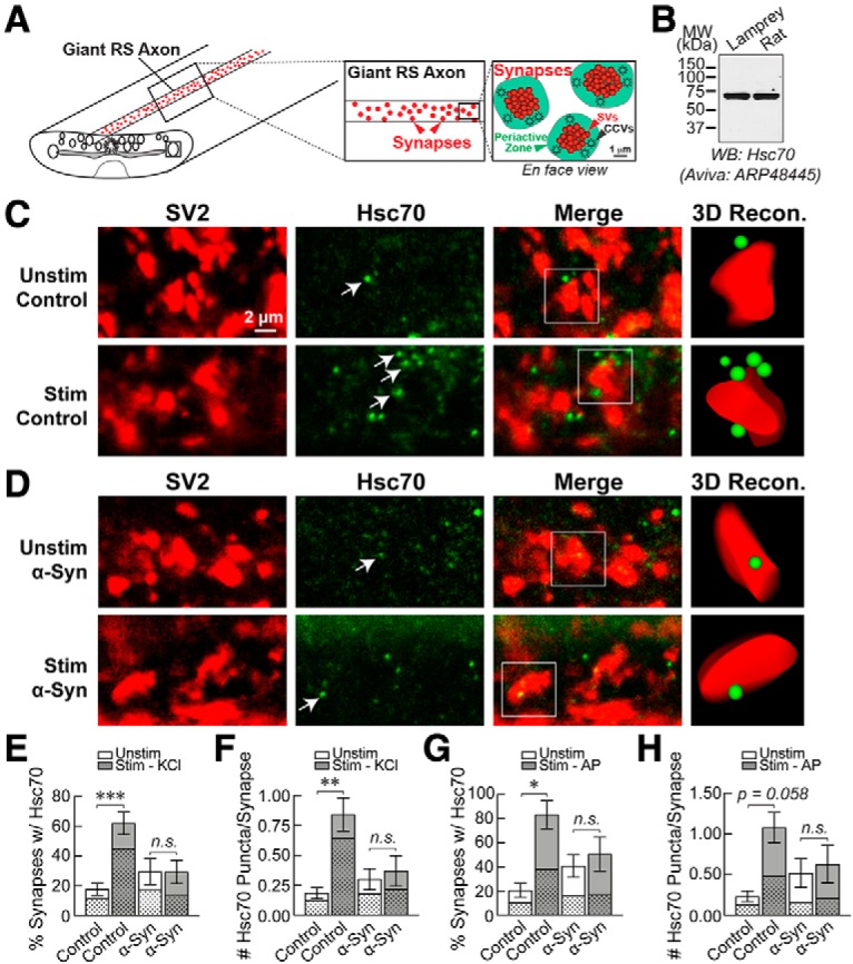
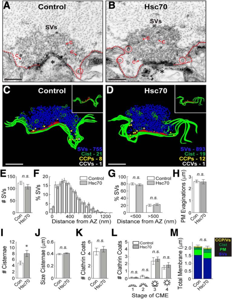
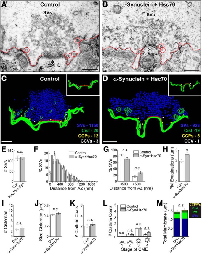
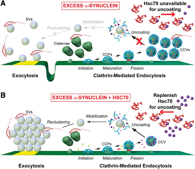
α-Synuclein overexpression and aggregation are linked to Parkinson's disease (PD), dementia with Lewy bodies (DLB), and several other neurodegenerative disorders. In addition to effects in the cell body, α-synuclein accumulation occurs at presynapses where the protein is normally localized. While it is generally agreed that excess α-synuclein impairs synaptic vesicle trafficking, the underlying mechanisms are unknown. We show here that acute introduction of excess human α-synuclein at a classic vertebrate synapse, the lamprey reticulospinal (RS) synapse, selectively impaired the uncoating of clathrin-coated vesicles (CCVs) during synaptic vesicle recycling, leading to an increase in endocytic intermediates and a severe depletion of synaptic vesicles. Furthermore, human α-synuclein and lamprey γ-synuclein both interact in vitro with Hsc70, the chaperone protein that uncoats CCVs at synapses. After introducing excess α-synuclein, Hsc70 availability was reduced at stimulated synapses, suggesting Hsc70 sequestration as a possible mechanism underlying the synaptic vesicle trafficking defects. In support of this hypothesis, increasing the levels of exogenous Hsc70 along with α-synuclein ameliorated the CCV uncoating and vesicle recycling defects. These experiments identify a reduction in Hsc70 availability at synapses, and consequently its function, as the mechanism by which α-synuclein induces synaptic vesicle recycling defects. To our knowledge, this is the first report of a viable chaperone-based strategy for reversing the synaptic vesicle trafficking defects associated with excess α-synuclein, which may be of value for improving synaptic function in PD and other synuclein-linked diseases.

Keywords: auxilin; chaperone; clathrin; clathrin-coated vesicles; endocytosis; lamprey.


Figures

Figure 1.
Figure 1.
Project goal and lamprey model. A, Diagram showing the major stages of clathrin-mediated synaptic vesicle endocytosis and several molecular players. The goal of the study is to determine how excess α-synuclein affects this process and the underlying molecular mechanisms. Graphics generated by Jack Cook and Tim Silva (Woods Hole Oceanographic Institution) using Cinema 4D. B, Diagram of the lamprey spinal cord showing microinjection strategy and location of RS synapses. All electron microscopy experiments were internally controlled such that the control synapses were taken from a region of the injected axon beyond which the reagents (e.g., α-synuclein) had diffused. D = dorsal; V = ventral.
Figure 2.
Figure 2.
Excess α-synuclein impairs CCV uncoating during synaptic vesicle recycling. A, B, Electron micrographs of untreated, control lamprey synapses stimulated at 20 Hz for 5 min. After stimulation, control synapses have large synaptic vesicle (SV) clusters, shallow plasma membrane (PM) evaginations (dotted lines), and only a few CCP/Vs (circles). In contrast, synapses treated with excess human α-synuclein had smaller SV clusters, larger PM evaginations, and greater numbers of CCP/Vs, indicative of a vesicle recycling defect. Asterisks mark postsynaptic spines. Scale bars = 500 nm. C, Insets show clusters of free CCVs at synapses after treatment with α-synuclein (arrows). Scale bars = 200 nm. D, E, 3D reconstructions comparing control and α-synuclein treated synapses. α-Synuclein caused severe endocytic defects, including a striking increase in CCVs (white spheres) and cisternae (dark green traces). Insets show the distributions of CCPs (yellow spheres) and CCVs (white spheres). While CCP/Vs at control synapses are sparse and localized near the plasma membrane at control synapses (D, inset), α-synuclein treated synapses exhibited large numbers of free CCVs throughout the synaptic area with little change in CCPs (E, inset). Active zone is shown in red. Scale bars = 500 nm. F–N, The SV recycling defect induced by α-synuclein is demonstrated by a loss of SVs (F) and synaptic vesicle dispersion (G, H), which was compensated by larger PM evaginations (I) and greater numbers/size of cisternae (J, K) as well as CCP/Vs (L). The selective increase in CCVs (stage 4) indicates a clathrin uncoating defect (M). Total membrane analysis reveals a redistribution of synaptic membranes reflecting the endocytic defect. Bars represent mean ± SEM (per section, per synapse) from n = 30–33 synapses, n = 2 axons, n = 2 animals/condition. Asterisks denote significance (p < 0.05) by Student’s t test (F, I–L, N) or ANOVA (H, M). n.s. = not significant by ANOVA.
Figure 3.
Figure 3.
α-Synuclein interacts directly with Hsc70, the chaperone protein that uncoats CCVs during synaptic vesicle recycling. A, Domain diagrams of full-length human α-synuclein, lamprey γ-synuclein and their respective NTDs. B, GST pull downs from rat brain lysates revealed no detectable interactions between α-synuclein and several major components of CME including AP2, clathrin, dynamin, synaptojanin, and auxilin. SM = starting material. Blots shown are representative of n = 3 experiments. C, In contrast, human α-synuclein and its NTD pulled down Hsc70 from rat brain and lamprey CNS lysates. D, Similarly, lamprey γ-synuclein pulled down Hsc70 from rat brain lysates, demonstrating conservation of the interaction. E, Domain diagrams of bovine Hsc70 and several truncations used in the experiments. F, G, In direct binding assays, both human α-synuclein and lamprey γ-synuclein, and their NTDs, pulled down recombinant bovine Hsc70. No interactions were detected with either NBD or Hsc70ΔC, indicating a role for the C terminus. In panels C, D, F, G, bars represent mean ± SEM from n = 3 independent experiments. n.s. = not significant by ANOVA (p > 0.05).
Figure 4.
Figure 4.
PD-associated α-synuclein mutants also interact directly with Hsc70. A, Diagram showing the locations of PD-linked point mutations A30P, E46K, and A53T, which occur within the alpha helical NTD of α-synuclein. B–D, In direct binding assays, GST-tagged A30P, E46K, and A53T all pulled down Hsc70 to a similar degree as wild-type α-synuclein. Only binding to E46K was slightly reduced. Bars represent mean ± SEM from n = 3–5 experiments. Asterisk indicates significance (p = 0.04); n.s. = not significant by ANOVA.
Figure 5.
Figure 5.
α-Synuclein does not affect Hsc70-mediated clathrin disassembly in vitro. A, left, Clathrin cages were assembled in vitro using recombinant clathrin heavy chain and auxilin, as described in Sousa et al. (2016). Right, An in vitro light scattering assay showed that addition of 2 μM Hsc70 exponentially reduced the light scattering intensity as clathrin cages were disassembled (blue trace). Addition of 10 μM α-synuclein did not alter the rate of Hsc70-mediated clathrin disassembly (red trace). No change in light scattering was observed in baseline control measurements after addition of buffer (yellow trace) or 10 μM α-synuclein alone (green trace). B, left, CCVs, which contain the underlying endocytic vesicle membranes, were freshly purified from bovine brains. Right, α-Synuclein alone slightly increased light scattering (green trace), which is likely due to its binding to the endocytic vesicles. Similar to the results with clathrin cages, introduction of 10 μM α-synuclein did not significantly affect the dynamics of Hsc70-mediated CCV uncoating (blue vs red trace).
Figure 6.
Figure 6.
Excess α-synuclein inhibits Hsc70 availability at lamprey synapses. A, Diagram showing the basic organization of the giant RS synapses within lamprey spinal cord. The large synaptic vesicle (SV) clusters (red) are surrounded by a distinct periactive zone where CME occurs (green). B, By Western blotting, the Hsc70 antibody used for these experiments (Aviva ARP48445) specifically recognized a single band at 70 kDa in both lamprey CNS and rat brain lysates, consistent with the expected molecular weight of Hsc70. C, Confocal images showing clusters of giant synapses immunostained for the synaptic vesicle-associated protein SV2 (red) and Hsc70 (green). Compared to unstimulated conditions, stimulation with high K+ increased Hsc70 availability at synapses, as evidenced by an increase in the number of visible Hsc70 puncta (arrows). D, In contrast, α-synuclein inhibited the stimulation-dependent increase in Hsc70 at synapses. E, F, Graphs showing the percentage of synapses (per axon) with associated Hsc70 puncta, as well as the average number of Hsc70 puncta per synapse. Bars represent mean ± SEM from n = 76–135 synapses, n = 5–13 axons, n = 5–7 animals/condition; *p < 0.05, **p < 0.01, ***p < 0.001, ****p < 0.0001; n.s. = not significant by ANOVA. G, H, Similar results were obtained using action potential stimulation (20 Hz, 5 min) and a different Hsc70 antibody (Enzo Life Sciences SPA815). Bars represent mean ± SEM from n = 31–47 synapses, n = 3–4 axons, n = 2–3 animals/condition; *p < 0.05, **p < 0.01; n.s. = not significant by ANOVA. In panels E–H, the stippled regions of the bars represent the proportion of Hsc70 puncta overlapping or touching the SV cluster, while the clear regions indicate the proportion localized within the periactive zone.
Figure 7.
Figure 7.
Increasing Hsc70 alone has no effect on CME at synapses. A, B, Hsc70 does not dramatically alter synaptic morphology, as demonstrated by the large synaptic vesicle (SV) clusters, shallow plasma membrane evaginations (dotted lines), and few CCP/Vs (circles) in both control and Hsc70-treated synapses. Asterisks mark postsynaptic spines. C = cisternae. Scale bars = 500 nm. C, D, 3D reconstructions further highlight the similarities and show that CCPs (yellow spheres) and CCVs (white spheres) are in normal numbers and clustered around the plasma membrane (green slabs; see insets). Active zone is shown in red. Scale bars = 500 nm. E–M, There is little effect of exogenous Hsc70 on clathrin-mediated synaptic vesicle endocytosis, as illustrated by normal number and distribution of SVs, PM evaginations, and CCPs/CCVs. Only the number of cisternae was greater, however, their size remains normal. Bars represent mean ± SEM (per section, per synapse) from n = 27–28 synapses, n = 2 axons, n = 2 animals/condition. Asterisk indicates significance (p < 0.05); n.s. = not significant (p > 0.05) by Student’s t test (E, H–K, M) or ANOVA (F, G, L).
Figure 8.
Figure 8.
Increasing exogenous Hsc70 largely reverses the α-synuclein-induced synaptic defects. A, B, Unlike synapses treated with α-synuclein alone (Fig. 2), those co-treated with Hsc70 and α-synuclein appear similar to control synapses, with large SV clusters and few cisternae or CCP/Vs (circles). C, D, 3D reconstructions reveal that synapses treated with Hsc70 and α-synuclein appear normal. Insets show the distributions of CCPs (yellow spheres) and CCVs (white spheres), which are sparse and clustered around the plasma membrane (green slabs). Active zone is shown in red. E–M, The CCV uncoating and vesicle recycling defects caused by α-synuclein were largely ameliorated by co-injection of Hsc70 as evidenced by normal numbers of SVs (E), cisternae (J), and CCPs/CCVs (K–L). Only the PM evaginations were larger (H). Notably, there was no longer any difference in the number of free CCVs after co-injection of Hsc70+α-synuclein, indicating a reversal of the uncoating defects (L, stage 4). Bars represent mean ± SEM (per section, per synapse) from n = 22–30 synapses, n = 2 axons, 2 n = animals/condition. Asterisk indicates significance (p < 0.05); n.s. = not significant (p > 0.05) by Student’s t test (E, H–K, M) or ANOVA (F–G, L).
Figure 9.
Figure 9.
Working model for α-synuclein-induced endocytic defects and amelioration by Hsc70. A, In the presence of excess α-synuclein, endogenous Hsc70 becomes depleted at synapses, leading to impaired CCV uncoating and an inhibition of synaptic vesicle recycling. B, Addition of exogenous Hsc70 restores CCV uncoating and leads to more normal synaptic vesicle recycling. (Graphics by Jack Cook and Tim Silva, Woods Hole Oceanographic Institution.)

References

    1. Antonny B, Burd C, De Camilli P, Chen E, Daumke O, Faelber K, Ford M, Frolov VA, Frost A, Hinshaw JE, Kirchhausen T, Kozlov MM, Lenz M, Low HH, McMahon H, Merrifield C, Pollard TD, Robinson PJ, Roux A, Schmid S (2016) Membrane fission by dynamin: what we know and what we need to know. EMBO J 35:2270–2284. 10.15252/embj.201694613 - DOI - PMC - PubMed
    1. Atias M, Tevet Y, Sun J, Stavsky A, Tal S, Kahn J, Roy S, Gitler D (2019) Synapsins regulate α-synuclein functions. Proc Natl Acad Sci USA 116:11116–11118. 10.1073/pnas.1903054116 - DOI - PMC - PubMed
    1. Auluck PK, Chan HY, Trojanowski JQ, Lee VM, Bonini NM (2002) Chaperone suppression of alpha-synuclein toxicity in a Drosophila model for Parkinson’s disease. Science 295:865–868. 10.1126/science.1067389 - DOI - PubMed
    1. Ben Romdhan S, Sakka S, Farhat N, Triki S, Dammak M, Mhiri C (2018) A novel SYNJ1 mutation in a Tunisian family with juvenile Parkinson’s disease associated with epilepsy. J Mol Neurosci 66:273–278. 10.1007/s12031-018-1167-2 - DOI - PubMed
    1. Bendor JT, Logan TP, Edwards RH (2013) The function of α-synuclein. Neuron 79:1044–1066. 10.1016/j.neuron.2013.09.004 - DOI - PMC - PubMed

Publication types

Substances