. 2020 Mar 15;80(6):1268-1278.
doi: 10.1158/0008-5472.CAN-19-2295. Epub 2020 Jan 15.

Increased Tumor Penetration of Single-Domain Antibody-Drug Conjugates Improves In Vivo Efficacy in Prostate Cancer Models

Affiliations

Increased Tumor Penetration of Single-Domain Antibody-Drug Conjugates Improves In Vivo Efficacy in Prostate Cancer Models

Ian Nessler et al. Cancer Res. .

Abstract

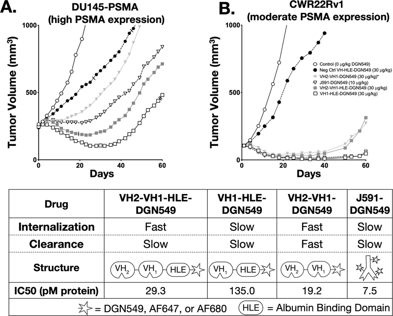
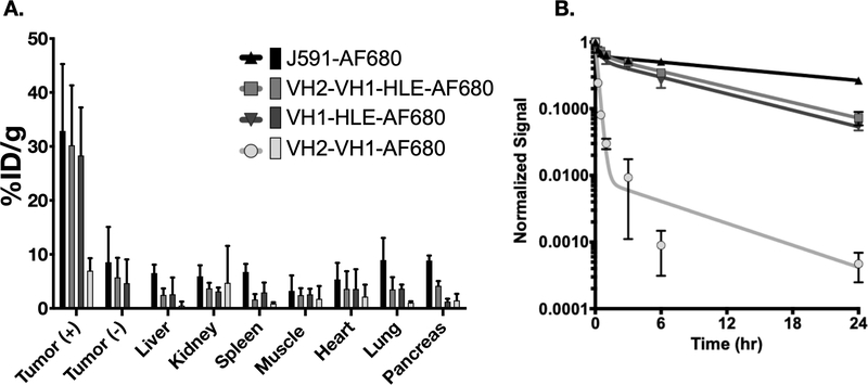
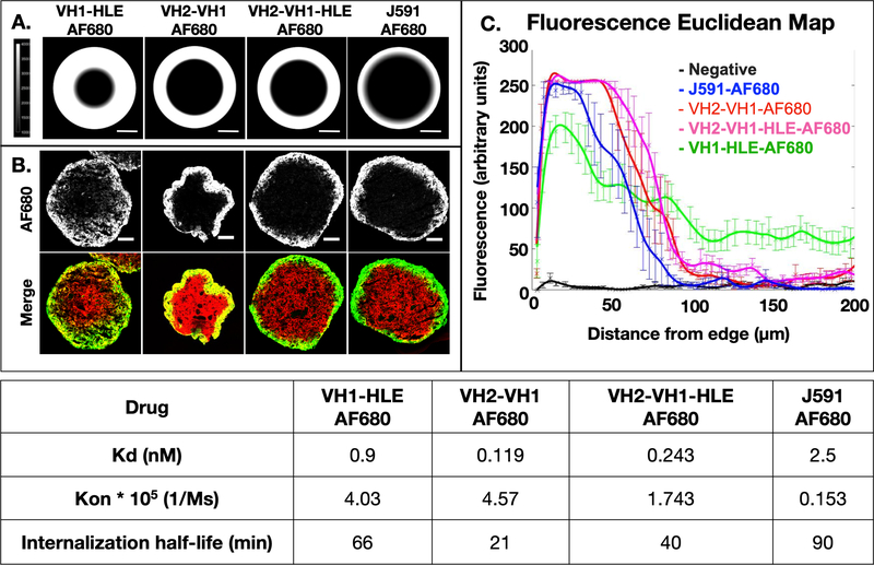
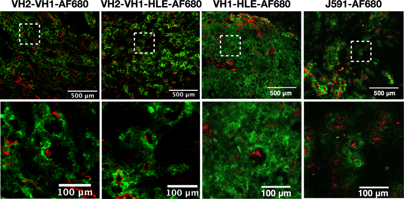
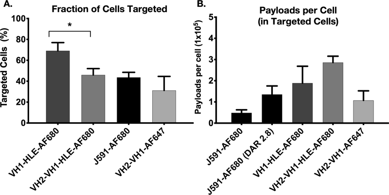
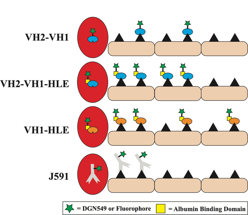
Targeted delivery of chemotherapeutics aims to increase efficacy and lower toxicity by concentrating drugs at the site-of-action, a method embodied by the seven current FDA-approved antibody-drug conjugates (ADC). However, a variety of pharmacokinetic challenges result in relatively narrow therapeutic windows for these agents, hampering the development of new drugs. Here, we use a series of prostate-specific membrane antigen-binding single-domain (Humabody) ADC constructs to demonstrate that tissue penetration of protein-drug conjugates plays a major role in therapeutic efficacy. Counterintuitively, a construct with lower in vitro potency resulted in higher in vivo efficacy than other protein-drug conjugates. Biodistribution data, tumor histology images, spheroid experiments, in vivo single-cell measurements, and computational results demonstrate that a smaller size and slower internalization rate enabled higher tissue penetration and more cell killing. The results also illustrate the benefits of linking an albumin-binding domain to the single-domain ADCs. A construct lacking an albumin-binding domain was rapidly cleared, leading to lower tumor uptake (%ID/g) and decreased in vivo efficacy. In conclusion, these results provide evidence that reaching the maximum number of cells with a lethal payload dose correlates more strongly with in vivo efficacy than total tumor uptake or in vitro potency alone for these protein-drug conjugates. Computational modeling and protein engineering can be used to custom design an optimal framework for controlling internalization, clearance, and tissue penetration to maximize cell killing. SIGNIFICANCE: A mechanistic study of protein-drug conjugates demonstrates that a lower potency compound is more effective in vivo than other agents with equal tumor uptake due to improved tissue penetration and cellular distribution.


Conflict of interest statement

Conflict of Interest

SV, TS, JL, LT, and NG were employed by Crescendo, QQ and TAK were employed by Immunogen, and AA was employed by Takeda during the study. GMT sits on the Scientific Advisory Board of Advanced Proteome Therapeutics.

Figures

Fig 1.
Fig 1.. In Vitro and In Vivo Efficacy of Antibody Drug Conjugates.
The ADCs were used in two separate tumor inhibition studies involving nude mice with either a high expression DU145-PSMA xenograft (A) or a moderate expressing CWR22Rv1 xenograft (B). The table displays relative internalization and clearance rates alongside ADC structures (all conjugations occurred at the C-terminus) and the in vitro potencies (C). The IC50 for monovalent and biparatopic antibody conjugates with and without an albumin binding ‘Half-Life Extension’ (HLE) were determined in DU145-PSMA cells. *VH2-VH1-DGN549 was dosed every other day for three total doses at a 10μg/kg DGN549 per dose for the CWR22Rv1 study and as a single bolus dose of 30 μg/kg DGN5459 in the DU145-PSMA study. Slashes mark mice removed early from the study (see Supplementary Methods).
Fig 2.
Fig 2.. Biodistribution and Plasma Clearance of Alexa Fluor 680 Antibody Constructs.
The ADC biodistribution %ID/g (A) and Plasma clearance normalized signal (B) are displayed as a mean value with error bars for each conjugate representing standard deviation. Sample sizes were as follows: VH2-VH1-HLE-AF680 (n=5), VH1-HLE-AF680 (n=5), VH2-VH1-AF680 (n=3), and J591-AF680 (n=3).
Fig 3.
Fig 3.. Computational and Experimental Tissue Penetration of Fluorescent Antibody Constructs.
Simulations based on the molecular weight, binding kinetics, affinity, and internalization rate predict penetration depths for antibody constructs (A). Tumor spheroids incubated with the Alexa Fluor 680 antibody constructs represent experimental penetration depths for each construct in 50% mouse serum while ex vivo staining of PSMA (red) displays available antigen (B). These images were analyzed with a Euclidean map to semi-quantitatively depict penetration depths (C, n = 6 – 13 spheroids per group). The table displays binding affinity, on rates and the net internalization rate for these antibody constructs (which accounts for trafficking effects such as recycling and down-regulation).
Fig 4.
Fig 4.. In Vivo Tissue Penetration of Fluorescent Antibodies.
24 hrs after tail vein administration of Alexa Fluor 680 antibody constructs dosed at the same level as the efficacy studies, DU145-PSMA xenografts were frozen in OCT and processed for histology. Blood vessels, shown in red, were ex vivo labeled with Alexa Fluor 555 anti-CD31 antibody while penetration of Alexa Fluor 680 antibody constructs is shown in green.
Fig 5.
Fig 5.. Single-Cell Payload Measurements.
A fraction of the tumor resected 24 hrs after antibody fluorophore conjugate administration was processed into a single cell suspension and used with flow cytometry to determine conjugate distribution and payload uptake. J591-AF680 antibody was given at a dose of ~0.07nmols (with a DAR of 2.8, so the equivalent payload uptake is shown) while Humabodies were dosed at ~0.7nmols. Targeted cells (A) and payloads per cell (B) are represented as median values with standard deviation error bars. Payload quantification involved the single cell analysis of three separately treated and dissociated tumors for each treatment. * = p < 0.05
Fig 6.
Fig 6.
Conceptual schematic of the distribution for the different single-domain and monoclonal antibodies.

References

    1. Polakis P. Antibody Drug Conjugates for Cancer Therapy. Pharmacological Reviews 68, 3–19 (2016). - PubMed
    1. Diamantis N. & Banerji U. Antibody-drug conjugates-an emerging class of cancer treatment. British Journal of Cancer 114, 362–367 (2016). - PMC - PubMed
    1. Vasalou C, Helmlinger G. & Gomes B. A Mechanistic Tumor Penetration Model to Guide Antibody Drug Conjugate Design. Plos One 10 (2015). - PMC - PubMed
    1. Baxter LT & Jain RK TRANSPORT OF FLUID AND MACROMOLECULES IN TUMORS .4. A MICROSCOPIC MODEL OF THE PERIVASCULAR DISTRIBUTION. Microvascular Research 41, 252–272 (1991). - PubMed
    1. Lambert JM & Chari RVJ Ado-trastuzumab Emtansine (T-DM1): An Antibody-Drug Conjugate (ADC) for HER2-Positive Breast Cancer. Journal of Medicinal Chemistry 57, 6949–6964 (2014). - PubMed

Publication types

MeSH terms

Substances