. 2020 Jan 15;10(1):367.
doi: 10.1038/s41598-019-57291-0.

Multifunctional Gas and pH Fluorescent Sensors Based on Cellulose Acetate Electrospun Fibers Decorated with Rhodamine B-Functionalised Core-Shell Ferrous Nanoparticles

Affiliations

Multifunctional Gas and pH Fluorescent Sensors Based on Cellulose Acetate Electrospun Fibers Decorated with Rhodamine B-Functionalised Core-Shell Ferrous Nanoparticles

Afroditi Petropoulou et al. Sci Rep. .

Abstract

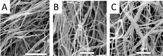
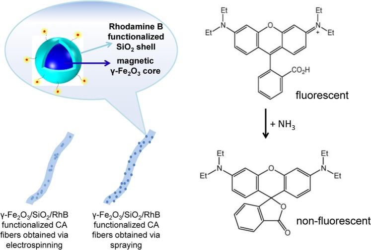
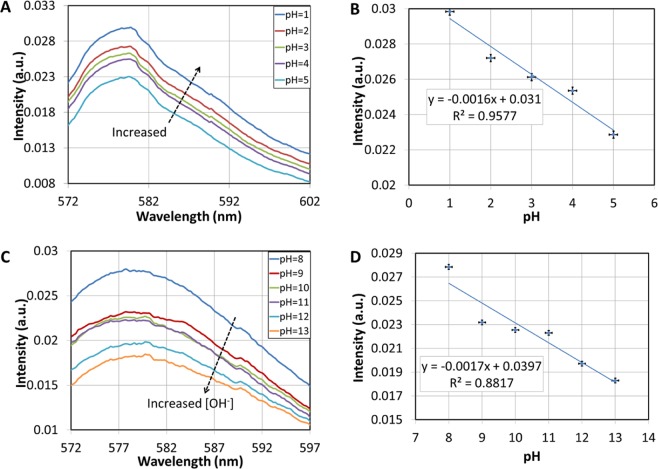
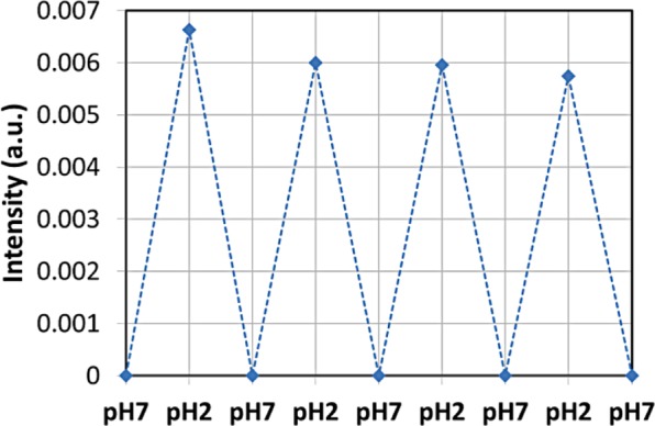
Ferrous core-shell nanoparticles consisting of a magnetic γ-Fe2O3 multi-nanoparticle core and an outer silica shell have been synthesized and covalently functionalized with Rhodamine B (RhB) fluorescent molecules (γ-Fe2O3/SiO2/RhB NPs). The resulting γ-Fe2O3/SiO2/RhB NPs were integrated with a renewable and naturally-abundant cellulose derivative (i.e. cellulose acetate, CA) that was processed in the form of electrospun fibers to yield multifunctional fluorescent fibrous nanocomposites. The encapsulation of the nanoparticles within the fibers and the covalent anchoring of the RhB fluorophore onto the nanoparticle surfaces prevented the fluorophore's leakage from the fibrous mat, enabling thus stable fluorescence-based operation of the developed materials. These materials were further evaluated as dual fluorescent sensors (i.e. ammonia gas and pH sensors), demonstrating consistent response for very high ammonia concentrations (up to 12000 ppm) and fast and linear response in both alkaline and acidic environments. The superparamagnetic nature of embedded nanoparticles provides means of electrospun fibers morphology control by magnetic field-assisted processes and additional means of electromagnetic-based manipulation making possible their use in a wide range of sensing applications.


Conflict of interest statement

The authors declare no competing interests.

Figures

Figure 1
Figure 1
Schematic presentation of the experimental setup employed in ammonia gas sensing and pH sensing experiments (A) and of the ammonia gas sensing set-up (B).
Figure 2
Figure 2
TEM images of γ-Fe2O3/SiO2/RhB NPs at low (A) and high (B) magnification, room-temperature measurement of the magnetization as a function of magnetic field strength (C), and intensity-weighted distribution of the γ-Fe2O3/SiO2/RhB NPs hydrodynamic diameters obtained from the DLS measurements in ethanol-based suspension at concentration 1 mgmL−1 (D). Emission spectra of γ-Fe2O3/SiO2/RhB NPs with high (9.33 µmol of RhB) and low (3.11 µmol of RhB) RhB loading (E). Emission spectra of the γ-Fe2O3/SiO2/RhB NPs and of the aqueous solution containing free RhB molecules (F).
Figure 3
Figure 3
Left: Schematic of the electrospinning set-up used in the fabrication of electrospun CA fibers. Right: Indicative photographs of the γ-Fe2O3/SiO2/RhB NPs-functionalized CA fibers obtained via spray deposition of the γ-Fe2O3/SiO2/RhB NPs onto the fibers’ surfaces – sprayed magnetic fibers – (up) and by mixing of the γ-Fe2O3/SiO2/RhB NPs dispersion with the polymer solution prepared in acetone, followed by electrospinning – electrospun magnetic fibers – (down).
Figure 4
Figure 4
SEM images of the as prepared CA fibers (A), the sprayed magnetic fibers (B) and the electrospun magnetic fibers (C).
Figure 5
Figure 5
TEM images of the electrospun magnetic fibers (AC) and the sprayed magnetic fibers (EG). Fibers diameter distributions were determined by analysis of the TEM images corresponding to the electrospun magnetic fibers (D) and the sprayed magnetic fibers (H).
Figure 6
Figure 6
Fluorescence microscopy image of the sprayed magnetic fibers (A) and the electrospun magnetic fibers (B).
Figure 7
Figure 7
Photoluminescence spectra of the γ-Fe2O3/SiO2/RhB NPs-functionalized CA fibers obtained via spray deposition (via spray deposition and electrospinning) (excitation wavelength: 520 nm).
Figure 8
Figure 8
A room-temperature measurement of the magnetization as a function of magnetic field strength of the γ-Fe2O3/SiO2/RhB NPs-functionalized CA fibers obtained via spray deposition (A) and electrospinning (B).
Figure 9
Figure 9
Sensing (fluorescence quenching) mechanism of RhB molecules undergoing structural changes when exposed to NH3 vapours, resulting to the generation of the non-fluorescent lactone form.
Figure 10
Figure 10
(A) Fluorescence spectra of the electrospun magnetic fibers for different concentrations of ammonia gas. (B) Response of the electrospun magnetic fibers at 577 nm for different ammonia concentrations. (C) Fluorescence spectra of the sprayed magnetic fibers for different concentrations of ammonia gas. (D) Response of the sprayed magnetic fibers at 577 nm for different ammonia concentrations.
Figure 11
Figure 11
(A) Fluorescence spectra of the electrospun magnetic fibers for acidic aqueous solutions with different pH values. (B) Fluorescence intensity at 580 nm versus pH. (C) Fluorescence spectra of the electrospun magnetic fibers for basic aqueous solutions with different pH values. (D) Fluorescence intensity at 580 nm versus pH.
Figure 12
Figure 12
Reversibility of the pH-dependent on-off-on fluorescence intensity profile of electrospun magnetic fibers. The intensity was normalised to the intensity measured at pH = 7. The response time of the pH sensor is 30 s as estimated by the fluorescence measurements instrumentation.

References

    1. Bigall NC, Parak WJ, Dorfs D. Fluorescent, magnetic and plasmonic-Hybrid multifunctional colloidal nano objects. Nano Today. 2012;7:282–296. doi: 10.1016/j.nantod.2012.06.007. - DOI
    1. Kralj S, et al. Effect of surface charge on the cellular uptake of fluorescent magnetic nanoparticles. J. Nanopart. Res. 2012;14:1151. doi: 10.1007/s11051-012-1151-7. - DOI
    1. Kim J, et al. Multifunctional Uniform Nanoparticles Composed of a Magnetite Nanocrystal Core and a Mesoporous Silica Shell for Magnetic Resonance and Fluorescence Imaging and for Drug Delivery. Angew. Chem.-Int. Edit. 2008;47:8438–8441. doi: 10.1002/anie.200802469. - DOI - PubMed
    1. Kralj S, Potrc T, Kocbek P, Marchesan S, Makovec D. Design and Fabrication of Magnetically Responsive Nanocarriers for Drug Delivery. Curr. Med. Chem. 2017;24:454–469. doi: 10.2174/0929867323666160813211736. - DOI - PubMed
    1. Le X, et al. Palladium nanoparticles immobilized on core-shell magnetic fibers as a highly efficient and recyclable heterogeneous catalyst for the reduction of 4-nitrophenol and Suzuki coupling reactions. J. Mater. Chem. A. 2014;2:19696–19706. doi: 10.1039/C4TA04919E. - DOI

Publication types