Comparative Study
. 2020 Jan 15;10(1):420.
doi: 10.1038/s41598-019-57354-2.

Comparative analysis of pathophysiological parameters between emphysematous smokers and emphysematous patients with COPD

Affiliations
Comparative Study

Comparative analysis of pathophysiological parameters between emphysematous smokers and emphysematous patients with COPD

Shuang Bai et al. Sci Rep. .

Abstract

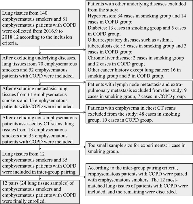
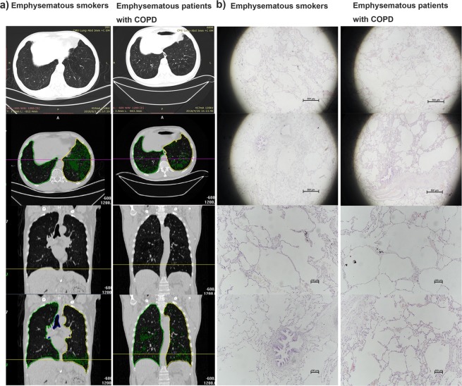
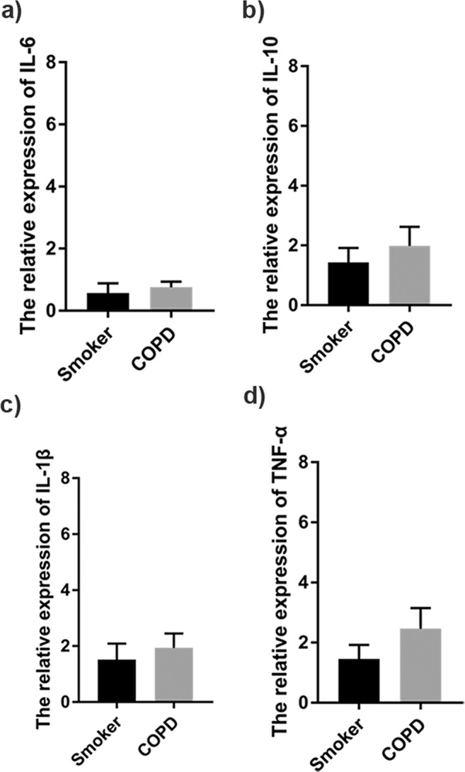
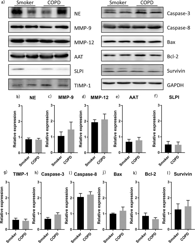
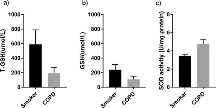
Emphysematous smokers with normal spirometry form a considerable proportion of the clinical population. However, despite presenting with respiratory symptoms and activity limitation, they cannot be diagnosed with chronic obstructive lung disease (COPD) according to current criteria. Thus, we aimed to determine whether emphysema in smokers has a different pathogenesis from that in patients with COPD. We compared 12 pairs of lung tissue samples from emphysematous patients with normal spirometry and COPD, and determined the degree of emphysema using computed tomography. With a focus on COPD-related pathogenesis, we independently assessed inflammatory response, protease-antiprotease balance, oxidative stress, and apoptosis in both groups. Both groups showed similar pathological changes at a comparable degree of emphysema; the expression of inflammatory factors was comparable, with overexpression of proteases and decreased levels of antiproteases. Moreover, there was no significant difference in the activities of glutathione and superoxide dismutase, and expression of apoptosis-related factors. In conclusion, emphysema in smokers with normal spirometry and in patients with COPD had similar pathogenesis. Forced expiratory volume in 1 second cannot be used as the sole diagnostic criterion in patients with COPD; early intervention is of great importance to such patients.


Conflict of interest statement

The authors declare no competing interests.

Figures

Figure 1
Figure 1
Process flowchart of screening and inter-group pairing.
Figure 2
Figure 2
Representative chest CT images and HE images of lung sections. (a) Representative CT analysis images of emphysema in subjects. The left and right lungs are distinguished by surrounding lines of different colours (yellow, for left lung and green for right lung). Emphysema is represented by the green area in lungs. (b) Representative haematoxylin and eosin images of lung sections. Lung sections were stained with HE and examined under a light microscope. The magnification scale is indicated in each figure panel.
Figure 3
Figure 3
Expression of inflammatory factors in lung tissues. (a) IL-6 (n = 4 in each group), (b) IL-10 (n = 7 in each group), (c) IL-1b (n = 8 for smokers and 7 for patients with COPD), and (d) TNF-α (n = 7 for each group) mRNA expression in lung tissues was measured using real-time PCR. Data represent the mean ± SEM from three independent experiments.
Figure 4
Figure 4
Comparison of protease-antiprotease balance and expression of apoptotic factors between both groups. Western blot analysis of (a,b) NE (n = 11 in each group), (a,c) MMP-9 (n = 11 in each group), (a,d) MMP-12 (n = 11 in each group), (a,e)AAT (n = 11 in each group), (a,f) SLPI (n = 11 in each group), (a,g) TIMP-1 (n = 11 in each group), (a,h) Caspase-3 (n = 11 in each group), (a,i) Caspase-8 (n = 11 in each group), (a,j) Bax (n = 11 in each group), (a,k) Bcl-2 (n = 11 in each group) and (a,l) Survivin (n = 11 in each group) expression in lung tissues of emphysematous smokers and emphysematous patients with COPD. There was no significant difference between both. Data represent the mean ± SEM from three independent experiments. Full-length blots are presented in Supplementary Fig. S1.
Figure 5
Figure 5
Measurement of GSH level and SOD activity. (a) T-GSH expression (n = 4 in each group), (b) GSH expression (n = 4 in each group), and (c) SOD activity (n = 4 in each group) in lung tissues of both groups are similar. Data represent the mean ± SEM from three independent experiments.

References

    1. Vogelmeier CF, et al. Global strategy for the diagnosis, management, and prevention of chronic obstructive lung disease 2017 Report. GOLD executive summary. Am. J. Respir. Crit. Care Med. 2017;195:557–582. doi: 10.1164/rccm.201701-0218PP. - DOI - PubMed
    1. Mohamed Hoesein FA, et al. CT-quantified emphysema in male heavy smokers: association with lung function decline. Thorax. 2011;66:782–787. doi: 10.1136/thx.2010.145995. - DOI - PubMed
    1. Qaseem A, et al. Diagnosis and management of stable chronic obstructive pulmonary disease: a clinical practice guideline update from the American College of Physicians, American College of Chest Physicians, American Thoracic Society, and European Respiratory Society. Ann. Intern. Med. 2011;155:179–191. doi: 10.7326/0003-4819-155-3-201108020-00008. - DOI - PubMed
    1. Jetmalani K, et al. Peripheral airway dysfunction and relationship with symptoms in smokers with preserved spirometry. Respirology. 2018;23:512–518. doi: 10.1111/resp.13215. - DOI - PubMed
    1. Regan EA, et al. Clinical and radiologic disease in smokers with normal spirometry. JAMA Intern. Med. 2015;175:1539–1549. doi: 10.1001/jamainternmed.2015.2735. - DOI - PMC - PubMed

Publication types