. 2019;7(2):167-182.
doi: 10.1080/21680396.2019.1666752. Epub 2019 Nov 4.

Lipid-based Liquid Crystalline Films and Solutions for the Delivery of Cargo to Cells

Affiliations

Lipid-based Liquid Crystalline Films and Solutions for the Delivery of Cargo to Cells

Marilyn Porras-Gomez et al. Liq Cryst Rev. 2019.

Abstract

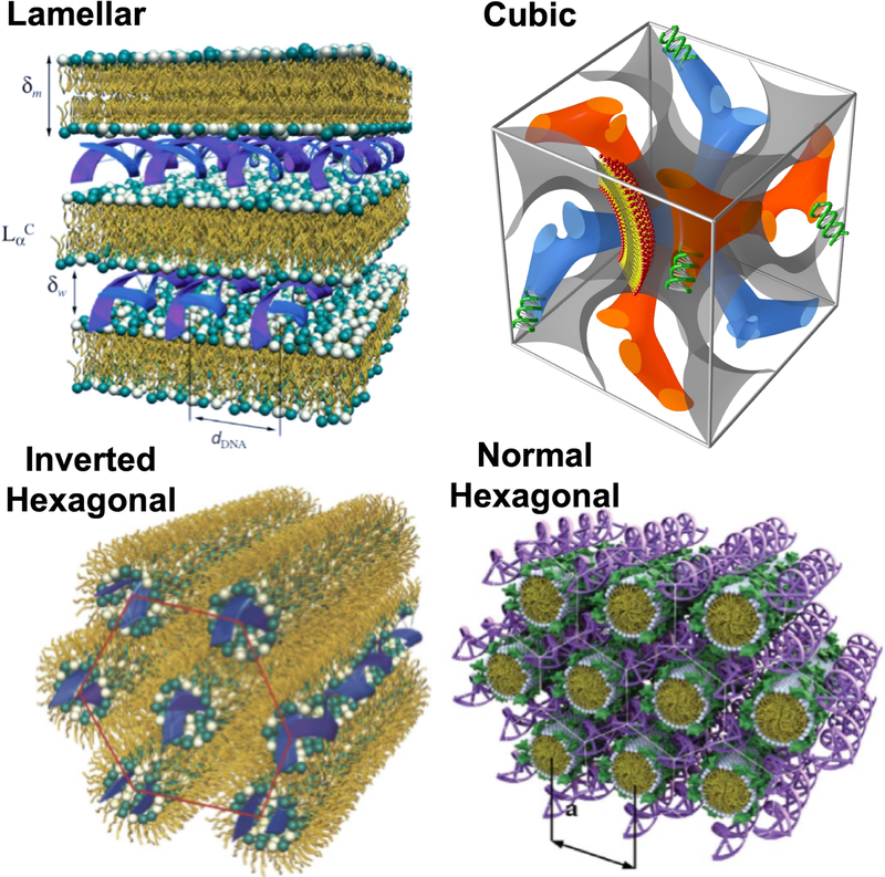
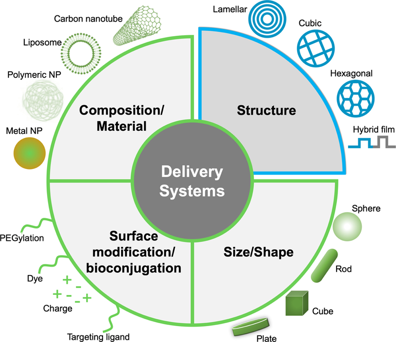
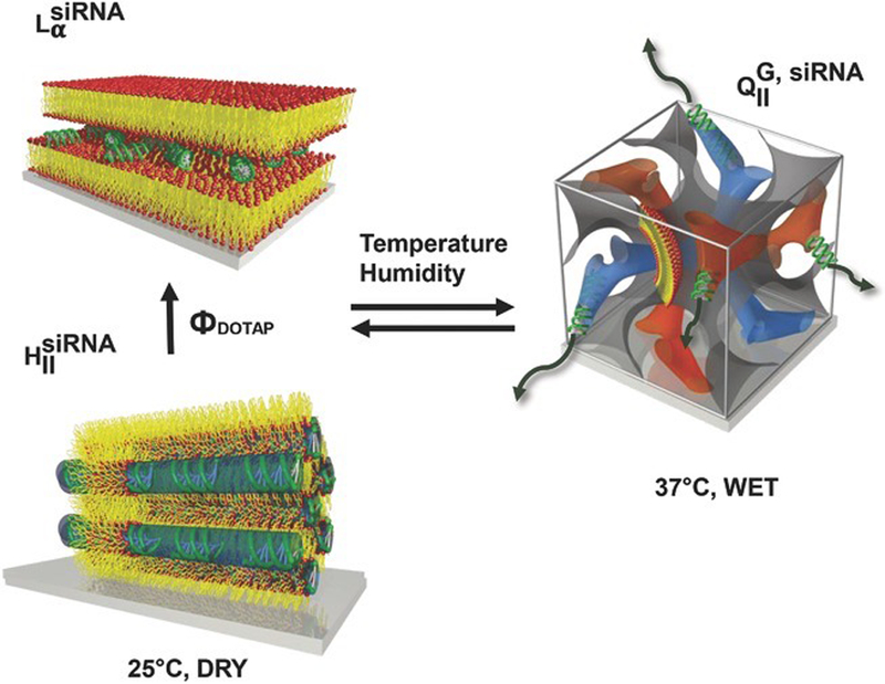
A major challenge in the delivery of cargo (genes and/or drugs) to cells using nanostructured vehicles is the ability to safely penetrate plasma membranes by escaping the endosome before degradation, later releasing the payload into the cytoplasm or organelle of interest. Lipids are a class of bio-compatible molecules that self-assemble into a variety of liquid crystalline constructs. Most of these materials can be used to encapsulate drugs, proteins, and nucleic acids to deliver them safely into various cell types. Lipid phases offer a plethora of structures capable of forming complexes with biomolecules, most notably nucleic acids. The physichochemical characteristics of the lipid molecular building blocks, one might say the lipid primary structure, dictates how they collectively interact to assemble into various secondary structures. These include bilayers, lamellar stacks of bilayers, two-dimensional (2D) hexagonal arrays of lipid tubes, and even 3D cubic constructs. The liquid crystalline materials can be present in the form of aqueous suspensions, bulk materials or confined to a film configuration depending on the intended application (e.g. bolus vs surface-based delivery). This work compiles recent findings of different lipid-based liquid crystalline constructs both in films and particles for gene and drug delivery applications. We explore how lipid primary and secondary structures endow liquid crystalline materials with the ability to carry biomolecular cargo and interact with cells.

Keywords: drug delivery; gene delivery; lipid films; lipid particles; lipid-based liquid crystals; small molecules.


Figures

Figure 1.
Figure 1.
Schematic representation of the lamellar, cubic, and hexagonal complexes for delivery of nucleic acids. [9]. Reprinted by permission of the publisher (Taylor & Francis Ltd)
Figure 2.
Figure 2.
Schematic representation of engineered physicochemical properties of drug delivery systems. Typically, size, shape and surface of nanoparticles (NPs) have concentrated the attention for the design of novel nanomedicines. The internal structure of particles and/or films is now known to play a pivotal role in the process internalisation of small molecules by cells.
Figure 3.
Figure 3.
Schematic representation of different lipid layers. A phospholipid bilayer unit represents the building block of the lipid membrane.
Figure 4.
Figure 4.
A) Reciprocal space patterns of different space groups simulated by NANOCELL. B) 2D GISWAXD patterns for selfassembled lipid films of a bicontinuous cubic gyroid (QG), an inverted hexagonal (HII), and a lamellar (Lα) structure from left to right. Films have highly ordered structures as indicated by spot patterns instead of rings. C) NANOCELL simulation overlaid onto the GISWAXD data. The fact that simulated patterns perfectly match with actual data proves that lipid self-assembled films have well-organised structures. D) Polarisation light microscopy images of lipid films with distinct textures. Optically isotropic cubic phases appear as a black background under the polariser while an inverted hexagonal phase shows focal conical texture and a lamellar phase (Lα) represents oily streaks with uniaxial texture. A nanostructure of each film is illustrated at the top of the figure. Reprinted by permission of the publisher (John Wiley and Sons) [13].
Figure 5.
Figure 5.
Schematic of different nanostructures as oriented onto solid substrates as lipid composition and environmental conditions change. This highlights the potential of using lipid film transformations for actuated surface-based cellular delivery. The diffusion of biomolecules is enhanced from 1D in lamellar phases to 3D in bicontinuous cubic phases by substrate hydration at 37 °C. The phase transitions are reversible demonstrating the equilibrium nature of these systems. Reprinted by permission of the publisher (John Wiley and Sons) [13].
Figure 6.
Figure 6.
A) Schematic illustration of hybrid films for paclitaxel incorporation and delivery: lipid (red) and polymer (blue) domains. B) Radial average intensity versus q profiles of hybrid, lipid and polymer films. C) AFM phase contrast image overlaid onto 3D height (topography) image of hybrid films. Note the colour contrast between different regions, indicative of the presence of different domains. D) Cumulative release profiles of paclitaxel from the hybrid films. Reprinted (adapted) with permission from [10]. Copyright 2017 American Chemical Society.
Figure 7.
Figure 7.
Structure of three different lipid particles and their respective Cryo-EM images. A) Schematic of a liposome and the correspondent Cryo-EM image indicating a size of ca 100 nm in diameter. B) Hexosome representattion and the correspondent Cryo-EM image of a ca 200 nm particle. The Cryo-EM data is reprinted with permission from [86]. Copyright 2005 American Chemical Society. C) Cubosome schematics having a primitive bicontinuous cubic structure enclosed by a single lipid leaflet, where yellow represents the midplane of the lipid bilayer and blue the water channels. A single unit cell is also represented to the right of the cubosome schematic. The Cryo-EM shows a ca 100 nm cubosome. Reprinted with permission from [19]. Copyright 2018 American Chemical Society.
Figure 8.
Figure 8.
siRNA loading. A) and C) Cryo-EM images of cubosomes having A) siRNA and C) siRNA–gold nanoparticles conjugates. siRNA–gold nanoparticles conjugates provide much higher contrast to locate siRNA molecules within the cubosomes. B) and D) 2D electron density maps of selected boxes (70 × 70 nm) shown in A) and C) (red for low and blue for high electron density). B) The well-ordered lipid membrane (green colour) is forming a square lattice aligned in the [100] direction. The red colour in the map represents less electron dense regions, the water channels. On the other hand, D) shows additional blue and purple regions (high electron density) corresponding to gold nanoparticles locations and hence the approximate siRNA location. This is a direct proof of siRNA incorporation into a cubosome. Schematics of cuboplexes are also shown in the middle of the figure. Reprinted with permission from [19]. Copyright 2018 American Chemical Society.
Figure 9.
Figure 9.
Gene silencing cuboplexes. A) Luciferase gene knockdown in HeLa-Luc cells of cuboplexes (green bar) and lipoplexes (red bar) at different charge ratios. For cuboplexes, the maximum knockdown efficiency was of 73.6% versus 35.8% from lipoplexes. B) Membrane integrity test of HeLa-Luc cells when incubated with cuboplexes and lipoplexes. C) Representative Cryo-EM images of a lipoplex and a cuboplex. Reprinted with permission from [19]. Copyright 2018 American Chemical Society.
Figure 10.
Figure 10.
A schematic representation of the cellular pathway and transfection mechanisms of different lipid-nucleic acid complexes. Due to its pore-like nature, the hexagonal phase, fuses directly with the cell membrane, crossing it rapidly avoiding endocytosis, but leading to cytotoxic effects. On the other hand, both, the lamellar and the cubic phases, enter the cell mostly via endocytosis. Lamellar phase toxicity depends on membrane charge density (regime I and II) while the cubic phase endocytosis does not depend on membrane charge density and does not present cytotoxic effects (regime III). Adapted with permission [18]. Copyright 2016 Elsevier.

References

    1. Verkleij AJ. Lipidic intramembranous particles. Biochimica et Biophysica Acta (BBA) - Reviews on Biomembranes. 1984;779(1):43–63. - PubMed
    1. Yorke M, Dickson D. Lamellar to tubular conformational changes in the endoplasmic reticulum of the retinal pigment epithelium of the newt, notophthalmus viridescens. Cell Tissue Res. 1985;241(3):629–637. - PubMed
    1. Almsherqi ZA, Landh T, Kohlwein SD, et al. Chapter 6 cubic membranes In: International review of cell and molecular biology. Vol. 274 Elsevier; 2009. p. 275–342. - PMC - PubMed
    1. Evans DF, Wennerström H. The colloidal domain: Where physics, chemistry, biology, and technology meet. Wiley-Vch New York; 1999.
    1. Radler JO. Structure of DNA-cationic liposome complexes: Dna intercalation in multilamellar membranes in distinct interhelical packing regimes. Science. 1997; 275(5301):810–814. - PubMed

LinkOut - more resources